


Introduction
Knee surgeries are among the most frequently performed orthopedic procedures and are often associated with moderate to severe post-operative pain. Effective analgesia is essential not only for patient comfort but also for early ambulation, participation in physiotherapy, prevention of pain-related complications, and overall functional recovery. Inadequate analgesia can delay rehabilitation, intensify the surgical stress response, increase opioid consumption, and reduce patient satisfaction [1].
Multimodal analgesia is now central to perioperative pain management for knee surgery [2,3]. Ultrasound-guided regional anesthesia has gained prominence because real-time visualization of anatomical landmarks, needle trajectory, and local anesthetic spread improves technical precision and may reduce procedure-related complications. Traditional femoral nerve block provides effective analgesia but may impair quadriceps strength, thereby increasing concern about delayed ambulation and fall risk. Consequently, motor-sparing regional techniques are increasingly preferred [4].
The adductor canal block predominantly targets sensory components, including the saphenous nerve and nerve to vastus medialis, while attempting to preserve quadriceps motor function. It provides effective anteromedial knee analgesia but may be insufficient for posterior capsular pain [5,6]. The infiltration between the popliteal artery and capsule of the posterior knee (IPACK) block was developed to target sensory articular branches supplying the posterior capsule by depositing local anesthetic between the popliteal artery and posterior femoral cortex, with the goal of avoiding tibial and common peroneal motor blockade [7,8].
Genicular nerve block (GNB) targets articular sensory branches around the knee, typically the superomedial, superolateral and inferomedial genicular nerves. Because these nerves are primarily sensory, GNB may provide analgesia without clinically meaningful motor impairment. The complex innervation of the knee joint means that no single block is universally effective for all pain generators. Direct comparison of adductor canal block (ACB), IPACK block and GNB is therefore clinically relevant, particularly in settings where safe, opioid-sparing and motor-sparing analgesia is desirable [9,10].
This study was designed to compare the post-operative analgesic efficacy of ultrasound-guided ACB, IPACK block and GNB in patients undergoing knee surgeries under subarachnoid block.
Materials and Methods
Study design and setting
This prospective, randomized, single-blind comparative study was conducted in the Department of Anaesthesiology, Orthopaedics Operation Theatre, N.S.C.B. Medical College and Hospital, Jabalpur, Madhya Pradesh, over 18 months. The study compared three ultrasound-guided motor-sparing blocks – ACB, IPACK block, and GNB – for post-operative analgesia in elective knee surgeries performed under subarachnoid block.
Ethics and consent
Institutional Ethics Committee approval was obtained before commencement of the study (IEC/2023/7335–1). Written informed consent was obtained from all participants after explanation of the anesthetic procedure, study objectives, expected benefits and possible complications. All patients underwent pre-anesthetic evaluation and observed a minimum fasting period of 6 h before surgery.
Participants
Patients aged 18–60 years, of ASA physical status I or II and scheduled for elective knee surgery were eligible. Exclusion criteria were refusal to participate, age outside the specified range, ASA physical status higher than II, known hypersensitivity to study drugs, infection at the block or spinal anesthesia site, diabetes mellitus, hypertension, psychiatric illness, bleeding disorder, hepatic dysfunction, renal dysfunction, pregnancy and lactation.
Sample size and randomization
Sample size was calculated using the Power and Sample Size module for one-way analysis of variance (ANOVA) in R version 4.4.3 for Windows, with delta 0.3209, variance 0.103 in each group, alpha error 0.05, confidence interval 95%, power 80%, and three study groups. The calculated sample size was 33 patients per group; therefore, 99 patients were included. Patients were allocated on the day of surgery into three equal groups using the sealed-envelope technique.
Interventions
Group A received ultrasound-guided ACB with 20 mL of 0.375% ropivacaine. Group I received ultrasound-guided IPACK block with 20 mL of 0.375% ropivacaine. Group G received ultrasound-guided GNB with a total of 20 mL of 0.375% ropivacaine distributed across the superomedial, superolateral and inferomedial genicular nerve targets.
Block techniques
On arrival in the operating theatre, the operative limb was verified and standard monitoring was applied, including pulse oximetry, non-invasive blood pressure and 3-lead electrocardiography. Baseline heart rate, systolic and diastolic blood pressure, mean arterial pressure and oxygen saturation were recorded. Intravenous access was secured, and Ringer lactate infusion was started at 10 mL/kg/h. All blocks were performed under aseptic precautions with the patient in the supine position unless posterior access was required.
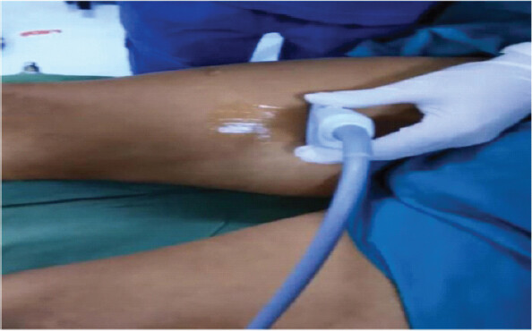
For ACB, a high-frequency linear ultrasound transducer was placed over the anterior thigh at the midpoint between the inguinal crease and medial femoral condyle (Fig. 1). The femoral artery, sartorius muscle and adductor canal were identified. Under continuous ultrasound guidance and after negative aspiration, 20 mL of 0.375% ropivacaine was deposited in the adductor canal [11,12].
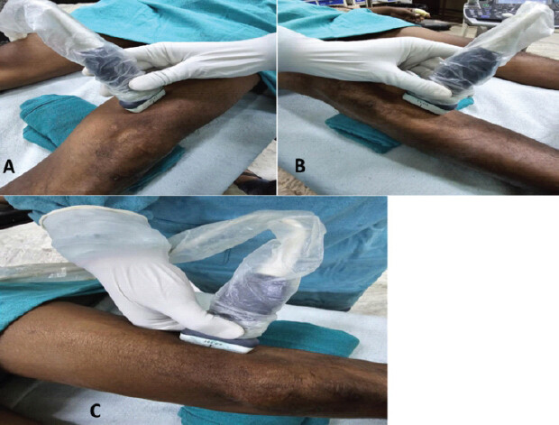
For GNB, a high-frequency linear transducer was used to identify the superomedial, superolateral, and inferomedial genicular nerve regions using vascular and periosteal landmarks (Fig. 2). The superomedial and superolateral genicular nerves were targeted near the corresponding femoral epicondylar regions, and the inferomedial genicular nerve was targeted near the anteromedial tibial region. After negative aspiration, 7 mL, 7 mL and 6 mL of 0.375% ropivacaine were deposited at the superomedial, superolateral and inferomedial targets, respectively, for a total volume of 20 mL [13,14,15].
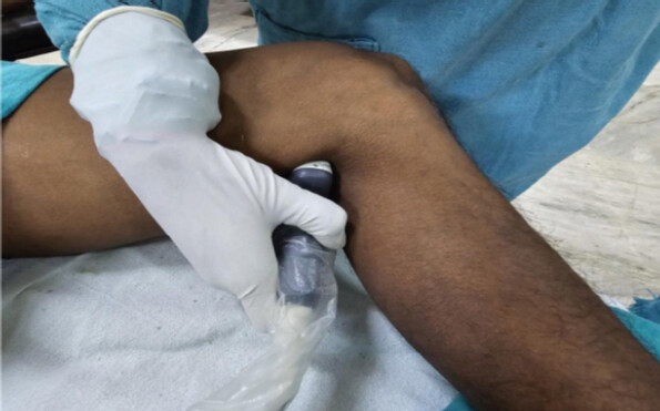
For the IPACK block, the posterior distal femoral surface and popliteal artery were identified using a low-frequency curvilinear transducer to optimize depth penetration (Fig. 3). An echogenic needle was advanced in-plane into the tissue plane between the popliteal artery and posterior femoral cortex. After negative aspiration, 20 mL of 0.375% ropivacaine was injected incrementally to achieve satisfactory spread in the target plane [16,17].
Spinal anesthesia and post-operative assessment
Thirty minutes after block placement, all patients received a subarachnoid block with 3 mL of 0.5% hyperbaric bupivacaine through a 23 G Quincke spinal needle, after which surgery commenced. After regression of spinal anesthesia, the operative limb was assessed for analgesic coverage and motor power during mobilization. Post-operative pain was assessed using a 10-cm Visual Analog Scale (VAS) at pre-defined intervals over 24 h. Intravenous paracetamol 1 g was administered as first-line rescue analgesia when VAS exceeded 4. Intravenous tramadol 50 mg was used when analgesia remained inadequate despite paracetamol [18,19]. Block-related complications, including block failure, vascular puncture, local anesthetic systemic toxicity and hypersensitivity reactions, were documented [20,21].
Outcomes
The primary outcome was post-operative VAS score over the first 24 h. Secondary outcomes were time to first and second rescue intravenous paracetamol dose, tramadol requirement and block-related complications.
Statistical analysis
Data were entered, validated, and analyzed using the IBM Statistical Package for the Social Sciences version 27.0 for Windows. Quantitative variables are presented as mean ± standard deviation and categorical variables as frequency and percentage. Baseline demographic variables were compared descriptively. Continuous outcomes were compared using one-way ANOVA or appropriate pairwise tests according to distribution, while categorical variables were analyzed using the Chi-square test or Fisher exact test when required. VAS scores were analyzed at each time point and interpreted with attention to the repeated-measure nature of the data. A P < 0.05 was considered statistically significant.
Results
A total of 99 patients were enrolled, with 33 patients in each group. Overall, 57 patients (57.58%) were male, and 42 patients (42.42%) were female. Sex distribution was comparable across groups (Table 1). The maximum number of patients belonged to the 51–60 years age group (33.33%), followed by 31–40 years (30.30%), 21–30 years (21.21%), and 41–50 years (15.15%).
Age, sex and surgery distribution of patients
| Block | Male patients | Female patients | 21–30 years age | 31–40 years age | 41–50 years age | 51–60 years age | ACL tear repair | Close fracture patella | Osteoarthritis knee |
|---|---|---|---|---|---|---|---|---|---|
| Adductor canal block | 20 | 13 | 7 | 8 | 6 | 12 | 7 | 12 | 14 |
| IPACK block | 19 | 14 | 7 | 11 | 5 | 10 | 11 | 9 | 13 |
| Genicular nerve block | 18 | 15 | 7 | 11 | 4 | 11 | 10 | 11 | 12 |
IPACK: Infiltration between the popliteal artery and capsule of the posterior knee, ACL: Anterior cruciate ligament
Baseline VAS scores before block placement were high and comparable among groups (Table 2). Group G showed lower mean VAS scores than the other two groups’ at most post-operative intervals. At 16, 20 and 24 h, Group G continued to demonstrate lower pain scores, particularly compared with Group I. The time to first and second rescue paracetamol doses was longest in Group G (Table 3). Tramadol requirement was lowest in Group G (6.06%), compared with Group A (21.21%) and Group I (30.30%). No major block-related complication was documented in the available dataset.
Post-operative VAS scores and pairwise P-values
| Time point | ACB mean±SD | IPACK mean±SD | GNB mean±SD | P: ACB versus IPACK | P: ACB versus GNB | P: IPACK versus GNB |
|---|---|---|---|---|---|---|
| Before block | 9.61±0.50 | 9.79±0.42 | 9.76±0.44 | 0.111 | 0.192 | 0.773 |
| 10 min | 6.55±0.51 | 6.67±0.48 | 6.36±0.49 | 0.321 | 0.142 | 0.013 |
| 30 min | 3.36±0.65 | 3.79±0.65 | 3.33±0.48 | 0.01 | 0.83 | 0.002 |
| 2 h | 0.39±0.50 | 0.48±0.51 | 0.15±0.36 | 0.465 | 0.027 | 0.003 |
| 4 h | 0.64±0.78 | 0.85±0.67 | 0.52±0.67 | 0.241 | 0.501 | 0.047 |
| 6 h | 2.61±0.83 | 3.00±0.71 | 2.52±0.71 | 0.042 | 0.634 | 0.007 |
| 8 h | 1.97±0.73 | 2.09±0.63 | 1.48±0.76 | 0.472 | 0.01 | 0.001 |
| 12 h | 4.18±0.39 | 4.24±0.44 | 4.12±0.33 | 0.554 | 0.5 | 0.208 |
| 16 h | 2.45±0.51 | 2.71±0.42 | 2.12±0.33 | 0.027 | 0.002 | <0.0001 |
| 20 h | 3.09±0.29 | 3.28±0.36 | 2.95±0.24 | 0.021 | 0.037 | <0.0001 |
| 24 h | 1.42±0.50 | 1.78±0.50 | 1.12±0.50 | 0.005 | 0.018 | <0.0001 |
VAS: Visual Analog Scale, ACB: Adductor canal block, IPACK: Infiltration between popliteal artery and capsule of posterior knee, GNB: Genicular nerve block, SD: Standard deviation. Pairwise P values are presented as reported in the dataset and should be interpreted in the context of multiple time-point comparisons
Time to rescue intravenous paracetamol
| Variable | ACB meant SD (h) | IPACK meant SD (h) | GNB meant SD (h) | P: ACB versus IPACK | P: ACB versus GNB | P: IPACK versus GNB |
|---|---|---|---|---|---|---|
| First dose of IV paracetamol 1 g | 5.62±0.48 | 5.42±0.49 | 5.87±0.43 | 0.104 | 0.029 | 0.0002 |
| Second dose of IV paracetamol 1 g | 11.29±0.94 | 10.74±0.87 | 11.79±0.83 | 0.017 | 0.025 | <0.0001 |
| Tramadol 50 mg requirement out of 33 patients | 7 | 10 | 2 |
*The group-wise counts indicate that 19 patients required tramadol. This corrects an internal inconsistency in the source draft, where the total was written as 10 despite group counts of 7, 10, and 2. ACB: Adductor canal block, IPACK: Infiltration between popliteal artery and capsule of posterior knee, GNB: Genicular nerve block, SD: Standard deviation
Discussion
This prospective randomized comparative study found that all three ultrasound-guided regional techniques provided useful post-operative analgesia after knee surgery. However, GNB was associated with lower VAS scores at most post-operative time points, longer time to rescue paracetamol, and the lowest tramadol requirement. These findings support the analgesic value of directly targeting articular sensory branches of the knee joint [22,23].
The demographic distribution was balanced across groups, reducing the likelihood that age or sex differences substantially influenced analgesic outcomes. Baseline VAS scores were also comparable, supporting the internal validity of post-operative comparisons. The early reduction in pain after all three blocks confirms that ultrasound-guided regional techniques can form an effective component of multimodal analgesia for knee procedures.
The relative advantage of GNB may be explained by its direct targeting of the superomedial, superolateral, and inferomedial genicular sensory branches, which contribute substantially to articular nociception. In contrast, ACB predominantly covers anteromedial sensory pathways and may not consistently address posterior knee pain. The IPACK block targets posterior capsular sensory branches, but its effect may vary according to spread of local anesthetic, anatomical variability, and whether the dominant post-operative pain generator is anterior, posterior, periosteal, or capsular.
The present results are consistent with contemporary literature suggesting that GNB can reduce post-operative pain and opioid requirement after knee arthroplasty, particularly when used as part of motor-sparing multimodal analgesia. Trials comparing IPACK-based strategies have reported variable analgesic benefit, which may reflect differences in technique, local anesthetic volume and concentration, concomitant blocks and outcome timing. The present study adds clinically relevant comparative data by evaluating ACB, IPACK block and GNB as separate ultrasound-guided interventions using the same local anesthetic volume and concentration.
The opioid-sparing finding is clinically important. Tramadol was required by only 6.06% of patients in the GNB group, compared with 21.21% in the ACB group and 30.30% in the IPACK group. Reduced opioid exposure may decrease nausea, vomiting, sedation, urinary retention, and delayed mobilization, although these outcomes were not formally analyzed in the available dataset [24,25].
No major block-related complication was documented. This supports the feasibility and apparent safety of ultrasound-guided motor-sparing blocks when performed by trained anesthesiologists using standard monitoring, incremental injection and aspiration precautions. Nevertheless, all local anesthetic techniques require vigilance for vascular puncture, local anesthetic systemic toxicity, nerve injury and block failure.
Strengths and limitations
Strengths of the study include prospective design, random allocation, equal group size, ultrasound-guided technique in all groups, standardized ropivacaine volume and concentration, and pre-defined rescue analgesic criteria.
This study has limitations. First, the relatively small sample size of 99 patients, with 33 participants in each group, may have limited the statistical power to detect smaller between-group differences and uncommon adverse events, thereby restricting the generalizability of the results. Second, the single-center setting may limit the applicability of the findings to other institutions and clinical populations. The single-blind design may have introduced potential observer or assessment bias. Furthermore, the inclusion of different knee surgical procedures, including anterior cruciate ligament (ACL) repair, patellar fracture fixation, and surgery for knee osteoarthritis, may have introduced clinical heterogeneity, as post-operative pain and analgesic requirements can vary according to the procedure performed. The follow-up period was limited to the first 24 post-operative h, precluding assessment of longer-term analgesic efficacy, functional recovery and rehabilitation outcomes. Objective measures of quadriceps strength, range of motion, ambulation and rehabilitation progress, as well as patient satisfaction, quality of recovery and health-related quality of life, were not adequately evaluated. In addition, incomplete reporting of perioperative variables, including tourniquet use, surgical duration and intraoperative analgesic supplementation, may have limited the assessment of potential confounding factors. Although post-operative pain scores were assessed at multiple time points, a comprehensive repeated-measures statistical analysis was not reported, and the available analysis may not have fully accounted for within-patient correlations or multiple comparisons. Finally, the limited sample size and short observation period constrain the ability to establish the safety profile of the blocks or detect rare block-related complications. Future multicenter, adequately powered, double-blind randomized controlled trials incorporating procedure-specific subgroup analyses, comprehensive statistical methods, longer follow-up and patient-cent red functional outcomes are warranted to validate these findings.
Conclusion
In patients undergoing orthopedic knee surgery (ACL tear repair, closed patella fracture fixation and knee osteoarthritis procedures), ultrasound-guided adductor canal, IPACK and GNBs all gave effective post-operative analgesia without motor blockade. GNB gave lower pain scores at most intervals over 24 h, delayed the first rescue analgesic request and had the lowest tramadol use. By controlling pain while sparing quadriceps strength, it appears to be a promising opioid-sparing option to support early mobilization and rehabilitation after knee surgery. Larger, adequately powered trials with procedure-specific subgroups and functional outcomes are needed before definitive recommendations can be made for individual orthopedic procedures.
Clinical Message
Post-operative pain after knee surgery can hold back early mobilization, physiotherapy and rehabilitation, which are central to orthopedic recovery. In this study, all three motor-sparing blocks gave useful analgesia, and no major complications were recorded. GNB gave the best result, with lower pain scores at most time points through 24 h, a later first rescue analgesic request and lesser tramadol use. Because it spares the quadriceps, it is a reasonable opioid-sparing option for knee surgery, where early quadriceps activation and ambulation matter.
Conflict of Interest:
Nil
Source of Support:
Nil
Consent
The authors confirm that informed consent was obtained from the patient for publication of this article
How to Cite this Article
Rath A, Himabindu M, Painkra A, Kakodia PS, Verma A, Mamtani B. Optimizing Post-operative Analgesia for Early Rehabilitation after Orthopedic Knee Surgery: A Randomized Comparison of Adductor Canal, Infiltration between the Popliteal Artery and Capsule of the Posterior Knee and Genicular Nerve Blocks. J Orthop Case Rep. 2026 November;16(11):450-456. doi: 10.13107/jocr.2026.v16.i11.8522.
References
- Batistaki C, Saranteas T, Chloros G, Savvidou O. Ultrasound-guided saphenous nerve block for saphenous neuralgia after knee surgery: Two case reports and review of literature. Indian J Orthop 2019;53:208-12. [Google Scholar] | [PubMed]
- Schug SA, Palmer GM, Scott DA, Halliwell R, Trinca J. Acute pain management: Scientific evidence, fourth edition, 2015. Med J Aust 2016;204:315-7. [Google Scholar] | [PubMed]
- Huynh MP, Yagiela JA. Current concepts in acute pain management. J Calif Dent Assoc 2003;31:419-27. [Google Scholar] | [PubMed]
- Laurent DB, Peng P, Girón Arango L, Niazi AU, Chan VW, Agur A. The nerves of the adductor canal and the innervation of the knee: An anatomic study. Reg Anesth Pain Med 2016;41:321-7. [Google Scholar] | [PubMed]
- Kertkiatkachorn W, Kampitak W, Tanavalee A, Ngarmukos S. Adductor canal block combined with iPACK (interspace between the popliteal artery and the capsule of the posterior knee) block vs periarticular injection for analgesia after total knee arthroplasty: A randomized noninferiority trial. J Arthroplasty 2021;36:122-9.e1. [Google Scholar] | [PubMed]
- Kandil MA, Elsheikh NA, Mostafa SF, Abdelghany MS. Analgesic effect of adductor canal block combined with infiltration of the interspace between popliteal artery and the capsule of the knee (IPACK) block versus genicular nerves block in knee arthroscopy, a prospective randomized controlled study. Int J Med Anesthesiol 2024;7:41-9. [Google Scholar] | [PubMed]
- Dündar A, İpek D, Yalvaç M, Kaya Ş. Comparison of the popliteal artery and the capsule of the posterior knee (IPACK) block and the genicular nerve block in primary total knee arthroplasty: A prospective randomized trial. Saudi Med J 2024;45:279-87. [Google Scholar] | [PubMed]
- Tayfun ET, Korkusuz M, Basaran B, Yarımoğlu R, Toprak H, Bilge A. Comparison of iPACK and periarticular block with adductor block alone after total knee arthroplasty: A randomized clinical trial. J Anesth 2022;36:276-86. [Google Scholar] | [PubMed]
- Wong J, Bremer N, Weyker PD, Webb CA. Ultrasound-guided genicular nerve thermal radiofrequency ablation for chronic knee pain. Case Rep Anesthesiol 2016;2016:8292450. [Google Scholar] | [PubMed]
- Rathore R, Joshi M, Saharan A, Agrawal H. Effect of ultrasound-guided genicular nerve block in knee osteoarthritis with neuropathic pain. Indian J Phys Med Rehabil 2022;32:72-8. [Google Scholar] | [PubMed]
- Hanson NA, Derby RE, Auyong DB, Salinas FV, Delucca C, Nagy R. Ultrasound-guided adductor canal block for arthroscopic medial meniscectomy: A randomized, double-blind trial. Can J Anaesth 2013;60:874-80. [Google Scholar] | [PubMed]
- Cicekci F, Yildirim A, Önal Ö, Celik JB, Kara I. Ultrasound-guided adductor canal block using levobupivacaine versus periarticular levobupivacaine infiltration after totalknee arthroplasty: A randomized clinical trial. Sao Paulo Med J 2019;137:45-53. [Google Scholar] | [PubMed]
- Tan YL, Neo EJ, Wee TC. Ultrasound-guided genicular nerve blockade with pharmacological agents for chronic knee osteoarthritis: A systematic review. Pain Physician 2022;25:E489-502. [Google Scholar] | [PubMed]
- Kesikburun S, Yaşar E, Uran A, Adigüzel E, Yilmaz B. Ultrasound-guided genicular nerve pulsed radiofrequency treatment for painful knee osteoarthritis: A preliminary report. Pain Physician 2016;19:E751-9. [Google Scholar] | [PubMed]
- Yasar E, Kesikburun S, Kılıç C, Güzelküçük Ü, Yazar F, Tan AK. Accuracy of ultrasound-guided genicular nerve block: A cadaveric study. Pain Physician 2015;18:E899-904. [Google Scholar] | [PubMed]
- Caballero-Lozada AF, Gómez JM, Ramírez JA, Posso M, Zorrilla-Vaca A, Lasso LF. IPACK block: Emerging complementary analgesic technique for total knee arthroplasty. Colomb J Anesthesiol 2020;48:78-84. [Google Scholar] | [PubMed]
- D'Souza RS, Langford BJ, Olsen DA, Johnson RL. Ultrasound-guided local anesthetic infiltration between the popliteal artery and the capsule of the posterior knee (IPACK) block for primary total knee arthroplasty: A systematic review of randomized controlled trials. Local Reg Anesth 2021;14:85-98. [Google Scholar] | [PubMed]
- Przybyła GW, Szychowski KA, Gmiński J. Paracetamol - an old drug with new mechanisms of action. Clin Exp Pharmacol Physiol 2021;48:3-19. [Google Scholar] | [PubMed]
- Graham GG, Davies MJ, Day RO, Mohamudally A, Scott KF. The modern pharmacology of paracetamol: Therapeutic actions, mechanism of action, metabolism, toxicity and recent pharmacological findings. Inflammopharmacology 2013;21:201-32. [Google Scholar] | [PubMed]
- Chen J. History of pain theories. Neurosci Bull 2011;27:343-50. [Google Scholar] | [PubMed]
- Sabatowski R, Schäfer D, Kasper SM, Brunsch H, Radbruch L. Pain treatment: A historical overview. Curr Pharm Des 2004;10:701-16. [Google Scholar] | [PubMed]
- Ismail AK, Abdul Ghafar MA, Shamsuddin NS, Roslan NA, Kaharuddin H, Nik Muhamad NA. The assessment of acute pain in pre-hospital care using verbal numerical rating and visual analogue scales. J Emerg Med 2015;49:287-93. [Google Scholar] | [PubMed]
- Bahreini M, Jalili M, Moradi-Lakeh M. A comparison of three self-report pain scales in adults with acute pain. J Emerg Med 2015;48:10-8. [Google Scholar] | [PubMed]
- Minami K, Ogata J, Uezono Y. What is the main mechanism of tramadol?. Naunyn Schmiedebergs Arch Pharmacol 2015;388:999-1007. [Google Scholar] | [PubMed]
- Grond S, Sablotzki A. Clinical pharmacology of tramadol. Clin Pharmacokinet 2004;43:879-923. [Google Scholar] | [PubMed]
© 2026 Journal of Orthopaedic Case Reports - Published by Indian Orthopaedic Research Group
About the Authors
-

Abhilash Rath
-

Mannasamudram Himabindu
-

Anjali Painkra
-

Pradyumna Singh Kakodia
-

Aditi Verma
-

Bhawna Mamtani
Related Articles in Journal of Orthopaedic Case Reports
 January 1, 2026 Evaluation of Post-operative Analgesic Efficacy of Adductor Canal Block Compared to Conventional Intravenous Analgesics in Knee Arthroscopy Surgery
 November 1, 2026 Concomitant Achilles Tendon Rupture and Ankle Fracture in a Snowboarder Managed with Early Rehabilitation: A Case Report
 November 1, 2026 Comparison of immediate post-operative pain and functional outcomes of surgeon-administered adductor canal block through subvastus versus medial parapatellar approach in total knee arthroplasty: A retrospective comparative study
 November 1, 2026 Pain Management in Orthopedic Surgery: A Procedure-Specific Orthopedic–Anesthesia Approach to Perioperative Analgesia