[box type=”bio”] Learning Point for this Article: [/box]
The posterior approach should be avoided when performing hip hemiarthroplasty in the neurologic or cognitively impaired due to the increased risk of dislocation.
Case Report | Volume 8 | Issue 1 | JOCR Jan – Feb 2018 | Page 18-22 | Robert Pearse Piggott, Emmett Karl Smithwick, Colin Gerard Murphy. DOI: 10.13107/jocr.2250-0685.980
Authors: Robert Pearse Piggott[1], Emmett Karl Smithwick[1], Colin Gerard Murphy[1]
[1]Department of Trauma and Orthopaedic Surgery, Galway University Hospitals, Saolta Healthcare Group, HSE, Galway, Ireland.
Address of Correspondence:
Dr. Robert Pearse Piggott,
Department of Trauma and Orthopaedic Surgery, Galway University Hospitals, Saolta Healthcare Group, HSE, Galway, Ireland.
E-mail: Robpiggott1@gmail.com
Abstract
Introduction: Hemiarthroplasty is the operation of choice for displaced intracapsular neck of femur fracture in elderly patients with low physical demands. Dislocation in this frail patient cohort can have devastating consequences. The patients with neurological and cognitive impairment are at additional risk secondary to imbalance of muscle tone and a reduced ability to engage with rehabilitation.
Case Report: We present three cases of early post-operative dislocation of hip hemiarthroplasties, all of whom suffered from neurological and cognitive impairment, and highlight the uncontrollable patient factors that contributed to dislocation.
Conclusion: The posterior approach was associated with all cases of dislocation in patients who also were neurologic or cognitively impaired. Posterior approach is safe to perform in the general population for hip hemiarthroplasty; however, the surgeon should consider avoiding the use of the posterior approach in this high-risk group.
Keywords: Hemiarthroplasty, Dislocation, Elderly, Neurologic impairment, Cognitive impairment.
Introduction
With the increased activity of elderly people and greater life span, the incidence of hip fracture is increasing and represents a significant workload for the practicing orthopedic surgeon. The goal of treatment is early operative intervention and a multidisciplinary rehabilitation to return patients to their pre-injury independence level and minimize the associated mortality. Arthroplasty has been shown to be superior to internal fixation in patients with displaced intracapsular neck of femur fractures. Hemiarthroplasty is the operation of choice in patients with relatively low physical demands and is one of the most commonly performed orthopedic operations with approximately over 1 million being performed annually worldwide [1]. Dislocation of a hemiarthroplasty is a devastating event in this frail patient group. It has a significant effect on patients’ morbidity and quality of life following surgery and may also affect the overall mortality rate [2]. The optimal surgical approach to the hip has long divided the orthopedic community. Studies have shown that the anterolateral approach has a lower incidence of dislocation than the posterior approach [3]. However, the advent of modern posterior structure repair has significantly reduced this risk. The Scottish intercollegiate guidelines network recommendation reflects the lack of consensus on the issue – “While the trend is in favor of the anterior approach, the use of an approach with which the surgeon is familiar is most likely to lead to lower complications” [4]. Despite these guidelines, special consideration must be given to patients with neurological disease of various etiologies. Neurological conditions are associated with paresis, spasticity, contractures, and tremors which affect the muscle balance around the hip, while cognitive impairment leads to a lack of understanding of post-operative hip precautions and decreased response to pain. These factors increase the rate of dislocation in this patient population, and thus every effort should be made to reduce the modifiable risk factors of dislocation such as the type of surgical approach. We present the following three cases in which the combination of a femoral neck fracture, which underwent uncemented hemiarthroplasty through the posterior approach and various neurological conditions lead to early post-operative dislocations, to highlight this terrible triad of circumstances.
Case report
We interrogated our prospectively maintained trauma database in a tertiary referral trauma center over a 6-month period from October 2015 to March 2016. We extracted patient demographics, fracture details, type of surgery, and approach used. We used dislocation as an endpoint to identify patients for detailed retrospective review to identify potential risk factors for dislocation. We identified 58 patients who underwent hip hemiarthroplasty for a displaced intracapsular neck of femur fracture during the time period. The average age of the patients was 81.4 years (range 59–95 years) and the gender ratio was 47 females to 11 males.
Of these, 17 patients underwent hip hemiarthroplasty through a posterior approach, in keeping with the senior surgeons preferred approach for hip arthroplasty. The rest underwent an anterolateral approach; no cases of dislocation were reported in patients with anterolateral approach. In our 6-month series, we present three cases of early post-operative dislocation of hip hemiarthroplasties in neurologic or cognitively impaired patients, all of which underwent surgery through a posterior approach. Two patients were male and the average age of the dislocated group was 79.6 years (range 76–83 years). The average time to dislocation was 16 days following surgery (range 10–19 days). All patients suffered a mechanical fall in their place of primary resident and brought by ambulance to the emergency department of our tertiary trauma center.
The patients were managed through a standardized local hip fracture protocol based on the blue book standards of hip fracture care [5]. All three patients had significant medical comorbidities which led to an increased risk of dislocation (Table 1). All three patients had a layered posterior repair, with capsule and piriformis repaired with transosseous sutures and a watertight repair of the fascia lata.
Case 1
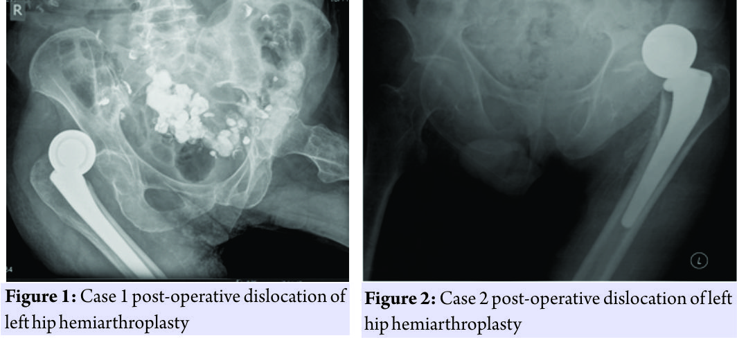
An 80-year-old male suffered a displaced left hip intracapsular neck of femur fracture. He underwent left hip hemiarthroplasty with the following components: Corail uncemented femoral stem size 15 with collar and self-centering bipolar head size 22.225 mm/50 mm (DePuy Ltd., Ringsakiddy, Cork, Ireland). His medical background was significant for previous cerebrovascular accident with residual hemiparesis and resulting vascular dementia. Post-operative course was uncomplicated; however, the patient’s poor cognition resulted in challenges while rehabilitation. The patient was non-compliant with hip precautions. On the 19th day following surgery, the patient complained of the left hip pain with a shortened internally rotated leg. No trauma was witnessed and it is theorized that the patient attempted to get out of bed and caused excessive flexion of his hip joint. X-ray confirmed dislocation of the hip hemiarthroplasty (Fig. 1). The patient underwent open reduction of the left hip hemiarthroplasty. Intraoperative findings demonstrated that there was a failure of posterior repair. Acetabulum was cleared of debris and the hip was reduced. Hip was stable through a physiological range of motion confirmed under intraoperative X-ray. The offset and soft tissue tension was adequately restored. Following reduction, the patient was transferred to another medical facility for ongoing inpatient rehabilitation. On the 52nd day after his original operation, the patient was transferred back to our facility after suffering a second dislocation while being transferred through a hoist in the rehabilitation facility. The patient progress with rehabilitation to date had been limited and multidisciplinary decision was that patient would not return to independent living. As a result of his limited rehabilitation potential and recurrent dislocations, the patient underwent a Girdlestone procedure and was discharged without further complication.
Case 2
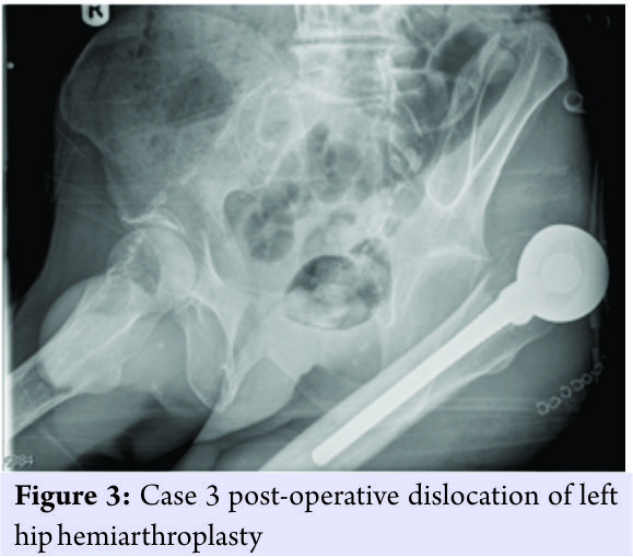
A 79-year-old male, nursing home resident, suffered a displaced left hip intracapsular neck of femur fracture. He underwent left hip hemiarthroplasty with the following components: Corail uncemented femoral stem size 12 with collar and self-centering bipolar head size 22.225 mm/53 mm (DePuy Ltd., Ringsakiddy, Cork, Ireland). His medical background was significant for dementia and Wernicke’s encephalopathy secondary to alcohol excess. Preoperatively, the patient had an ataxic gait and was non-compliant with falls prevention strategies. These difficulties continued postoperatively with non-compliance with physiotherapy with marked choreoathetoid movements and were deemed high risk for recurrent falls. On day 10 postsurgery, his leg was noted to be internally rotated, adducted, and shorted. X-ray confirmed posterior dislocation (Fig. 2) and the patient was brought to theater for a closed hip reduction, which was stable post-reduction and application of a brace. He was discharged back to his nursing home without further incident. The patient was readmitted 1 week following discharge with signs of acute limb ischemia secondary to embolic event from pre-existing atherosclerotic disease of his common iliac artery. He underwent attempted limb salvage procedure of an embolectomy and femoral popliteal bypass by our vascular colleagues. This was unsuccessful and patient proceeded to above knee amputation. He required no further treatment with regards his hip.
Case 3
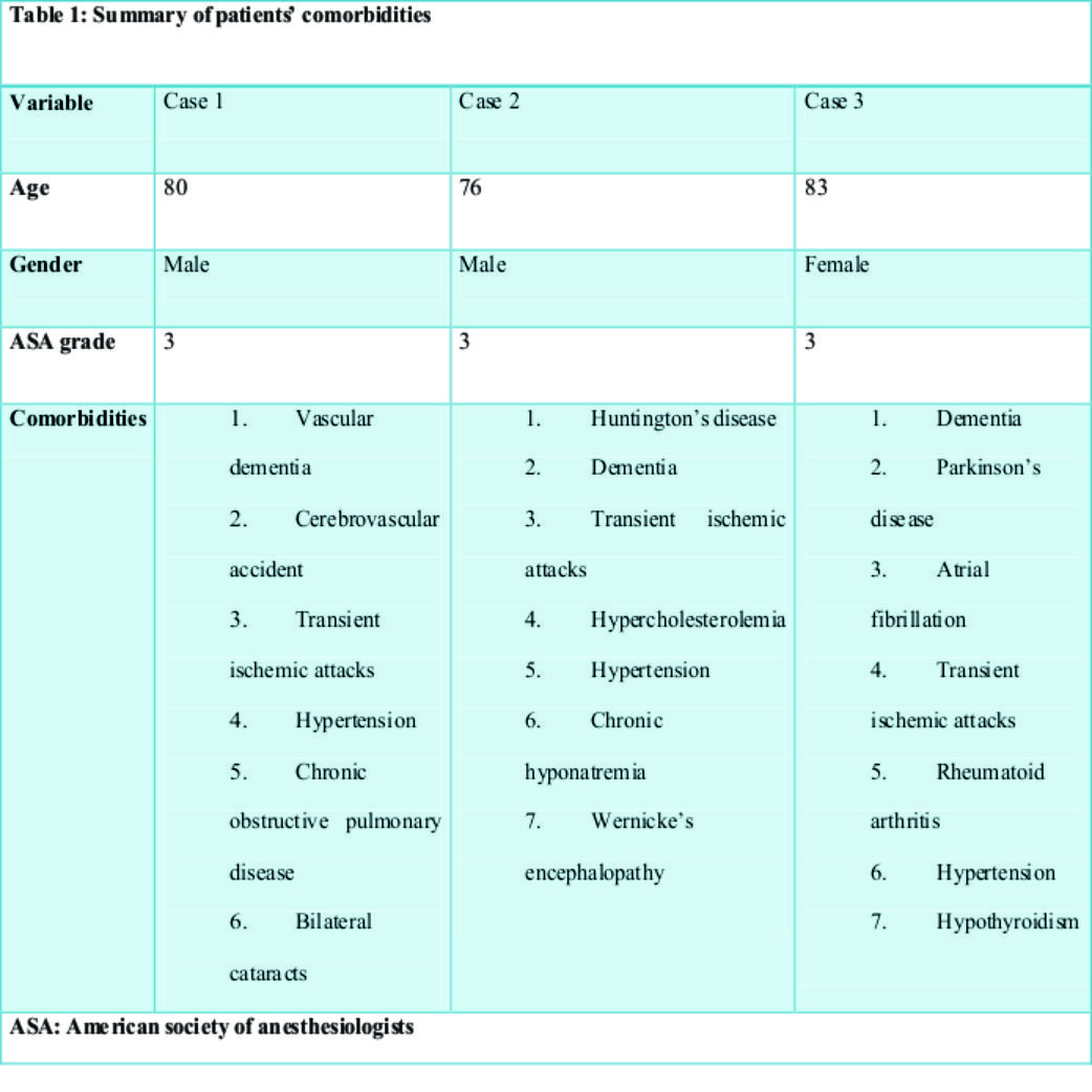
An 83-year-old female suffered a displaced right hip intracapsular neck of femur fracture. She underwent right hip hemiarthroplasty with the following components: Corail uncemented femoral stem size 14 with collar and self-centering bipolar head size 22.225 mm/46 mm (DePuy Ltd., Ringsakiddy, Cork, Ireland). Her medical background was significant for severe Parkinson’s disease and dementia. Preadmission patient was a non-ambulatory and need full hoist transfer. Post-operative course was complicated by pneumonia. Day 19 postsurgery, following hoisting from bed to wheelchair the right lower limb was noted to be shorted and rotated and patient was subjectively in pain. X-ray confirmed dislocation (Fig. 3). The patient underwent Girdlestone procedure given her preadmission status and complicated post-operative course. The patient was transferred to long-term care for palliative care after deterioration in her condition and died 35 days after initial surgery.
Discussion
Hip fracture is a devastating event in the elderly with significant morbidity and mortality, and hip hemiarthroplasty is usually reserved for the oldest patients with the lowest physical demands. Early operative intervention is essential for timely multidisciplinary rehabilitation with a view to maintaining independence and minimizing complications. Early dislocation can be a devastating complication in this frail group of patients with mortality rates of 65%, rising to 75% with recurrent dislocations [6]. A systematic review of 14,846 hip hemiarthroplasties reported an overall dislocation rate of 3.4% [7]. Currently, the most common approaches used for hip hemiarthroplasty are the posterior and the anterolateral. According to the same review, they have a dislocation rate of 5% and 2.1%, respectively [7]. A multivariate analysis of 739 consecutive hip hemiarthroplasties demonstrated that the posterior approach was the only factor associated with a significantly increased risk of dislocation [3]. The importance of the posterior repair was also highlighted with the odds ratio (OR) of dislocation was decrease from OR 6.9 (confidence interval [CI]: 2.6–19) to OR 3.9 (CI: 1.6–10) with the addition of a posterior repair [3]. Due to incomplete data, however, cognitive impairment was not included as a variable in the regression analyses, and thus its effect is not fully known. Although the anterolateral approach has been shown to have a lower dislocation risk, posterior approach leads to less adductor weakness and blood loss. With the modern reattachment of short external rotators [8] or indeed a piriformis sparing posterior approach [9], the risk of dislocation in the posterior group is reduced, and thus the surgeon should select the approach which they are most familiar with. A recent randomized trial comparing the two approaches demonstrated that there was no statistically difference between the approaches with regard to mortality, residual pain, and ability to regain walking ability [10]. Special consideration, however, should be given to the neurologic or cognitively impaired patients who undergo hip hemiarthroplasty. In our series, our rate of dislocation was disproportionally high in patients who underwent a posterior approach secondarily to uncontrollable neurologic patient factors. Sierra et al. observed that a neurological condition was present in 45% of bipolar hemiarthroplasty dislocations in their unit over a 25-year period [11]. Neurological conditions affect the hip joint by creating an imbalance of muscle tone across the hip joint which the operating surgeon must consider before embarking on arthroplasty surgery. Neurological conditions can be subdivided into three categories depending on their effect: (1) Decreased muscle tone (e.g., poliomyelitis, down syndrome, and spina bifida), (2) increased muscle tone (e.g., cerebral palsy, Parkinson’s disease, and stroke), and (3) not associated with a change of muscle tone (e.g., dementia, confusion, and psychoses) [12]. Altered muscle tone across the hip can lead to imbalance of the dynamic hip stabilizers resulting in abnormal forces which can cause dislocation. These can also result in contractures or abnormal muscle movements. Case 2 in our series suffered from Huntington’s disease and Case 3 suffered from severe Parkinson’s disease and was wheelchair bound before suffering a neck of femur fracture. Both patients were hypertonic and this muscle imbalance resulted in the early dislocation in these patients, which led to a significant morbidity in Case 2 and contributed to the mortality of an already frail patient with regard to Case 3. Cognitive impairment can also increase the risk of dislocation but by a different mechanism. Muscle tone is not affected and there is no resting imbalance across the hip joint. Instead, dislocation is because of patients’ lack of understanding, decreased response to pain, difficulties in communicating with doctors and an overall decline in walking ability [8]. In addition, it is sometimes difficult to maintain posture, both while sitting and standing, in cognitively impaired patients, which places the leg in an at-risk position for dislocation [13]. All three of our cases suffered from significant cognitive impairment, which played a role in their dislocations. Li et al. found that dislocation was associated with cognitive decline including dementia and lower MMSE score [13]. Dislocation following hip hemiarthroplasty is multifactorial, and though the association between surgical approach and neurological comorbidities is well documented in the literature, other factors need to be considered. Restoration of femoral offset is a crucial element of a successful total hip arthroplasty to reduced post-operative dislocation and must also be restored in the hip hemiarthroplasty patient. An improved functional outcome has been demonstrated in these patients [14], but little work has been done to date on its impact on dislocation. Regardless intraoperative consideration must be given to restoring offset and adjusting femoral stem anteversion to reduce the risk of dislocation. Digital templating is a useful adjunct to the surgeon in pre-operative planning to restore leg length and femoral offset in hip hemiarthroplasty patients; however, it has been shown to less accurate than its use in elective surgery [15].
Conclusion
Dislocation is a devastating complication of hip hemiarthroplasty and every effort should be made to reduce its incidence. When the dislocation risk is increased secondary to irreversible patient factors such as neurological conditions and cognitive impairment, careful consideration should be given to the choice of surgical approach. Both the anterolateral and posterior approaches are acceptable in the general population; however, surgeons should strongly consider using the anterolateral approach to reduce the risk of dislocation in patients with these high-risk comorbidities.
Clinical Message
The posterior approach should be avoided when performing hip hemiarthroplasty in the neurologic or cognitively impaired due to the increased risk of dislocation.
References
1. Gullberg B, Johnell O, Kanis JA. World-wide projections for hip fracture. Osteoporos Int 1997;7:407-13.
2. Petersen MB, Jørgensen HL, Hansen K, Duus BR. Factors affecting postoperative mortality of patients with displaced femoral neck fracture. Injury 2006;37:705-11.
3. Enocson A, Tidermark J, Tornkvist H, Lapidus LJ. Dislocation of hemiarthroplasty after femoral neck fracture: Better outcome after the anterolateral approach in a prospective cohort study on 739 consecutive hips. Acta Orthop 2008;79:211-7.
4. Scottish Intercollegiate Guidelines Network (SIGN). Management of Hip Fracture in Older People. Edinburgh: SIGN; 2009.
5. British Orthopaedic Association (BOA), British Geriatrics Society (BGS). The Care of Patients with Fragility Fracture; 2007. Available between: http://www.bgs.org.uk/pdf_cms/pubs/Blue Book on fragility fracture care.pdf. Last accessed: 11/12/2017
6. Blewitt N, Mortimore S. Outcome of dislocation after hemiarthroplasty for fractured neck of the femur. Injury 1992;23:320-2.
7. Varley J, Parker MJ. Stability of hip hemiarthroplasties. Int Orthop 2004;28:274-7.
8. Kim Y, Kim JK, Joo IH, Hwang KT, Kim YH. Risk factors associated with dislocation after bipolar hemiarthroplasty in elderly patients with femoral neck fracture. Hip Pelvis 2016;28:104-11.
9. Han SK, Kim YS, Kang SH. Treatment of femoral neck fractures with bipolar hemiarthroplasty using a modified minimally invasive posterior approach in patients with neurological disorders. Orthopedics 2012;35:e635-40.
10. Parker MJ. Lateral versus posterior approach for insertion of hemiarthroplasties for hip fractures: A randomised trial of 216 patients. Injury 2015;46:1023-7.
11. Sierra RJ, Schleck CD, Cabanela ME. Dislocation of bipolar hemiarthroplasty: Rate, contributing factors, and outcome. Clin Orthop Relat Res 2006;442:230-8.
12. Hernigou P, Filippini P, Flouzat-Lachaniette CH, Batista SU, Poignard A. Constrained liner in neurologic or cognitively impaired patients undergoing primary THA. Clin Orthop Relat Res 2010;468:3255-62.
13. Li L, Ren J, Liu J, Wang H, Sang Q, Liu Z, et al. What are the risk factors for dislocation of hip bipolar hemiarthroplasty through the anterolateral approach? A Nested case-control study. Clin Orthop Relat Res 2016;474:2622-9.
14. Buecking B, Boese CK, Bergmeister VA, Frink M, Ruchholtz S, Lechler P, et al. Functional implications of femoral offset following hemiarthroplasty for displaced femoral neck fracture. Int Orthop 2016;40:1515-21.
15. Kwok IH, Pallett SJ, Massa E, Cundall-Curry D, Loeffler MD. Pre-operative digital templating in cemented hip hemiarthroplasty for neck of femur fractures. Injury 2016;47:733-6.
 |
 |
 |
| Dr. Robert Pearse Piggott | Dr. Emmett Karl Smithwick | Dr. Colin Gerard Murphy |
| How to Cite This Article: Piggott R. P, Smithwick E. K, Murphy C. G. Hip Hemiarthroplasty in Neurologic or Cognitively Impaired Patients: A Case Series of Post-operative Dislocations. Journal of Orthopaedic Case Reports 2018 Jan-Feb; 8(1): 18-22. |
[Full Text HTML] [Full Text PDF] [XML]
[rate_this_page]
Dear Reader, We are very excited about New Features in JOCR. Please do let us know what you think by Clicking on the Sliding “Feedback Form” button on the <<< left of the page or sending a mail to us at editor.jocr@gmail.com


