
1.Atraumatic fractures of osteochondroma may be caused secondary to strong muscle contraction
2.Such presentations are acutely symtomatic and surgical excision of the lesion gives prompt relief
Dr Pankaj Kumar Mishra, 530/2F New Dhyanchand Colony, Sipri Bazar Jhansi, U.P. India. Email: drpankajv@yahoo.com
Abstract
Introduction: Non-traumatic fracture of pedunculated osteochondroma is a known, but rare complication. Treatment protocols for such complication are debatable. However, surgical intervene have been advocated.
Case Report: It is a case report of a 14 years old male patient of atraumatic fracture of solitary osteochondroma. On radiographic examination, it showed fracture through base of pedunculated osteochondroma.
Conclusion: By pathology and clinico-radiological examination, it was a benign diagnosis. We recommend operative intervention for this rare complication.
Keywords: Pedunculated osteochondroma; fracture; non-traumatic.
Among all skeletal tumors, osteochondroma constitutes about 10-15 % [1] Non-traumatic fracture of osteochondroma through the stalk is a known, but rare complication.Treatment protocols for such complication are debatable. However, surgical intervene have been advocated [2]. It is going to be case reported as a perusal of rare entity.
A 14 years old male patient presented to our outpatient department with pain, redness, and swelling over distal and posterolateral aspect of left thigh with 4 days. On history, taking patient stated that he had a gradual progressive painless mild swelling over that area for 6 month. For 4 days patient increased pain, redness, along with increments in size of previous. He did not reveal episode of recent significant trauma. On physical examination, there was tender, palpable mild swelling over posterolateral aspect of distal thigh. Hip joint was normal but knee joint was painful and restricted due to pain. Distal neurovascular status was intact. Past medical history was insignificant. On radiographic examination, it showed fracture through base of pedunculated osteochondroma, which was situated in distal left thigh postreolaterally (fig-1). On meticulous questioning patient gave the history of mild intermittent pain over osteochondroma site after moderate to severe exertion. For excision posterolatearl approach used, vastus lateralis reflected anteriorly and plane developed between it and lateral intermuscular septum covering hastring muscles ( internervous plane of femoral and sciatic nerve) and extraperiosteally reached to the base of growth. With the help of chisel, extra-periosteal excision done (with cartilaginous cap) and raw surface filed,(fig-2) and confirmation was done after histopathological examination. Recuperating period was uneventful and patient resume to previous activity status after two week.
Most common presenting age of osteochondroma is childhood or in the second decade [3]. Most of the time it is symptomless, and beings diagnosed incidentally [4]. Fracture, bursa formation, malignant transition and neurovascular compromise are mentioned complications of osteochondroma which entail it for being symptomatic [5,6]. Malignant transition in solitary lesion of osteochondroma is very less (1%) in comparison to autosomal dominant hereditary multiple exostoses, in which it may be up to 5% [1,8,9,10].Usually pathognonomonic radiological features are sufficient for delineating it, and CT scan is further needed for diagnostic confirmation and pre-op management. MRI is preferable diagnostic tool to detect malignant change. It also measures the cartilaginous cap, which (≥ 1.5cm) may be the herald of malignant changes [1,2,11]. Biopsy usually not used, until other diagnostic tools fail, due to chance of spreading.
Commonest site of fracture of osteochondroma is around knee [1,2,12,13].In little number of cases, non-traumatic fracture of pedunculated osteochondroma has been reported [4,7,13,16,17]. Fracture of osteochondroma may occur due to direct or indirect injury. Growth of stalked osteochondroma leads to weakness at its stalk, so the violent muscle contraction or relative frictions of muscle may cause the fracture and pain in the absence of history of trauma [14,15].Carpintero et al. noticed fracture of osteochondroma occurred due to indirect violent muscle contraction [2]. Our patient did not give the history of significant trauma so it was the probable cause of fracture. Observation and conservative management has also been suggested for such a rare complication, but surgery gives quicker recovery and return to normalcy [2,7]. In our case report, it was also true in which patient got full recovery in two week.
In conclusion, it was an uncommon type of presentation of non-traumatic fracture of pedunculated osteochondroma. By pathology and clinico-radiological examination, it was a benign diagnosis. We also recommend operative intervention for this rare complication.
References
- 1. Murphey MD, Choi JJ, Kransdorf MJ, Flemming DJ, Gannon FH. Imaging of osteochondroma: variants and complications with radiologic-pathologic correlation. Radiographics. 2000 ; 20 :1407–1434. [Google Scholar] [PubMed]
- 2. Carpintero P, León F, Zafra M, Montero M, Berral FJ. Fractures of osteochondroma during physical exercise. Am J Sports Med. 2003;31:1003–1006. [Google Scholar] [PubMed]
- 3. Campanacci M, Bacchini P, Bertoni F. Bone and Soft Tissue Tumors: Clinical Features, Imaging, Pathology and Treatment. 2. Wien: Springer; 1999. [Google Scholar] [PubMed]
- 4. Davids JR, Glancy GL, Eilert RE. Fracture through the stalk of pedunculatedosteochondromas: a report of three cases. ClinOrthop. 1991;271:258–264. [Google Scholar] [PubMed]
- 5. Fiddian NJ, King RJ. The winged scapula. ClinOrthop. 1984;185:228–236. [Google Scholar] [PubMed]
- 6. Kwon OS, Kelly JI. Delayed presentation of osteochondroma on the ventral surface of the scapula. Int J Shoulder Surg. 2012;6:61–63. [Google Scholar] [PubMed]
- 7. OzkanKose,Abdullah Ertas, Mustafa Celiktas, and BulentKisin. Fracture of an osteochondroma treated successfully with total excision: two case reports. – Cases J. 2009; 2: 8062. [Google Scholar] [PubMed]
- 8. Kivioja A, Ervasti H, Kinnunen J. Chondrosarcoma in a family with multiple hereditary exostoses. Journal of Bone & Joint Surgery (Br). 2000 ; 82:261-6. [Google Scholar] [PubMed]
- 9. Hendel HW, Daugaard S, Kjaer A. Utility of planar bone scintigraphy to distinguish benign osteochondromas from malignant chondrosarcomas. Clinical Nuclear Medicine. 2002 ; 27:622-4. [Google Scholar] [PubMed]
- 10. Willms R, Hartwig C, Bohm P. Malignant transformation of a multiple cartilaginous exostosis a case report. International Orthopaedics. 1997; 21:133-6. [Google Scholar] [PubMed]
- 11. Fageir MM, Edwards MR, Addison AK. The surgical management of osteochondroma on the ventral surface of the scapula. J PediatrOrthop B. 2009;18:304–307. [Google Scholar] [PubMed]
- 12. Lee K, Davies A, Cassar-Pullicino V. Imaging the Complications of Osteochondroma. Clinical Radiology. 2002 ; 57:18-28. [Google Scholar] [PubMed]
- 13. Prakash U, Court-Brown C.M. Fracture through an osteochondroma. Injury. 1996 ; 27:357-8. [Google Scholar] [PubMed]
- 14. Woertler K, Lindner N, Gosheger G. Osteochondroma: MR imaging of tumor-related complications. European Radiology. 2000; 10:832-40. [Google Scholar] [PubMed]
- 15. Krieg JC, Buckwalter JA, Peterson KK, el Khoury GY, Robinson RA. Extensive growth of an osteochondroma in a skeletally mature patient. A case report. Journal of Bone & Joint Surgery (Am). 1995;77:269-73. [Google Scholar] [PubMed]
- 16. Tanigawa N, Kariya S, Kojima H, Komemushi A, Fujii H, Sawada S. Lower limb ischaemia caused by fractured osteochondroma of the femur. Br J Radiol. 2007;80(952):78–80. doi: 10.1259/bjr/44678280. [Google Scholar] [PubMed] [CrossRef]
- 17. Alonso-Torres A, Bernabéu D, López-Barea F, Martín-Hervás C, González-López JM. Growth and fracture of an osteochondroma in an adult patient. Eur Radiol. 2004;14(12):2366–2367. doi: 10.1007/s00330-004-2397-y. [Google Scholar] [PubMed] [CrossRef]
Related Articles in Journal of Orthopaedic Case Reports
 March 10, 2020 Procalcitonin –Vital Tool to Differentiate Septic Progression of Spondylodiscitis from Drug Hypersensitivity: A Case Report and Brief Review of Literature
 August 1, 2025 Hidden in Plain Palm: A Rare Case of Incidental Thenar Schwannoma
 August 1, 2025 Surgical Management of Chronic Locked Posterior Dislocation of Shoulder using the Modified McLaughlin Procedure
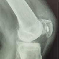 February 10, 2024 Patella Fracture Fixation Technique with Suture Anchors