
Custom-made monoflange acetabular components can provide reliable fixation and biomechanical restoration in Paprosky IIIA defects where conventional implants fail.
Dr Bharath Loganathan, Department of Joint Replacement, Dr Bharath Orthopaedics Hospital, Chennai, Tamil Nadu, India. E-mail: bharathmsortho@gmail.com
Abstract
Introduction: Severe acetabular bone loss in revision total hip arthroplasty (THA), particularly Paprosky type IIIA defects, remains a major reconstructive challenge. Conventional off-the-shelf implants often fail to achieve adequate primary stability and durable fixation in such cases. Custom-made acetabular components, designed using computed tomography (CT)-based three-dimensional (3D) planning, offer a defect-specific reconstructive option in selected cases by optimising implant fit, fixation, and restoration of hip biomechanics.
Case Report: We report the case of a 30-year-old male who presented with progressive hip pain and a limp 9 years after primary THA performed for a femoral neck fracture secondary to an aneurysmal bone cyst. Imaging revealed extensive periacetabular osteolysis with component migration, consistent with a Paprosky type IIIA acetabular defect. Infection and malignancy were excluded through laboratory investigations, biopsy, and advanced imaging. Due to the severity of bone loss and the inadequacy of standard revision options, acetabular reconstruction was performed using a CT-based, custom-made flange acetabular component with pre-planned screw trajectories and a cemented liner. Rigid primary fixation was achieved, followed by protected weight-bearing and gradual rehabilitation.
Results: Postoperative radiographs confirmed accurate implant positioning and stable fixation. At 6 months and 1-year follow-up, the patient demonstrated significant pain relief, independent ambulation without support, and stable radiographic fixation without evidence of migration or loosening. The custom implant provided effective restoration of the hip's centre of rotation with satisfactory short-term clinical and radiological outcomes.
Conclusion: Custom-made monoflange acetabular components represent a viable and effective treatment option for managing severe acetabular bone loss in revision THA. CT-based 3D planning is critical for precise implant design, safe fixation, and restoration of hip biomechanics. Although technically demanding and resource-intensive, this approach may provide reliable short-term reconstruction in carefully selected complex revision scenarios where conventional implants are unsuitable. Further studies with larger cohorts and longer follow-up are required.
Keywords: Paprosky IIIA, revision total hip arthroplasty, custom acetabular cup; monoflange implant, three-dimensional computed tomography planning.
Acetabular bone loss represents a major surgical challenge in revision total hip arthroplasty (THA), particularly in cases of re-revision surgery or following implant migration. Successful acetabular reconstruction with durable component fixation requires sufficient primary stability to facilitate subsequent osseointegration. A wide range of surgical strategies has been described, including antiprotrusio cages [1], hemispherical or asymmetrical cups with intra- or extramedullary fixation [2], and modular, highly porous acetabular revision systems with or without metal wedges, buttress augments, and cage options [3,4]. However, no single strategy has yet been established as the benchmark for the management of severe acetabular defects [5]. Although custom-made implants require substantial organisational effort and financial resources, they represent an important treatment option for extensive osseous defects that cannot be adequately addressed using standard implants. Based on computed tomography (CT) imaging, custom-made acetabular components enable defect-specific implant design, including the incorporation of metal augments to compensate for hemipelvic bone loss, customisation of flanges to optimise fixation to the remaining bone stock, and preoperative planning of hip centre of rotation (COR) restoration [6]. Most custom-made acetabular components are designed as triflange constructs. These custom-made triflange acetabular components (CTACs) are intended to “span the gap” by bridging periacetabular defects and providing fixation at the ilium, pubis, and ischium. However, these implants were originally developed for use through a posterior approach, which typically requires an extensile exposure to allow accurate positioning of all three flanges. Consequently, reported clinical outcomes of CTACs have been highly variable [7]. High-grade acetabular bone defects are primarily treated using various off-the-shelf acetabular reconstruction systems. One such system combines extra- and intramedullary iliac fixation through an iliac flange with an optional intramedullary press-fit stem and has demonstrated favourable results in several studies [8]. Nevertheless, certain defect patterns – such as extensive loss of supportive bone at the anterior or posterior acetabular rim, often accompanied by dome resorption – may render off-the-shelf implants inadequate. In these situations, a custom-made monoflange acetabular component (CMAC) may be indicated. This implant features an iliac flange fixed to the gluteal surface of the ilium and can be augmented with an intramedullary press-fit stem to enhance fixation. In the following report, we present a case treated with this implant for acetabular reconstruction following complex revision THA.
A 30-year-old male presented to our outpatient department with complaints of progressive pain in the hip and an associated limp for the past 6 months. The pain was insidious in onset, gradually progressive, aggravated by weight-bearing, and partially relieved with rest and analgesics. There was no history of recent trauma, fever, night pain, or constitutional symptoms such as weight loss or loss of appetite. The patient had a significant past medical and surgical history. Nine years earlier, he had sustained a fracture of the neck of the femur secondary to an underlying aneurysmal bone cyst and subsequently underwent a primary total hip replacement (THR). The postoperative course following the index surgery was uneventful, and the patient had remained asymptomatic with satisfactory functional outcomes for several years. There was no history of wound complications, prolonged antibiotic use, or prior revision procedures. On clinical examination, the patient walked with an antalgic gait. Local examination of the hip revealed a healed surgical scar with no signs of erythema, warmth, or sinus formation. There was localised tenderness around the hip, with a painful and restricted range of motion, particularly terminal flexion and internal rotation. Limb length discrepancy was not clinically significant. Distal neurovascular examination was normal. Routine laboratory investigations, including complete blood count, erythrocyte sedimentation rate, and C-reactive protein, were within normal limits, making periprosthetic joint infection unlikely. Plain radiographs of the pelvis and affected hip demonstrated periacetabular osteolysis with evidence of component migration (Figure 1). Further evaluation with computed tomography (CT) revealed extensive retroacetabular osteolysis and associated proximal femoral osteolysis (Figure 2). A CT-guided biopsy was performed to exclude infection or recurrent tumour. Histopathological examination showed chronic inflammatory infiltrate with osteoclast-like giant cells and areas of necrotic tissue, without evidence of malignancy or acute infection. Magnetic resonance imaging demonstrated a large periprosthetic soft-tissue mass consistent with osteolysis. A positron emission tomography scan further confirmed extensive periacetabular osteolysis. Based on the clinical, radiological, and histopathological findings, a diagnosis of aseptic failure of THR with severe acetabular bone loss was made. Given the extent of bone loss and the unsuitability of standard off-the-shelf revision implants, a decision was made to proceed with acetabular reconstruction using a CMAC. In this case of revision hip arthroplasty with a Paprosky 3A acetabular defect, three-dimensional (3D) planning was essential due to the presence of superior bone loss and loss of anterior and posterior column support (Figure 3). In such complex defects, conventional two-dimensional radiographs are inadequate to accurately assess the quantity and quality of remaining host bone, plan safe and effective screw trajectories, or reliably predict initial implant stability. Therefore, CT-based 3D reconstruction became mandatory to enable precise preoperative assessment and surgical planning. A thin-slice CT scan of the pelvis with metal artefact reduction was performed to generate a detailed 3D digital pelvic model. This allowed accurate mapping of the bone defects, identification of the remaining ilium, ischium, and pubis, assessment of the size and location of osteolytic cavities, and evaluation of column integrity. Using this 3D model, virtual implant positioning was carried out, enabling optimal placement of a custom flange cup to restore the hip COR, maximise host bone contact, and avoid injury to surrounding neurovascular structures. During preoperative planning, various Trabecular Metal Augmented Revision System options, such as jumbo cups, cup-with-augment constructs, and cages with cemented liners, were evaluated (Figure 4). However, these standard options were deemed insufficient because of the large uncontained defect, inadequate potential for achieving reliable initial stability, and the high risk of implant migration. Consequently, a custom monoflange acetabular cup was selected because the standard options were deemed insufficient to achieve reliable initial stability in the presence of extensive bone loss (Figure 4). Using the 3D pelvic reconstruction, the custom cup was designed with flanges contoured to rest precisely on the ilium, thereby maximising surface contact, improving load transfer, and enhancing construct stability (Figures 5 and 6). A major advantage of 3D planning in this context was the ability to predefine screw number, direction, and length (Figure 7). Virtual drilling paths were planned to ensure engagement of the best-quality bone while avoiding pelvic organs and major vessels, with all screw parameters finalised before surgery, thereby reducing intraoperative guesswork, risk of malposition, and operative time. The 3D planning process also highlighted several critical challenges. Bone bed preparation had to precisely replicate the preplanned 3D geometry, since over-reaming could lead to loss of press-fit while under-preparation could prevent proper seating of the implant (Figure 8). In addition, thorough soft-tissue clearance was required, particularly removal of fibrotic tissue, as inadequate clearance could obstruct seating of the custom implant (Figure 8). Accurate intraoperative matching of the custom cup to the prepared bone bed was essential, as even minor mismatches could compromise fixation (Figure 9). The implant fixation strategy involved achieving rigid primary fixation using multiple screws placed exactly as per the 3D plan to provide immediate mechanical stability, followed by secondary biological fixation through bone ingrowth facilitated by the porous implant surface. For the bearing surface, a cemented liner was used within the custom cup to allow fine control of version and inclination in situations where shell orientation was dictated by available bone, and a ceramic femoral head was chosen to reduce wear and minimise the risk of further inflammatory reaction. The postoperative protocol in this case was directly influenced by the use of a custom flange acetabular implant designed to provide rigid initial fixation with the potential for long-term biological ingrowth. In the immediate postoperative period, the construct was stable, allowing controlled rehabilitation. The patient was mobilised with partial weight-bearing for the first 6 weeks to protect the bone–implant interface and facilitate early osseointegration, after which full weight-bearing was gradually introduced. In addition, denosumab therapy was initiated to suppress osteoclast activity, aiming to reduce further bone resorption and prevent progression of osteolysis, which was particularly important given the extensive preoperative bone loss. Radiological and clinical outcomes demonstrated favourable progression over time. Immediate postoperative radiographs confirmed accurate implant positioning with stable fixation (Figure 10A). At 6 months, imaging showed maintenance of component position without evidence of migration. At 1-year follow-up, radiographs continued to demonstrate stable fixation with no signs of loosening, and the custom flange remained well seated against the host bone, suggesting sustained mechanical stability with progression toward biological osseointegration (Figure 10B and C). Clinically, the patient reported a significant reduction in hip pain and was able to perform activities of daily living without discomfort. He ambulated independently without support and exhibited a near-normal gait pattern. Hip range of motion was satisfactory, limb length was maintained, and there were no complications such as dislocation, infection, or neurovascular deficit during the follow-up period. Although formal functional outcome measures such as the Harris Hip Score or Western Ontario and McMaster Universities (WOMAC) scores were not recorded, the patient demonstrated substantial clinical improvement in pain, gait, and functional mobility.
Revision THA with severe acetabular bone loss remains a major reconstructive challenge. In contained defects with preserved column support, cementless hemispherical cups with supplemental screw fixation provide reliable outcomes [9,10]. However, in extensive uncontained defects such as Paprosky type IIIA, conventional options – including jumbo cups, augments, reinforcement rings, and cages – often fail to achieve durable primary stability [11]. Cup–cage constructs have been advocated for managing massive bone loss and pelvic discontinuity. This technique protects a porous cup with a cage until osseointegration occurs and has shown acceptable mid-term outcomes in Paprosky IIIA and IIIB defects [12,13,14]. Nevertheless, the construct depends on indirect mechanical support, and restoration of the hip COR can remain technically demanding in distorted anatomy. CTACs were developed to address catastrophic defects by providing fixation at the ilium, ischium, and pubis. Systematic reviews have demonstrated encouraging survivorship; however, outcomes are variable, and the procedure often requires extensive exposure with technically demanding implantation [7,12]. A CMAC offers a focused alternative by utilising iliac fixation, which frequently retains relatively better bone stock even in advanced defects. CT-based 3D planning enables accurate mapping of residual bone, restoration of the hip COR, and predefined screw trajectories, thereby improving safety and primary stability [6,15,16]. Preoperative virtual planning reduces intraoperative uncertainty and facilitates optimal screw purchase while minimising the risk to surrounding neurovascular structures. Compared with established reconstructive strategies such as cup–cage constructs, trabecular metal augments, and CTACs, the custom monoflange design used in the present case provided satisfactory primary fixation while avoiding the need for more extensile exposure. However, because this report represents a single-case experience without direct comparison, definitive conclusions regarding superiority or comparative effectiveness cannot be established. Further comparative studies are required to determine the optimal indications and long-term performance of this technique. In the present case, rigid primary fixation was achieved with a custom monoflange component and multiple preplanned screws, followed by satisfactory clinical and radiological outcomes at 1 year. Stable component position without migration suggests progression toward biological fixation. Early mechanical stability is critical for long-term success in complex revision scenarios. Despite these advantages, custom implants require meticulous preoperative planning, increased production time, and higher cost. Long-term survivorship data remain limited compared to established revision systems. In addition, intraoperative seating must precisely replicate the planned bone preparation to avoid compromise of fixation. This case demonstrates that in selected Paprosky type IIIA defects where off-the-shelf implants are unlikely to provide reliable fixation, a CT-based CMAC can offer stable reconstruction with restoration of hip biomechanics.
CMACs represent a viable reconstructive option for managing severe Paprosky type IIIA acetabular defects in complex revision THA. CT-based 3D planning plays a crucial role in achieving accurate implant positioning, restoration of hip biomechanics, and safe screw fixation. Although favourable short-term radiological and clinical outcomes were observed in this case, larger studies with longer follow-up are necessary to determine long-term survivorship, reproducibility, and comparative effectiveness of this technique.
Limitations:
This report has several important limitations. First, it represents a single-case experience, which inherently limits the generalisability and external validity of the findings. The indication for a CMAC was highly specific, introducing potential selection bias, and outcomes may vary depending on surgeon expertise, institutional resources, implant availability, and experience with advanced 3D planning technologies. Second, the follow-up duration of 1 year is relatively short to assess long-term implant survivorship, durability, biological fixation, and sustained osseointegration. Although radiographs demonstrated stable fixation without migration, radiological stability alone may not necessarily confirm long-term biological integration. Furthermore, complications such as aseptic loosening, screw loosening, stress shielding, implant breakage, or the need for re-revision cannot yet be adequately evaluated. Another limitation is the absence of comparative analysis with other established reconstructive strategies for severe acetabular bone loss, including cup–cage constructs, trabecular metal augments, and CTACs. Therefore, conclusions regarding the comparative effectiveness or superiority of this technique cannot be established from the present report. Standardised functional outcome measures such as the Harris Hip Score or WOMAC score were not included, limiting objective quantification of postoperative functional improvement. In addition, no quantitative biomechanical analysis or stress-distribution assessment was performed to validate the theoretical mechanical advantages of the implant design. The use of custom implants is also associated with substantial manufacturing costs, resource-intensive production, and increased preoperative planning requirements, which may restrict widespread applicability, particularly in low-resource settings. The time required for implant design and fabrication may further limit utility in urgent or semi-urgent revision scenarios. Finally, the procedure is highly dependent on accurate CT-based imaging, 3D modelling, and meticulous intraoperative execution. Errors in imaging acquisition, implant planning, or bone preparation could compromise implant seating and fixation quality. As with many reports describing novel reconstructive techniques, the possibility of reporting bias toward favourable outcomes cannot be excluded. Larger multicentric studies with longer follow-up and standardised outcome assessment are required to determine reproducibility, long-term survivorship, and broader clinical applicability of this approach.
In Paprosky type IIIA acetabular defects, standard revision implants may not achieve adequate fixation. Computed tomography-based three-dimensional planning with a custom-made monoflange acetabular component allows precise screw placement, improved primary stability, and restoration of hip biomechanics in complex revision total hip arthroplasty.
References
- 1. Makita H, Kerboull M, Inaba Y, Tezuka T, Saito T, Kerboull L. Revision total hip arthroplasty using the Kerboull acetabular reinforcement device and structural allograft for severe defects of the acetabulum. J Arthroplasty 2017;32:3502-9. [Google Scholar] [PubMed]
- 2. Hoberg M, Holzapfel BM, Steinert AF, Kratzer F, Walcher M, Rudert M. Treatment of acetabular bone defects in revision hip arthroplasty using the Revisio system. Orthopade 2017;46:126-32. [Google Scholar] [PubMed]
- 3. Wassilew GI, Janz V, Perka C, Muller M. Treatment of acetabular defects with the trabecular metal revision system. Orthopade 2017;46:148-57. [Google Scholar] [PubMed]
- 4. Frenzel S, Horas K, Rak D, Boelch SP, Rudert M, Holzapfel BM. Acetabular revision with intramedullary and extramedullary iliac fixation for pelvic discontinuity. J Arthroplasty 2020;35:3679-85.e1. [Google Scholar] [PubMed]
- 5. Burastero G, Cavagnaro L, Chiarlone F, Zanirato A, Mosconi L, Felli L, et al. Clinical study of outcomes after revision surgery using porous titanium custom-made implants for severe acetabular septic bone defects. Int Orthop 2020;44:1957-64. [Google Scholar] [PubMed]
- 6. Von Lewinski G. Custom-made acetabular implants in revision total hip arthroplasty. Orthopade 2020;49:417-23. [Google Scholar] [PubMed]
- 7. DeMartino I, Strigelli V, Cacciola G, GU A, Bostrom MP, Sculco PK. Survivorship and clinical outcomes of custom triflange acetabular components in revision total hip arthroplasty: A systematic review. J Arthroplasty 2019;34:2511-8. [Google Scholar] [PubMed]
- 8. Prodinger PM, Lazic I, Horas K, Burgkart R, Von Eisenhart-Rothe R, Weissenberger M, et al. Revision arthroplasty through the direct anterior approach using an asymmetric acetabular component. J Clin Med 2020;9:3031. [Google Scholar] [PubMed]
- 9. Della Valle CJ, Shuaipaj T, Berger RA, Rosenberg AG, Shott S, Jacobs JJ, et al. Revision of the acetabular component without cement after total hip arthroplasty. A concise follow-up, at fifteen to nineteen years, of a previous report. J Bone Joint Surg Am 2005;87:1795-800. [Google Scholar] [PubMed]
- 10. Park DK, Della Valle CJ, Quigley L, Moric M, Rosenberg AG, Galante JO. Revision of the acetabular component without cement. A concise follow-up, at twenty to twenty-four years, of a previous report. J Bone Joint Surg Am 2009;91:350-5. [Google Scholar] [PubMed]
- 11. Issack PS. Use of porous tantalum for acetabular reconstruction in revision hip arthroplasty. J Bone Joint Surg Am 2013;95:1981-7. [Google Scholar] [PubMed]
- 12. Berasi CC 4th, Berend KR, Adams JB, Ruh EL, Lombardi AV Jr. Are custom triflange acetabular components effective for the reconstruction of catastrophic bone loss? Clin Orthop Relat Res 2015;473:528-35. [Google Scholar] [PubMed]
- 13. Kosashvili Y, Backstein D, Safir O, Lakstein D, Gross AE. Acetabular revision using an anti-protrusion (ilio-ischial) cage and trabecular metal acetabular component for severe acetabular bone loss associated with pelvic discontinuity. J Bone Joint Surg Br 2009;91:870-6. [Google Scholar] [PubMed]
- 14. Makinen TJ, Kuzyk P, Safir OA, Backstein D, Gross AE. Role of cages in revision arthroplasty of the acetabulum. J Bone Joint Surg Am 2016;98:233-42. [Google Scholar] [PubMed]
- 15. Horas K, Arnholdt J, Steinert AF, Hoberg M, Rudert M, Holzapfel BM. Acetabular defect classification in times of 3D imaging and patient-specific treatment protocols. Orthopade 2017;46:168-78. [Google Scholar] [PubMed]
- 16. Telleria JJ, Gee AO. Classifications in brief: Paprosky classification of acetabular bone loss. Clin Orthop Relat Res 2013;471:3725-30. [Google Scholar] [PubMed]


Related Articles in Journal of Orthopaedic Case Reports
 July 1, 2026 Bone Morphogenetic Proteins-2–Augmented Acetabular Bone Stock Restoration During Two-stage Revision for Periprosthetic Joint Infection: A Case Report
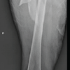 June 1, 2026 Two-Stage Reconstruction for Failed Subtrochanteric Femur Fixation with Peri-Implant Infection and Severe Proximal Femoral Bone Loss: A Case Report
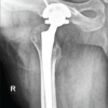 May 1, 2026 A Novel Failure Interface in Constrained Total Hip Arthroplasty: Dissociation at the Constrained Liner Insert-Bipolar Head Junction Following Periprosthetic Joint Infection
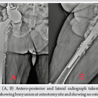 April 10, 2024 Cage Augmentation of the Retained Acetabular Shell with a Dual Mobility Cup using the Double Socket Technique