

Antibiotic cement-coated intramedullary nailing is a cost-effective single-stage treatment for infected nonunion (bone gap ≤2 cm), combining culture-guided local antibiotic delivery, meticulous debridement, and stable fixation with 6 weeks of systemic antibiotics to achieve infection control and bone union.
Dr Mohd Shahzad Ghazi, Department of Orthopaedics, Government Medical College, Chhatrapati Sambhajinagar, Maharashtra, India. E-mail: shahzadghazi321@gmail.com
Abstract
Introduction: Infected non-union of long bones represents one of the most challenging problems in orthopaedic surgery. Conventional two-stage approaches involving debridement, antibiotic spacers, and subsequent fixation are associated with high morbidity and prolonged treatment duration.
Materials And Methods: We present a prospective case series of 10 patients with infected non-union of various long bones (tibia, femur, humerus) treated with antibiotic cement-coated intramedullary nails (AC-CIMN). The nail was coated with polymethylmethacrylate mixed with culture-sensitive antibiotics in a single-stage procedure combining debridement and mechanical stabilisation.
Results: At a mean follow-up of 12 months, infection was successfully controlled in 80% of patients, and radiological union was achieved in 70%. The mean union time was 28 weeks for tibial fractures and 22 weeks for femoral fractures. Functional outcomes were satisfactory in 80% of cases. Two patients required secondary intervention for persistent infection.
Conclusion: AC-CIMN is an effective single-stage strategy for infected non-union of long bones with bone gaps ≤2 cm. It simultaneously addresses infection control and mechanical stability, enabling early mobilisation. Careful patient selection and culture-guided antibiotic loading are critical determinants of success.
Keywords: Infected non-union, antibiotic cement-coated nail, polymethylmethacrylate, intramedullary nail, long bone, osteomyelitis, limb salvage.

Infected non-union represents the confluence of two devastating orthopaedic complications – failure of fracture healing and persistent deep-seated infection. It is defined as failure of bone union beyond 6–8 months with concurrent evidence of infection at the fracture site, typically manifesting as chronic draining sinuses, mechanical instability, and profound physical disability [1,2]. The aetiology is multifactorial, encompassing patient-related factors (diabetes mellitus, immunosuppression, smoking, tobacco use, and damage to the nutrient vessel); fracture-related factors (open wound, bone loss, and severe comminution); and surgeon-related factors (inadequate immobilisation, faulty implants, and excessive periosteal stripping) [3]. Conventional management follows a two-stage protocol: The first stage involves radical debridement, removal of implants, and insertion of antibiotic-laden cement beads or spacers alongside systemic antibiotic therapy to convert an infected environment to a sterile one. The second stage, performed weeks to months later, achieves definitive skeletal stabilisation through internal or external fixation with bone grafting as required [4]. While effective, this approach carries significant morbidity, prolonged hospitalisation, patient compliance challenges, and substantial healthcare costs. External fixation methods, such as the Ilizarov ring fixator or limb reconstruction system (LRS), are frequently employed as definitive management, particularly for cases involving significant bone loss or poor soft tissue envelopes. However, these devices carry inherent complications, including pin tract infections, joint stiffness, and poor patient acceptance [5,6]. The antibiotic cement-coated intramedullary nails (AC-CIMNs) have emerged as an innovative single-stage approach that integrates mechanical stabilisation with local antimicrobial therapy. By coating a standard intramedullary nail with polymethylmethacrylate (PMMA) impregnated with high-dose antibiotics, the technique provides sustained high local antibiotic concentrations at the infection site while simultaneously restoring axial stability through load-sharing properties [7,8]. This single-stage strategy reduces total surgical interventions, enables earlier patient mobilisation, and may achieve cost-effectiveness compared to staged procedures or external fixation [9]. In this prospective case series, we present our institutional experience with AC-CIMN for infected non-union of long bones, including tibia, femur, and humerus, with particular emphasis on our surgical technique, patient selection criteria, and clinical outcomes.
Study design and ethics:
This prospective observational study was conducted in the Department of Orthopaedics, GMCH Chhatrapati Sambhajinagar, over 2 years. Institutional Ethics Committee approval was obtained. Written informed consent was obtained from all participants. The study was conducted in accordance with the Declaration of Helsinki.
Inclusion and exclusion criteria:
Inclusion criteria:
(1) Radiologically confirmed infected non-union of a long bone (tibia, femur, or humerus) with a pre-existing nail or plate; (2) bone gap ≤2 cm; (3) clinically draining sinus or positive intraoperative culture.
Exclusion criteria:
(1) Critical bone gap >2 cm; (2) known allergy to PMMA or the planned antibiotic; (3) moribund patients; (4) patients lost to follow-up before 6 months.
Classification:
Infected non-union was graded using the Weiland classification. Type I: No significant bone loss, good bone stock, healthy soft-tissue envelope. Type II: Segmental bone loss 2–6 cm, moderate soft-tissue damage, compromised bone stock. Type III: Bone loss >6 cm with poor soft-tissue envelope. Only Weiland Types I and II (bone gap ≤2 cm) were included.
Surgical technique – GMCH CSN protocol:
All surgeries were performed under spinal or general anaesthesia. The operative steps were:
- Step 1 – Implant removal: The index nail or plate was extracted. Reamings and granulation tissue were sent for aerobic and anaerobic culture and sensitivity.
- Step 2 – Canal reaming: The medullary canal was reamed 2 mm beyond the intended nail diameter using progressive flexible reamers to debride the endosteum and create an adequate cement mantle space.
- Step 3 – Medullary lavage: The canal was irrigated with 2–3 L of normal saline with betadine using an infant feeding tube or Ryle’s tube passed to the distal extent of the medullary cavity, mechanically reducing biofilm burden.
- Step 4 – Nail coating: A standard intramedullary nail was coated with low-viscosity PMMA cement mixed with a culture-sensitive antibiotic (vancomycin 2 g/40 g cement for methicillin-sensitive Staphylococcus aureus (MSSA)/methicillin-resistant aureus (MRSA); tobramycin or colistin for Gram-negative organisms). Cement was applied in the dough phase and allowed to partially cure around the nail before insertion (Fig. 1).
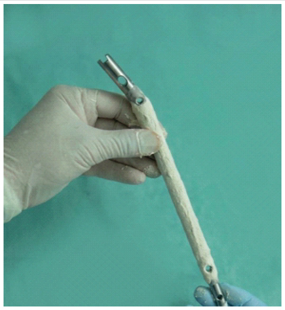
Figure 1: Antibiotic cement-coated intramedullary nail.
- Step 5 – Nail insertion and interlocking: The coated nail was inserted using the standard closed technique. Proximal and distal interlocking screws were placed where anatomically feasible.
- Step 6 – Closure: Wound closure was performed in layers over a suction drain.
Post-operative protocol:
Culture-directed intravenous antibiotics were administered for 3 weeks, followed by 3 weeks of oral antibiotics. Weight-bearing commenced as toe-touch and progressed to full weight-bearing based on clinical examination and radiological callus formation. Clinical and radiological assessment was performed every 4–6 weeks until union.
Outcome measures:
Primary outcome:
Radiographic union, defined as bridging callus across ≥3 cortices on anteroposterior and lateral radiographs.
Secondary outcome:
Infection control is defined as the absence of draining sinus and normalisation of erythrocyte sedimentation rate and C-reactive protein at final follow-up.
Case 1:
A 28-year-old male with an infected non-union of the right tibia following early implant removal after intramedullary nailing was treated with debridement and culture-directed antibiotics, resulting in infection control and complete radiological union at 1 year (Fig. 2).
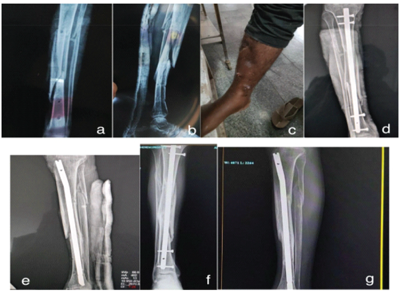
Figure 2: (a and b) Pre-operative AP and lateral X-ray of right tibia showing non-union with lucency at fracture site, (c) Pre-operative clinical photo of affected limb, (d and e) Early post-operative radiograph demonstrating satisfactory nail position with interlocking. (f) Follow-up radiograph at 1 year demonstrating solid bridging callus and complete radiological union, (g) Clinical photograph at 1-year follow-up showing healed wound and restored limb alignment.
Case 2:
A 50-year-old male presented with an infected non-union of the right distal femur following implant removal for infection after locking plate fixation. He underwent extensive debridement with antibiotic cement-coated retrograde femoral nail insertion, resulting in infection control, progressive callus formation at 3 months, and radiological union by 6 months (Fig. 3).
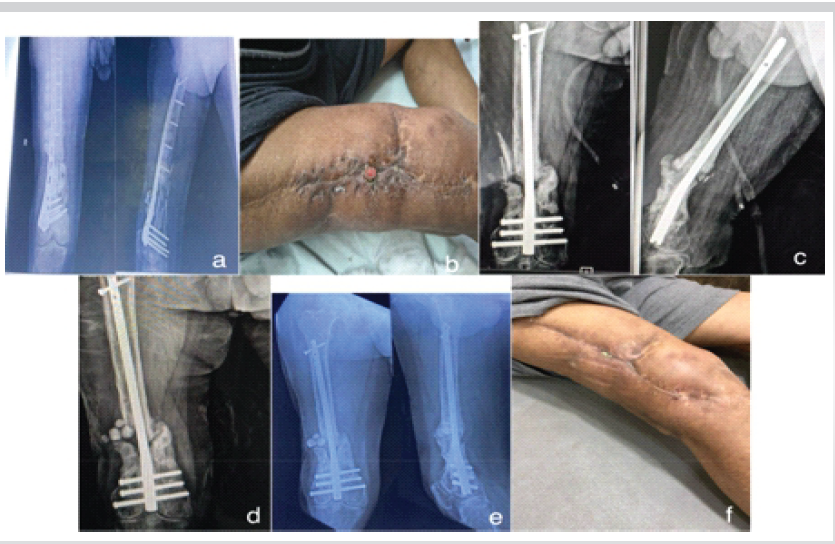
Figure 3: (a) Pre-operative radiograph of right distal femur showing failed plate fixation with non-union, (b) Clinical limb photo, (c) Post-operative radiograph following antibiotic cement-coated nail insertion in right distal femur, (d) Radiograph at 3 months showing early callus formation, (e) Radiograph at 6 months follow-up confirming solid radiological union, (f) Clinical photograph at 6-month follow-up showing healed sinus and restored function.
Case 3:
A 33-year-old male presented with an infected non-union of the right femur following intramedullary nailing for a road traffic accident (RTA) (Fig. 4).
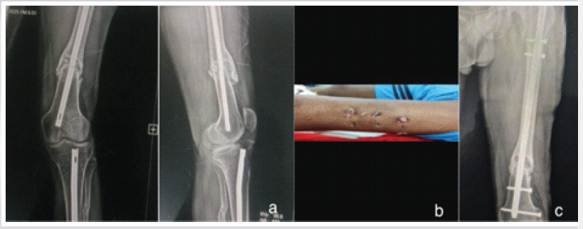
Figure 4: (a) Pre-operative radiograph of right femur showing infected non-union with sclerotic margins, (b) Clinical photo of limb, (c) Post-operative radiograph.
Case 4:
A 13-year-old female presented with an infected non-union of the left tibia following interlocking nailing for an RTA. Antibiotics-cement-coated V-nail done (Fig. 5).
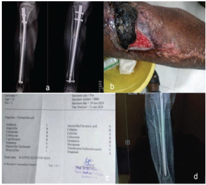
Figure 5: (a) Pre-operative radiograph of left tibia non-union, nail in situ, (b) clinical photo of limb, (c) Pus culture sensitivity report, (d) Follow-up radiograph demonstrating bridging callus and union.
Case 5:
A 50-year-old male presented with an infected non-union of the left distal femur despite prior plate fixation and subsequent Ilizarov ring fixator application. Following removal of the fixator, extensive debridement, and insertion of an AC-CIMN, infection was controlled, fracture union was achieved, and the patient regained independent ambulation (Fig. 6).
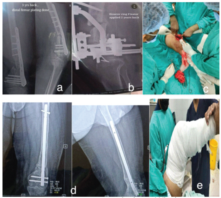
Figure 6: (a and b) Pre-operative radiograph showing left distal femur non-union with distal femur plating and ilizarov ring fixation, (c) Intraoperative photograph following fixator removal and debridement, (d) Case 5: Follow-up radiograph showing progressive callus and union, (e) On-table knee range of motion.
Case 6:
A 34-year-old male presented with an infected non-union of the left distal femur with osteomyelitis and a small bone defect following fracture fixation (Fig. 7).
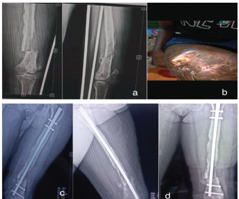
Figure 7: (a) Pre-operative radiograph of left distal femur showing infected non-union with osteomyelitic changes and bone loss, (b) Clinical photo of limb, (c) Post-operative radiograph showing antibiotic cement-coated nail in situ, (d) Follow-up radiograph at 12 months showing union.
Demographics and microbiological profile:
Ten patients (8 male, 2 female) with a mean age of 39.9 years (range 26–59 years) were enrolled. The most common mechanism of injury was RTA in 7 cases. Bones involved were the tibia in 6 patients, the femur in 3, and the humerus in 1. The mean duration from initial injury to presentation was 9 months (range 6–18 months). Table 1 summarises the demographic, microbiological, and outcome data for all 10 patients. Intraoperative cultures yielded a pathogen in 9 of 10 patients. MSSA was the most frequent isolate (4 cases), followed by MRSA (2 cases) and Pseudomonas aeruginosa, Klebsiella pneumoniae, and Escherichia coli (1 case each). One patient (Case 5, right femur) had no microbiological growth and was treated empirically.
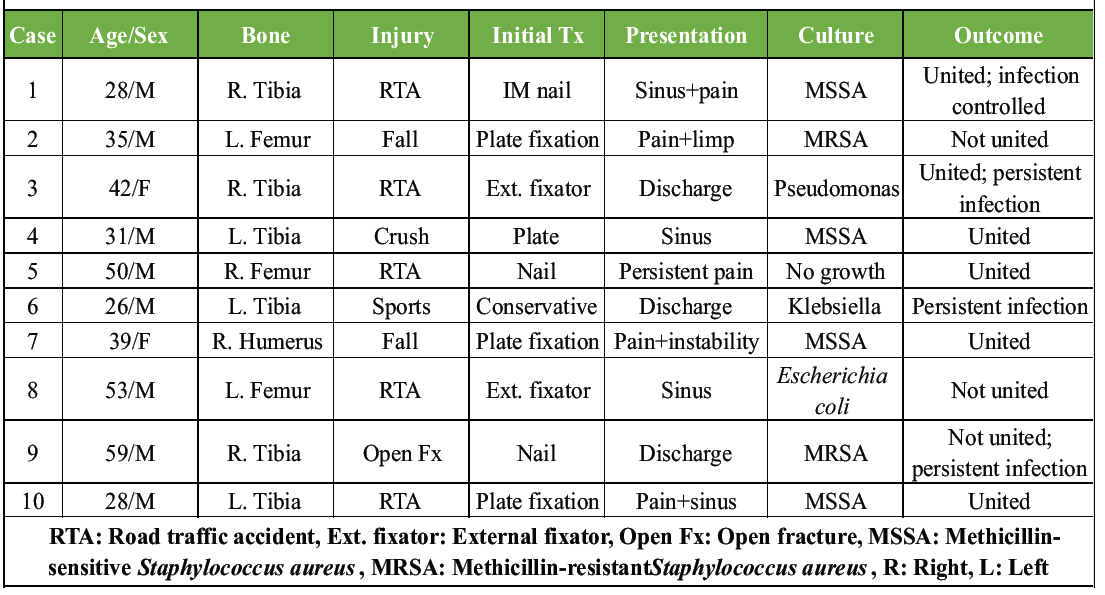
Table 1: Patient demographics, microbiological profile, and outcomes
Primary outcome: Bony union.
Bony union was achieved in 7 of 10 patients (70%). The mean time to union was 28 weeks for the tibia (range 24–32 weeks) and 22 weeks for the femur (range 18–28 weeks). The single humerus case (Case 7, right humerus, MSSA) united at 20 weeks. Three patients (Cases 2, 8, and 9) failed to achieve union. Cases 2 and 9 harboured MRSA; Case 8 had an E. coli infection. Both Cases 8 and 9 subsequently required secondary bone grafting and extended antibiotic therapy.
Secondary outcome: Infection control:
Infection was eradicated in 8 of 10 patients (80%). Persistent infection was observed in Cases 3, 6, 8, and 9. All four harboured multidrug-resistant or Gram-negative organisms. Case 3 (P. aeruginosa) achieved radiographic union despite persistent bacteriological positivity at 3 months, with eventual clinical resolution at 6 months.
Functional outcomes and complications:
Functional improvement – defined as resolution of sinus discharge, improved limb stability, and return to weight-bearing – was documented in 8 of 10 patients (80%). Early mobilisation (toe-touch weight-bearing within 6 weeks of surgery) was possible in 7 patients due to the load-sharing properties of the intramedullary construct. Complications encountered during the study period are detailed in Table 2.
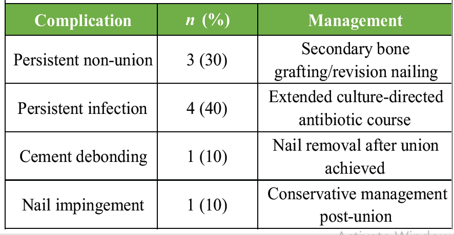
Table 2: Complications and their management
Infected non-union of long bones remains a formidable challenge in orthopaedic practice. Its management requires simultaneous addressing of three critical elements: eradication of deep-seated infection, achievement of skeletal stability, and biological stimulation of healing. Traditional two-stage approaches, while conceptually sound, impose a significant burden on patients through repeated surgical interventions, prolonged hospitalisation, and reliance on external fixation devices that are poorly tolerated [10,11]. The AC-CIMN technique has progressively gained acceptance as a single-stage alternative. The fundamental principle rests on combining load-sharing intramedullary stabilisation with local delivery of antibiotics at concentrations far exceeding systemic levels achievable through parenteral routes. PMMA-eluted antibiotics achieve local tissue concentrations 100–200 times higher than systemic administration, effectively penetrating the avascular zone around the infected non-union site and overcoming biofilm-associated resistance mechanisms [7,12]. In our series, the overall infection control rate of 80% and union rate of 70% are consistent with published literature. Shyam et al. reported union in 92.3% of their 13 cases using antibiotic cement-impregnated nails, while Zalikha et al. reported a 75% union rate in a larger multicentre cohort [2,3]. The slightly lower union rate in our series may reflect the heterogeneity of our case mix, including a complex distal femur case with prior Ilizarov failure and cases with no organism isolated on culture. The GMCH protocol employed in our series includes several critical technical steps that we believe contribute to outcomes. Medullary cavity irrigation with 2–3 L of normal saline with betadine through a paediatric nasogastric tube ensures thorough washout of debris and biofilm before nail insertion. Canal reaming 2 mm larger than the nail coating diameter ensures adequate canal preparation and prevents impaction. Reaming debris and granulation tissue are systematically sent for culture and sensitivity to guide post-operative antibiotic selection [13]. Antibiotic selection for PMMA impregnation is guided by pre-operative and intraoperative culture results wherever available. In culture-negative cases (as in Case 3 of our series), empirical broad-spectrum coverage is used. Heat-stable antibiotics suitable for PMMA mixing include vancomycin, tobramycin, and gentamicin, with combination regimens commonly employed for MRSA-positive cases. Post-operative systemic antibiotic therapy for 6 weeks (3 weeks intravenous followed by 3 weeks oral) parallels the protocol described by Solanki et al. [1]. Key selection criteria for this technique include: Infected non-union of a long bone suitable for intramedullary nailing, bone gap ≤2 cm, absence of critical-sized defects requiring segmental reconstruction, and absence of allergy to PMMA or the selected antibiotic. Cases with larger bone gaps, critical soft tissue deficiency, or multiple drug-resistant organisms refractory to PMMA-compatible antibiotics are better served by alternative strategies, such as LRS bone transport or induced membrane technique (Masquelet) [14,15]. Complications encountered in our series included two cases of persistent infection requiring extended antibiotic courses, one case of non-union requiring secondary intervention, and one case of cement debonding managed conservatively. Nail impingement at the fracture site in one patient was managed with post-union nail adjustment. These complication rates align with previously reported series and underscore the importance of meticulous patient selection and technique [16]. Limitations of this study include its small sample size (n = 10), single-institution design, heterogeneous case mix across different long bones and prior treatment modalities, and a variable follow-up period. A controlled comparative study against staged procedures or LRS bone transport would more definitively establish the superiority of this technique. Nevertheless, our series provides valuable real-world evidence from a resource-constrained setting where minimising surgical stages and implant cost carries particular importance.
AC-CIMN is a technically feasible and clinically effective single-stage strategy for infected non-union of long bones with bone gaps ≤2 cm. By simultaneously addressing infection control and mechanical stability, it reduces the total number of surgical interventions compared to conventional staged protocols and enables earlier patient mobilisation. Culture-guided antibiotic loading of PMMA, meticulous medullary debridement, and appropriate patient selection are critical determinants of success. Larger prospective studies comparing this technique with staged protocols and external fixation strategies are warranted to establish definitive evidence-based indications.
Antibiotic cement-coated intramedullary nailing offers a viable single-stage solution for infected non-union of long bones with bone gaps not exceeding 2 cm, combining effective local antimicrobial delivery with immediate mechanical stability and permitting early patient mobilization.
References
- 1. Solanki T, Maurya MK, Singh PK. Results of antibiotic-impregnated cement/polymer-coated intramedullary nails in the management of infected nonunion and open fractures of long bones. Cureus 2023;15:e43421. [Google Scholar] [PubMed]
- 2. Zalikha AK, Sayeed Z, Stine SA, Bray R, Vaidya R. Antibiotic cement-coated interlocked intramedullary nails for the treatment of infected nonunion after intramedullary nailing. J Orthop Trauma 2023;37:e1-6. [Google Scholar] [PubMed]
- 3. Shyam AK, Sancheti PK, Patel SK, Rocha S, Pradhan C, Patil A. Use of antibiotic cement-impregnated intramedullary nail in treatment of infected non-union of long bones. Indian J Orthop 2009;43:396-402. [Google Scholar] [PubMed]
- 4. Uikey S, Oddam VK, Verma R. To study the clinical, radiological and functional outcome of antibiotic mixed cement coated nails in infected long bone diaphyseal fractures. Surg Update Int J Surg Orthop 2017;3:169-74. [Google Scholar] [PubMed]
- 5. Jain AK, Sinha S. Infected nonunion of the long bones. Clin Orthop Relat Res 2005;431:57-65. [Google Scholar] [PubMed]
- 6. Patzakis MJ, Wilkins J, Wiss DA. Infection following intramedullary nailing of long bones. Diagnosis and management. Clin Orthop Relat Res 1986;212:182-91. [Google Scholar] [PubMed]
- 7. Weiland AJ, Moore JR, Hotchkiss RN. Soft tissue procedures for reconstruction of tibial shaft fractures. Clin Orthop Relat Res 1983;178:42-53. [Google Scholar] [PubMed]
- 8. Klemm KW. Antibiotic bead chains. Clin Orthop Relat Res 1993;295:63-76. [Google Scholar] [PubMed]
- 9. Ostermann PA, Seligson D, Henry SL. Local antibiotic therapy for severe open fractures. A review of 1085 consecutive cases. J Bone Joint Surg Br 1995;77:93-7. [Google Scholar] [PubMed]
- 10. Bose D, Kugan R, Stubbs D, McNally M. Management of infected nonunion of the long bones by a multidisciplinary team. Strategies Trauma Limb Reconstr 2015;10:27-34. [Google Scholar] [PubMed]
- 11. Conway JD. Osteomyelitis and malignant degeneration. Hand Clin 2004;20:489-501. [Google Scholar] [PubMed]
- 12. Tetsworth K, Cierny G 3rd. Osteomyelitis debridement techniques. Clin Orthop Relat Res 1999;360:87-96. [Google Scholar] [PubMed]
- 13. Lew DP, Waldvogel FA. Osteomyelitis. Lancet 2004;364:369-79. [Google Scholar] [PubMed]
- 14. Cierny G 3rd, Mader JT, Penninck JJ. A clinical staging system for adult osteomyelitis. Clin Orthop Relat Res 2003;414:7-24. [Google Scholar] [PubMed]
- 15. Lindsey RW, Probe R, Miclau T, Alexander JW, Perren SM. The effects of antibiotic-impregnated autogeneic cancellous bone graft on bone healing. Clin Orthop Relat Res 1993;291:303-12. [Google Scholar] [PubMed]
- 16. Paley D, Herzenberg JE. Intramedullary infections treated with antibiotic cement rods: Preliminary results in nine cases. J Orthop Trauma 2002;16:723-9. [Google Scholar] [PubMed]






Related Articles in Journal of Orthopaedic Case Reports
 October 1, 2026 Soft-Tissue Closure through Acute Deformation and Gradual Correction Using Taylor Spatial Frame in a Gustilo-Anderson Type IIIB Open Tibial Pilon Fracture: A Case Report
 September 1, 2026 Limb Salvage for Entire Tibial Ewing Sarcoma in a 4-year-Old Child Using Extracorporeal Irradiation and Free Flap Reconstruction: A Case Report and Surgical Technique
 September 1, 2026 Proximal Midfoot Amputation as a Functional Alternative to Major Amputation in Advanced Diabetic Foot Infection: A Case Report
 September 1, 2026 Proximal Humerus Ewing Sarcoma: Technical Challenges and Strategies for Bone Defect Reconstruction