

In patients with persistent unexplained anterior knee pain, intra-articular ganglion cysts should be considered, with MRI enabling accurate diagnosis and arthroscopic treatment providing excellent symptomatic relief in symptomatic cases.
Dr. Wasif Kawsar Qadri, Department of Orthopaedics, Lady Hardinge Medical College and Associated Hospitals, New Delhi - 110 001, India. E-mail: wasifkqadri14@gmail.com
Abstract
Introduction: Ganglionic cysts are mostly found around the wrists, but they can occur in any part of the body. They are also common around the knees as popliteal cysts and meniscal cysts. Intra-articular cysts of the knee, however, are much rarer, with only sporadic cases being reported till 1990. Most of these cysts are asymptomatic and detected during routine magnetic resonance imaging (MRI) and arthroscopy for other pathologies. Most of the time, they are reported in relation to the anterior cruciate ligament (ACL) and rarely in relation to the posterior cruciate ligament, menisci, and very rarely in relation to the anterior fat pad. These patients occasionally report with knee pain, mechanical locking, and restriction of terminal flexion/extension. The symptoms depend on the site and size of the cysts.
Materials and Methods: We report a case series of 10 patients with ganglionic cysts of the knee who were studied over a period of 4 years. The study included four males and six females with an average age of 34.8 years. All reported with persistent knee pain at the outset. Clinical examination, preliminary X-ray, and MRI were done in all patients. Seven patients were examined arthroscopically, and their findings were recorded. Of these, three patients were treated with radiofrequency ablation and four with arthroscopic debridement of the cyst. Histopathology examination was carried out for the specimen material taken out.
Results: Arthroscopic examination revealed two cystic masses originating from ACL, two from the anterolateral knee fat pad, one from the retropatellar fat pad, one from the anteromedial fat pad, and one from the anterior horn of the lateral meniscus. All were treated successfully with arthroscopic debridement or radiofrequency ablation. All were symptom-free at the latest follow-up. The mean follow-up duration for the surgically treated patients was 29 months (range, 24–34 months). Out of the three patients who did not undergo surgery, one had a spontaneous resorption of the cyst as evidenced by MRI taken at 3 years.
Conclusion: Although uncommon, intra-articular ganglion cysts should be considered in patients presenting with unexplained anterior knee pain. MRI facilitates early diagnosis, and arthroscopic management offers a safe and effective treatment with favorable clinical outcomes and low recurrence rates.
Keywords: Intra-articular ganglion cyst; Knee; Anterior knee pain; Magnetic resonance imaging; Arthroscopy; Anterior cruciate ligament.

Ganglionic cysts are mostly found around the wrists, but they can occur in any part of the body. They are also common around the knees as popliteal cysts and meniscal cysts. In 1877, Baker described the association of popliteal cysts with intra-articular lesions. Several authors have reported that 41–83% of joint disorders have associated popliteal cysts [1,2,3,4].
Intra-articular cysts of the knee, however, are much rarer. First intra articular cyst of the anterior cruciate ligament (ACL) was reported by Caan 1924 in a cadaveric specimen [5]. Only sporadic cases of intra-articular cysts of the knees were reported till 1990. An increasing number of these cases have been reported in the last three decades with the advent of magnetic resonance imaging (MRI) and arthroscopy. Most of these cysts are asymptomatic and detected during routine MRI and arthroscopy for other pathologies [5]. Intra-articular ganglionic cysts, especially those arising from the cruciate ligaments, have a reported prevalence of 0.2–1.3% on MRI and 0.1–0.6% on knee arthroscopy. Most of the time, they are reported in relation to the ACL and rarely in relation to the posterior cruciate ligament (PCL), menisci, and very rarely in relation to the anterior fat pad. These patients occasionally report with knee pain, mechanical locking, and restriction of terminal flexion/extension. The symptoms depend on the site and size of the cysts [5,6].
We report a case series of 10 patients with ganglionic cysts and describe their clinical features, radiology, morphology, and location.
Ten patients with ganglion cysts of the knee were studied over a 4-year period. The patients reported with persistent knee pain at the outset. Clinical examination was carried out to assess the knees for various correlative signs. MRI was carried out in all cases to provide radiological assessment after a preliminary X-ray. Seven of the 10 patients agreed to undergo surgery. Their arthroscopic findings and the procedure carried out were recorded. Histopathology examination was carried out for the specimen material taken out. The findings were tabulated (Table 1).
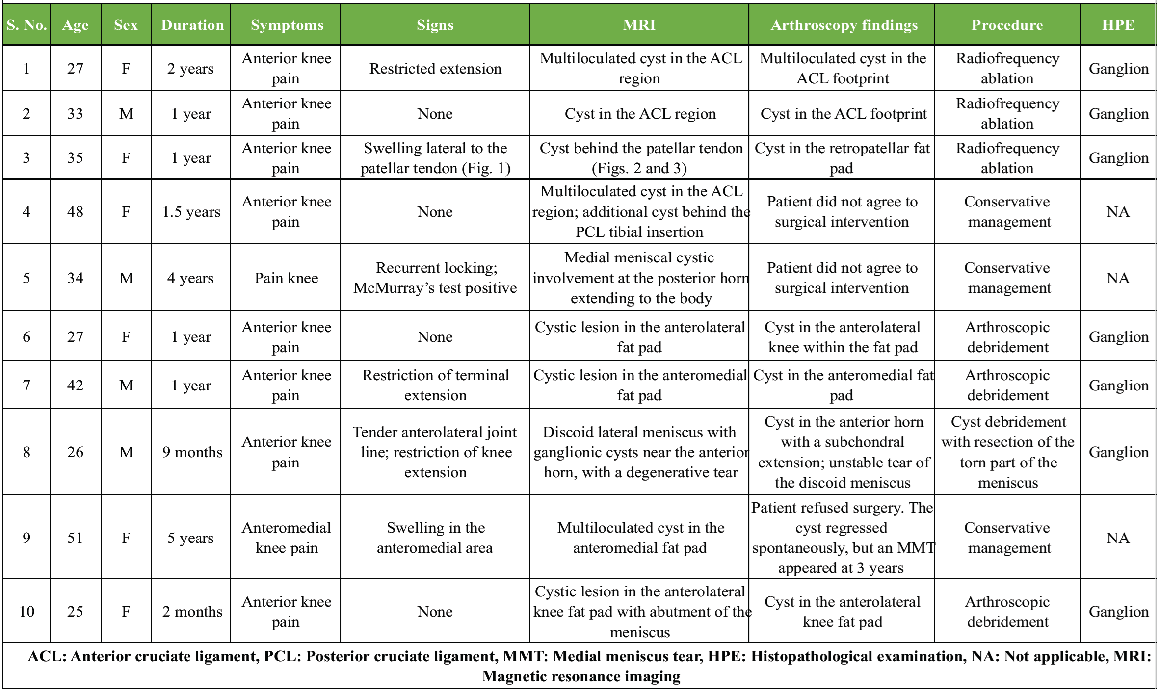
Table 1: Clinico-radiological and arthroscopic findings in patients under study
Ten patients with MRI-confirmed intra-articular cysts of the knee were included in the study. The mean age was 34.8 years (range, 25–51 years). There were four males and six females. Nine patients presented with anterior knee pain, whereas one reported generalized knee pain.
On clinical examination, four patients had no demonstrable physical signs. Two patients had a palpable swelling, one had joint line tenderness, three had restriction of knee extension, and one had a positive McMurray test.
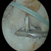
Seven patients underwent arthroscopic treatment. In all cases, arthroscopic findings correlated with the preoperative MRI. Radiofrequency ablation was performed in three patients, while four underwent arthroscopic debridement of the cyst. Three patients were managed non-operatively. One of these demonstrated spontaneous complete resorption of the cyst on follow-up MRI obtained 3 years after the initial diagnosis. Representative clinical, MRI, and arthroscopic images are shown in Fig. 1–6.
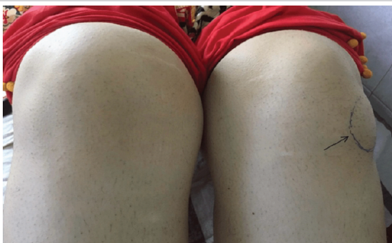
Figure 1: Cyst arising from retropatellar fat pad reaching up to proximal tibia (black arrow).
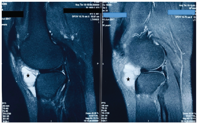
Figure 2: Ganglionic cyst in retropatellar fat pad (asterisk).
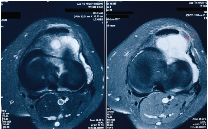
Figure 3: Retropatellar ganglionic cyst (arrow).
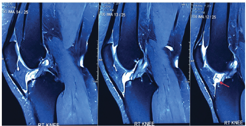
Figure 4: Intra-articular cyst of the knee at the base of the anterior cruciate ligament (arrow).
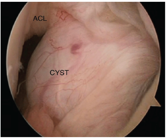
Figure 5: Arthroscopic image showing a ganglionic cyst at the base of the anterior cruciate ligament.
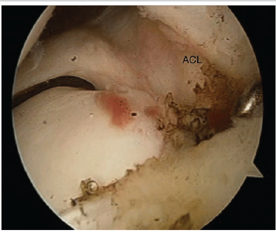
Figure 6: Post-radiofrequency ablation of cyst.
The mean follow-up duration for the surgically treated patients was 29 months (range, 24–34 months). At final follow-up, all operated patients had complete resolution of pain, swelling, and mechanical symptoms, with no evidence of recurrence. No procedure-related complications were observed.
Intra-articular cysts of the knee are mostly found around the ACL, but they have also been reported around the PCL, within the substance of the ACL, in relation to the medial or lateral menisci, within the infrapatellar fat pad, and as subchondral bone cysts [5,6,7,8,9,10,11,12,13,14,15,16,17].
They are mostly detected incidentally during MRI or arthroscopy performed for unrelated knee pathology. On MRI, the cysts appear as fluid-filled lobulated lesions with low signal intensity on T1-weighted images and high signal intensity on T2-weighted images [5,18]. They are mainly round or elliptical lobulated structures, and internal septa are visible in some cysts [17]. Ganglion cysts located anterior to the cruciate ligaments have been reported to restrict terminal knee extension, whereas cysts located posterior to the cruciate ligaments may restrict terminal knee flexion [19]. In the present study, the intra-articular cyst arising from the anterior horn of the lateral meniscus and the cysts located anterior to the cruciate ligaments were also associated with restriction of terminal knee extension. Ganglion cysts originating from the ACL have been reported to occur approximately three times more frequently than those arising from the PCL [5]. Cysts arising from the infrapatellar fat pad are rarely large enough to present as a palpable swelling, as was also observed in one of our cases [7]. In symptomatic cases, these intra-articular cysts most commonly present with anterior knee pain [5,11,13,20,21,22,23], whereas locking has only rarely been reported. Similarly, the majority of patients in our series presented with anterior knee pain, while the cyst arising from the infrapatellar fat pad presented as a giant ganglion cyst measuring approximately 9 cm vertically and 5 cm horizontally. Only a few cases of giant intra-articular ganglion cysts have been reported in the literature [7,14].
The pathogenesis of these intra-articular cysts still remains unclear. The proposed etiopathogenetic mechanisms include herniation of synovial tissue, post-traumatic connective tissue degeneration, mucinous degeneration of connective tissue, proliferation of pluripotent mesenchymal stem cells, and displacement of synovial tissue during embryogenesis [5,6,8,14]. The theory of cellular response to trauma involves liberation of mucin and hyaluronic acid, which become interspersed within the fibers of the ligament, causing fusiform dilatation. With joint movement, the mucinous material dissects between the ligament fibers and may subsequently be found at the ligament attachments or within the intercondylar notch of the knee [18,19].
Ultrasound-, computed tomography (CT)-, or arthroscopic-guided needle aspiration has been associated with a higher rate of recurrence [5]. Management options for intra-articular ganglion cysts include observation, image-guided aspiration, arthroscopic excision or debridement, radiofrequency ablation, and open excision. Asymptomatic lesions may be managed conservatively with observation, as spontaneous regression has been reported in some cases. Ultrasound- or CT-guided aspiration is minimally invasive but is associated with a higher recurrence rate because the cyst wall may not be completely removed. Arthroscopic excision or debridement allows direct visualization of the lesion and associated intra-articular pathology, facilitates complete cyst removal, enables early rehabilitation, and has been associated with excellent clinical outcomes and low recurrence rates. Open excision is generally reserved for large cysts with extra-articular extension or lesions that are technically difficult to access arthroscopically, although it may be associated with greater surgical morbidity. Mostly large cysts have been excised through an open approach to reduce the chances of recurrence, but many cases have also been successfully managed arthroscopically without recurrence. Cysts arising from the infrapatellar fat pad may be difficult to reach arthroscopically, especially if they are large or associated with subcutaneous extension [16,22]; however, all operated cases in our series, including the large cyst arising from the infrapatellar fat pad, were successfully managed arthroscopically, as also reported by Yang et al. [9,10].
Recent reports continue to support the importance of MRI in accurately diagnosing intra-articular ganglion cysts of the knee and delineating their relationship to surrounding structures, thereby facilitating appropriate surgical planning. These reports have also demonstrated excellent clinical outcomes following arthroscopic management of symptomatic lesions while emphasizing that giant ganglion cysts with extra-articular extension may still require an open approach in selected cases. Recent case reports of Hoffa’s fat pad ganglion cysts further highlight the importance of individualized treatment based on cyst size, location, and symptomatology [2,3].
Histologically, ganglion cysts show a fibrous cyst wall containing myxoid viscous material without an epithelial lining [8]. Not all ganglion cysts of the knee require excision; only those associated with symptoms require surgical treatment [11,24].
Seven cysts in our study were managed arthroscopically, of which three underwent radiofrequency ablation, and four underwent arthroscopic decompression and debridement. The use of the radiofrequency probe minimized damage to adjacent structures and reduced post-operative hemarthrosis. At the latest follow-up, all surgically treated patients remained asymptomatic without recurrence of pain, swelling, or mechanical symptoms. Although a repeat MRI was not routinely performed, the absence of recurrent clinical symptoms suggests that no clinically significant recurrence occurred during the follow-up period.
The strengths of the present study include preoperative MRI confirmation in all patients, arthroscopic correlation in surgically treated cases, and histopathological confirmation where tissue was obtained. The study is limited by its retrospective design, heterogeneous cyst locations, lack of standardized functional outcome scores, and absence of routine post-operative MRI to objectively assess recurrence.
Intra-articular ganglion cysts of the knee are uncommon but important causes of persistent anterior knee pain and mechanical symptoms. MRI is invaluable for the diagnosis and localization of these lesions. Arthroscopic debridement or radiofrequency ablation provides effective symptom relief with excellent clinical outcomes and low morbidity in symptomatic patients, while selected asymptomatic lesions may be managed conservatively.
Persistent anterior knee pain with inconclusive clinical examination should prompt consideration of an intra-articular ganglion cyst. MRI plays a pivotal role in diagnosis, and arthroscopic excision or radiofrequency ablation is a safe and effective treatment for symptomatic lesions, resulting in excellent functional outcomes and symptom resolution.
References
- 1. Baker WM. The formation of synovial cysts in the leg in connection with disease of the knee joint. St Bart Hosp Rep 1994;15:245-61. [Google Scholar] [PubMed]
- 2. Narkhede YD. Hoffa’s fat pad Ganglion cyst protruding from lateral retinacular rent: A case report. Int J Res Orthop 2024;10:833-5. [Google Scholar] [PubMed]
- 3. Khadka SK, Banmala S, Pandey A, Pandey R, Khadka SK. Hoffa’s fat pad Ganglion cyst of the knee: A case report of a rare cause of knee pain and swelling. Ann Med Surg (Lond) 2024;86:4913-7. [Google Scholar] [PubMed]
- 4. Vahvanen V. Popliteal cyst. A follow-up study on 42 operatively treated patients. Acta Orthop Scand 1973;44:303-10. [Google Scholar] [PubMed]
- 5. Krudwig WK, Schulte KK, Heinemann C. Intra-articular Ganglion cysts of the knee joint: A report of 85 cases and review of the literature. Knee Surg Sports Traumatol Arthrosc 2004;12:123-9. [Google Scholar] [PubMed]
- 6. Bui Mansfield LT, Youngberg RA. Intraarticular Ganglia of the knee: Prevalence, presentation, etiology, and management. AJR Am J Roentgenol 1997;168:123-7. [Google Scholar] [PubMed]
- 7. Vaishya R, Kansagra A, Agarwal AK, Vijay V. A giant Ganglion cyst arising from lateral Hoffa’s fat pad of the knee. J Orthop Case Rep 2019;9:36-9. [Google Scholar] [PubMed]
- 8. Yilmaz E, Karakurt L, Ozercan I, Ozdemir H. A Ganglion cyst that developed from the infrapatellar fat pad of the knee. Arthroscopy 2004;20:e65-8. [Google Scholar] [PubMed]
- 9. Yang JH, Kim TS, Lim HC, Kim HJ, Oh CH, Yoon JR, et al. Endoscopic excision of a Ganglion cyst in an infrapatellar fat pad extending into the subcutaneous layer. J Orthop Sci 2012;17:654-8. [Google Scholar] [PubMed]
- 10. Zantop T, Rusch A, Hassenpflug J, Petersen W. Intra-articular ganglion cysts of the cruciate ligaments: Case report and review of the literature. Arch Orthop Trauma Surg 2003;123:195-8. [Google Scholar] [PubMed]
- 11. Rakesh G, Rajat C, Shivendra S, Kamra P. Ganglion cyst of Hoffa’s fat pad of knee-a rare cause of knee pain and swelling-a case report and literature review. J Clin Orthop Trauma 2019;10:S215-7. [Google Scholar] [PubMed]
- 12. Abreu FG, De Abreu GV, Pioger C, Franck F, Vieira TD, Bulle S, et al. Excision of a posterior cruciate ligament cyst using an arthroscopic trans-septal approach. Arthrosc Tech 2020;9:e581-5. [Google Scholar] [PubMed]
- 13. Andreozzi V, Monaco E, Conteduca F, Iorio R, Mazza D, Drogo P, et al. Diagnosis and treatment of a symptomatic posterior cruciate Ganglion cyst in a child with Autism. Case Rep Orthop 2019;2019:192347. [Google Scholar] [PubMed]
- 14. Miljko M, Markotić V, Huseinbegović A, Kapur E, Marijanović I, Jurić I. Giant infrapatellar ganglion cyst of Hoffa’s fat pad. Eur J Radiol Open 2019;6:275-80. [Google Scholar] [PubMed]
- 15. Andrikoula SI, Vasiliadis HS, Tokis AV, Kosta P, Batistatou A, Georgoulis AD. Intra-articular Ganglia of the knee joint associated with the anterior cruciate ligament: A report of 4 cases in 3 patients. Arthroscopy 2007;23:800 e1-6. [Google Scholar] [PubMed]
- 16. Saha P, Bandyopadhyay U, Mukhopadhyay AS, Kundu S, Mandal S. Ganglion cyst of knee from Hoffa’s fat pad protruding anterolaterally through retinacular rent: A case report. J Orthop Case Rep 2015;5:69-71. [Google Scholar] [PubMed]
- 17. Mao Y, Dong Q, Wang Y. Ganglion Cysts of the cruciate ligaments: A series of 31 cases and review of the literature. BMC Musculoskelet Disord 2012;13:137. [Google Scholar] [PubMed]
- 18. Kakutani K, Yoshiya S, Matsui N, Yamamoto T, Kurosaka M. An intraligamentous Ganglion cyst of the anterior cruciate ligament after a traumatic event. Arthroscopy 2003;19:1019-22. [Google Scholar] [PubMed]
- 19. Bergin D, Morrison WB, Carrino JA, Nallamshetty SN, Bartolozzi AR. Anterior cruciate ligament Ganglia and mucoid degeneration: Coexistence and clinical correlation. AJR Am J Roentgenol 2004;182:1283-7. [Google Scholar] [PubMed]
- 20. Gupta R, Singhal A, Mehta R. Cyst between femoral attachments of cruciate ligaments: Unusual cause of knee pain and review of literature. Hindawi Case Rep Orthop 2020;2020:8833683. [Google Scholar] [PubMed]
- 21. Sloane J, Gulati V, Penna S, Pastides P, Baghla DP. Large intra-articular anterior cruciate ligament Ganglion cyst, presenting with inability to flex the knee. Case Rep Med 2010;2010:705919. [Google Scholar] [PubMed]
- 22. Takahashi T, Kimura M, Ohsawa T, Yamaguchi N, Takeshita K. A case of infrapatellar fat pad Ganglion of the knee. Open Orthop J 2017;11:1142-6. [Google Scholar] [PubMed]
- 23. Kodaira S, Nakajima T, Takahashi R, Moriya S, Nakagawa T, Ohtake H, et al. A case of intra-articular Ganglion cysts of the knee joint: Correlation between arthroscopic and magnetic resonance imaging. BMC Med Imaging 2016;16:36. [Google Scholar] [PubMed]
- 24. Feldman MD. Arthroscopic excision of a Ganglion cyst from the elbow. Arthroscopy 2000;16:661-4. [Google Scholar] [PubMed]





Related Articles in Journal of Orthopaedic Case Reports
 April 1, 2026 Bucket Handle Tear of the Medial Meniscus with a New Magnetic Resonance Imaging Sign: The “Head in the Sand Sign” – A Case Report
 August 6, 2024 A Comparative Study on Correlation between Magnetic Resonance Imaging and Arthroscopic Findings in the Knee Joint Injuries
 August 1, 2026 The Boba Tea Sign: A Novel Magnetic Resonance Imaging Finding in Synovial Chondromatosis of the Knee – A Case Report
 June 1, 2026 Functional Outcomes of Arthroscopic Anterior Cruciate Ligament Reconstruction Using Hamstring Tendon Autograft in a Predominantly Chronic Anterior Cruciate Ligament Injury Cohort: A Prospective Study