

Bilateral transient osteoporosis of the knee joint is a rare but important condition that should be considered in the differential diagnosis of knee pain in pregnant or postpartum women presenting with atraumatic bilateral knee pain.
Dr. M Azaharhussain, Department of Orthopaedics, Dr. Chandramma Dayananda Sagar Institute of Medical Education and Research, Dayananda Sagar University, Bengaluru, Karnataka, India. E-mail: azahar007@gmail.com
Abstract
Introduction: Transient osteoporosis (TO) is a rare, self-limiting condition characterized by the acute onset of joint pain and radiological evidence of periarticular osteopenia with bone marrow edema on magnetic resonance imaging (MRI). It most commonly affects the hip during the third trimester of pregnancy or the early postpartum period. Knee involvement is uncommon, and bilateral knee involvement is exceedingly rare, particularly in association with twin pregnancy. We report a rare case of bilateral TO of the knees in the early postpartum period following twin gestation, highlighting its clinical presentation, diagnostic workup, and management.
Case Report: A 34-year-old female presented in the early postpartum period after a twin pregnancy with severe bilateral knee pain and inability to bear weight, which began during the third trimester. There was no history of trauma, corticosteroid use, alcohol abuse, or systemic illness. Clinical examination revealed quadriceps wasting, joint line tenderness, and painful restriction of knee movements bilaterally. Laboratory investigations showed elevated ESR with normal rheumatoid markers. Plain radiographs demonstrated periarticular osteopenia of both knees. MRI revealed diffuse bone marrow edema involving bilateral femoral condyles and patellae, with minimal joint effusion, consistent with TO. The patient was managed conservatively with transnasal salmon calcitonin (200 IU daily for 3 months), calcium and Vitamin D supplementation, analgesics, and protected weight bearing. Significant symptomatic improvement was observed within 4 weeks, and a follow-up MRI at 6 months showed complete resolution of marrow edema. At the 1-year follow-up, the patient had regained a full, pain-free range of motion.
Conclusion: Bilateral TO of the knee in pregnancy is an extremely rare clinical entity that should be considered in the differential diagnosis of severe atraumatic knee pain in late pregnancy or the postpartum period. MRI plays a pivotal role in early diagnosis. Conservative management with symptomatic therapy and pharmacological support, including transnasal calcitonin, may provide effective pain relief and functional recovery. Early recognition is essential to prevent complications such as insufficiency fractures.
Keywords: Transient osteoporosis, bilateral knee, twin pregnancy, postpartum, bone marrow edema, calcitonin.

Transient osteoporosis (TO), first described by Curtish and Kinkaid in 1959, has since been documented in fewer than 200 cases [1]. TO is characterized by acute joint pain, often in the weight-bearing joints [2].
It most often occurs in women in the third trimester of pregnancy or the early postpartum period. While the hip joint is typically involved, reports of knee joint involvement are sparse, and bilateral knee involvement is even more uncommon [3]. The pathophysiology of TO during pregnancy is not fully understood, but hormonal changes, altered biomechanics, and metabolic disturbances associated with pregnancy may contribute [4,5].
This report describes a rare case of bilateral knee joint involvement in a woman with a twin pregnancy, providing further insight into the diagnosis and management of this uncommon condition. The condition is characterized by a gradual onset of pain and restricted movement, with radiographic findings of periarticular osteoporosis [6].
A 34-year-old female patient, in the early postpartum period following a twin pregnancy, presented to the orthopedic department with complaints of bilateral knee pain and inability to walk, which had started approximately 6 months into her pregnancy. The discomfort worsened around the 28th week of pregnancy, making mobility increasingly difficult. The patient reported no history of fever, weight loss, or appetite loss, and denied any trauma, alcohol abuse, or corticosteroid use.
Upon examination, the patient had quadriceps muscle wasting, shiny skin over the knee, and local redness. There was bilateral joint line tenderness, and both tibial and femoral condyle tenderness were noted. Range of motion at the knee was restricted due to pain. Blood tests revealed a total leukocyte count of 12.95 × 10³/µL, erythrocyte sedimentation rate (ESR) of 85 mm/h, C-reactive protein of 6 mg/L, and a serum calcium level of 7.9 mg%. Serum Vitamin D was found to be 28.5 ng/mL, and both rheumatoid arthritis (RA factor) and anti-cyclic citrullinated polypeptide were within normal limits.
Radiographs of the bilateral knees showed periarticular osteopenia and decreased bone mineral density in both the femoral condyles and patellae (Fig. 1a and b). Magnetic resonance imaging scans (MRI) (T1- and T2-weighted images) revealed bone marrow edema involving the femoral condyles and patella, with the lateral femoral condyles being more affected than the medial condyles (Fig. 2a and b). Minimal joint effusion was observed in the suprapatellar recess of both knees.
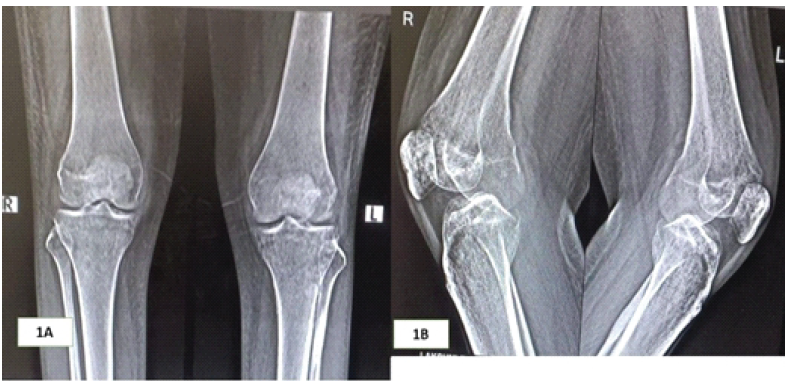
Figure 1: (a) Bilateral knee standing AP view: Periarticular osteopenia with patchy osteopenia. (b) Bilateral knee lateral view: Periarticular osteopenia with patchy osteopenia involving both medial and lateral femoral and tibial condyles and patella.
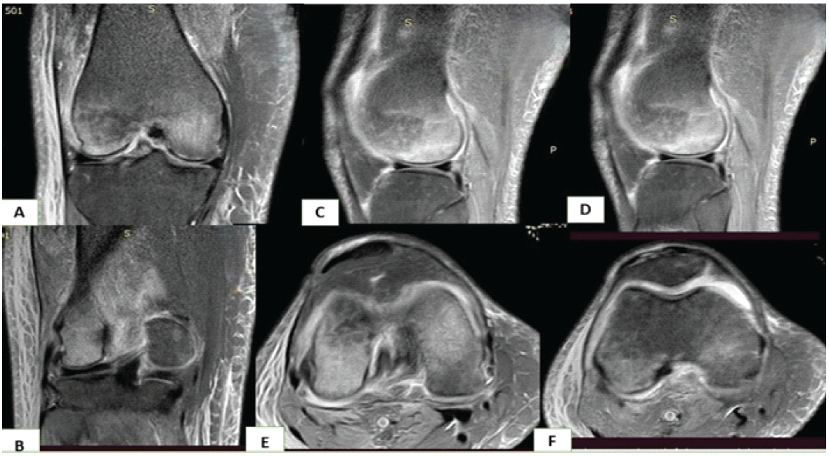
Figure 2: (a) Coronal sections show a hyperintense signal involving both lateral and medial femoral knee condyles. (b) Coronal section showing predominant marrow edema in the lateral femoral condyle. (c and d) Sagittal section showing diffuse marrow edema of the medial and lateral femoral condyles, respectively. (e and f) Axial section showing hyperintense signal changes involving both femur condyles.
The patient was treated with calcitonin nasal spray (200 IU daily inhaled via alternate nostrils for 3 months), along with oral calcium and Vitamin D supplements. Non-steroidal anti-inflammatory drugs (NSAIDs) were prescribed for short-term pain relief. The patient was closely monitored with regular follow-ups every month for the first 3 months, and subsequently every 2 months for up to a year. At her follow-up visits, the patient’s pain, range of motion, and functional mobility were assessed. The Visual Analog Scale pain scores were 10 at the start of treatment, but within 4 weeks, the score dropped to 2. Follow-up MRI at 6 months showed resolution of bone marrow edema (Fig 3a and b), and the improvement continued, with a full return of range of motion and pain-free mobility by the end of 12 months [7,8].
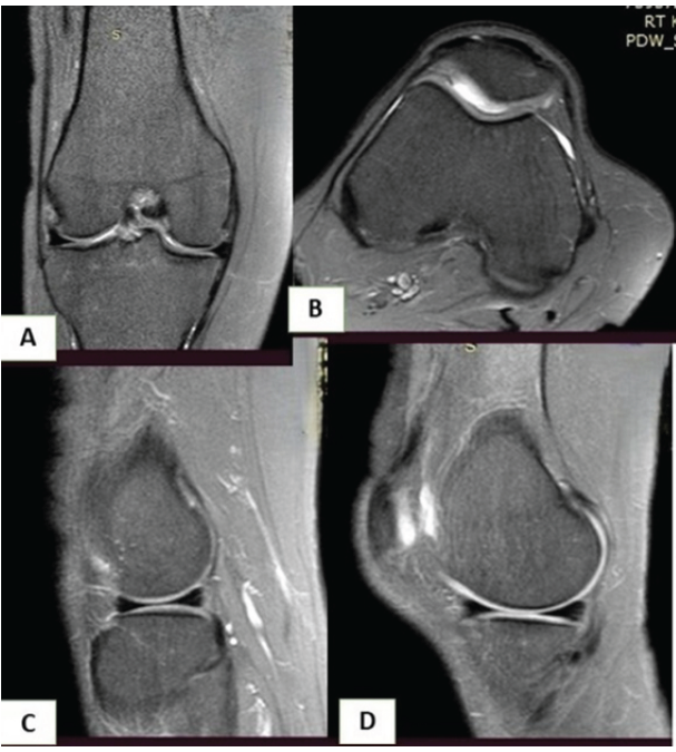
Figure 3: Proton density fat-saturated magnetic resonance image of the knee post-treatment. (a) Coronal section showing reduced marrow edema. (b) Axial section showing a normal intensity signal. (c and d) Sagittal section showing reduced marrow edema in the lateral and medial femoral condyles, respectively.
TO is a rare condition, particularly in pregnancy, and it most commonly affects the hip joint, with knee involvement being significantly less frequent [1,2,9]. The precise etiology remains unclear, but proposed mechanisms include nerve compression, demineralization due to functional impairment, and viral infections [4,5,10]. The increased demands on calcium metabolism during pregnancy may exacerbate bone resorption and contribute to the development of TO.
The clinical progression of TO typically follows a three-phase course: Phase 1: Characterized by a gradual onset of pain and stiffness, lasting approximately 1 month. Phase 2: Symptoms persist, with radiographic evidence of osteoporosis around the affected joint. Phase 3: Spontaneous restoration of bone density, which may continue for up to 8 months.
In this case, the diagnosis was made during Phase 2, when radiographic changes were already evident. Radiographs typically show moderate-to-severe periarticular osteoporosis, with joint space remaining unaffected [6]. MRI is the gold standard for diagnosing TO, as it can reveal characteristic bone marrow edema and joint effusion, which are hallmarks of the condition [3]. Our patient’s MRI revealed bone marrow edema in both femoral condyles, tibia, and patella, with more extensive involvement of the lateral condyles. The joint effusion observed in the suprapatellar recess further supported the diagnosis.
Management of TO generally involves conservative measures aimed at symptom relief, such as NSAIDs for short-term pain relief, weight-bearing restrictions, and avoidance of activities that place stress on the affected joints. Physiotherapy [11]: To prevent joint stiffness and muscle wasting. In this case, the patient was treated with calcitonin nasal spray, oral calcium, and Vitamin D supplementation. This is the first reported use of calcitonin nasal spray for TO, with previous studies primarily focusing on intramuscular calcitonin injections or bisphosphonates. Bisphosphonates were not used in this case, as their safety during pregnancy and the postpartum period remains uncertain [12]. The transnasal route avoids the discomfort of intramuscular or subcutaneous injections of calcitonin and appears to be equally effective in providing pain relief to the patient.
Bilateral TO of the knee joints during pregnancy is a rare but important condition that should be considered in the differential diagnosis of knee pain, particularly in cases of bilateral involvement. The diagnosis requires a high index of suspicion and detailed imaging, with MRI being the investigation of choice. Although medical treatment may not alter the natural course of the disease, symptomatic therapy and functional rehabilitation are crucial in preventing complications, such as fragility fractures.
Further studies are needed to better understand the pathophysiology of TO, particularly in pregnant women, and to establish optimal management strategies. This case report highlights the value of recognizing TO as a potential cause of knee pain in pregnancy, contributing to the growing body of medical literature on this rare condition.
Bilateral transient osteoporosis of the knee joints during pregnancy is a rare but important condition that should be considered in the differential diagnosis of knee pain in pregnancy, particularly in cases of bilateral involvement. Early MRI evaluation, weight-bearing restriction, and timely conservative management can prevent fractures and lead to complete clinical and radiological recovery.
References
- 1. Curtish JL, Kinkaid R. Transient osteoporosis of the hip in pregnancy. Am J Obstet Gynecol 1959;77:973-8. [Google Scholar] [PubMed]
- 2. Banu T, Gupta P. Pregnancy-related transient osteoporosis: Case report and review of literature. Eur J Orthop Surg Traumatol 2015;25:257-62. [Google Scholar] [PubMed]
- 3. Sen R, Aggarwal S. MRI findings in transient osteoporosis: A review. J Clin Orthop Trauma 2010;1:146-51. [Google Scholar] [PubMed]
- 4. Zahir M, Razi S. Transient osteoporosis of the knee joint during pregnancy: A rare and under-recognized condition. J Bone Joint Surg 2017;99:312-6. [Google Scholar] [PubMed]
- 5. Deshmukh RK, Shah PR. Transient osteoporosis of the hip joint in pregnancy: A case report and review of literature. Orthop J Trauma Surg 2013;3:45-9. [Google Scholar] [PubMed]
- 6. Möller H, Jakobsen T. The clinical course of transient osteoporosis in pregnancy: Case report and review of the literature. Rheumatol Int 2012;32:3599-602. [Google Scholar] [PubMed]
- 7. Rhee SM, Jang YS. Bilateral transient osteoporosis of the knees in a young woman: A case report. Knee Surg Sports Traumatol Arthrosc 2011;19:2060-3. [Google Scholar] [PubMed]
- 8. Uygur E, Aydogan A. Transient osteoporosis: A clinical challenge for the obstetrician and orthopaedist. J Clin Rheumatol 2016;22:234-8. [Google Scholar] [PubMed]
- 9. Varon C, Farge D. Transient osteoporosis of pregnancy: A review. J Orthop Surg Res 2010;5:36. [Google Scholar] [PubMed]
- 10. Takatori Y, Kokubo T, Ninomiya S, Nakamura T, Okutsu I, Kamogawa M. Transient osteoporosis of the hip. Magnetic resonance imaging. Clin Orthop Relat Res 1991;271:190-4. [Google Scholar] [PubMed]
- 11. Lidder S, Lang KJ, Lee HJ, Masterson S, Kankate RK. Bilateral hip fractures associated with transient osteoporosis of pregnancy. J R Army Med Corps 2011;157:176-8. [Google Scholar] [PubMed]
- 12. Brodell JD, Burns JE Jr., Heiple KG. Transient osteoporosis of the hip of pregnancy. two cases complicated by pathological fracture. J Bone Joint Surg Am 1989;71:1252-7. [Google Scholar] [PubMed]





Related Articles in Journal of Orthopaedic Case Reports
 January 1, 2026 Magnetic Resonance Imaging Evaluation of Temporomandibular Joint Involvement in Ankylosing Spondylitis Patients
 September 10, 2023 Percutaneous Management of Aneurysmal bone Cyst of Axis Vertebrae in a 12-year-old Child: A Case Report
 July 10, 2023 Regional Migratory Osteoporosis – A Rare Cause of Knee Pain (Case Report)
 April 10, 2023 A Rare Presentation of Transient Osteoporosis of the Talus: Case Report