

Acute neurological deficits after arthroscopic surgery should never be attributed solely to anesthesia and require urgent imaging and multidisciplinary assessment.
Dr. Rahul Kakran, Department of Orthopaedics, Yashoda Group of Hospitals, Ghaziabad, Uttar Pradesh, India. E-mail: rahul.kakran@yahoo.com
Abstract
Introduction: Acute ischemic stroke following arthroscopic knee surgery is extremely rare and can result in significant morbidity and mortality. Early recognition is crucial for timely management.
Case Report: A 44-year-old male with a multiligament injury of the left knee underwent arthroscopic anterior cruciate ligament reconstruction, meniscal repair, and posterior cruciate ligament avulsion fixation. In the immediate post-operative period, he developed altered sensorium and left-sided weakness. Neuroimaging revealed a massive right hemispheric infarction with complete occlusion of the right internal carotid artery. The exact etiology remained uncertain despite extensive evaluation.
Conclusion: Post-operative neurological deterioration following arthroscopic surgery warrants urgent evaluation. Early neurovascular imaging and multidisciplinary management are essential for the diagnosis and treatment of these rare but catastrophic events.
Keywords: Arthroscopic knee surgery, ischemic stroke, carotid artery occlusion, multiligament knee injury, post-operative complication, case report.

Knee arthroscopy is one of the most commonly performed orthopedic procedures and is generally considered safe, minimally invasive, and associated with a low complication rate. Reported complications are predominantly local and include infection, hemarthrosis, deep vein thrombosis, arthrofibrosis, and neurovascular injuries. Major cerebrovascular events occurring in the perioperative period following arthroscopic knee surgery are exceedingly uncommon [1].
Acute ischemic stroke in young and middle-aged adults can result from a variety of etiologies, including carotid artery dissection, thromboembolic disease, underlying vascular abnormalities, hypercoagulable states, and perioperative factors. Establishing the exact cause may be challenging, particularly when neurological deterioration occurs after trauma and surgical intervention within a short interval [2,3,4].
The occurrence of a large territorial cerebral infarction in the immediate post-operative period following an otherwise uneventful arthroscopic procedure presents a significant diagnostic and therapeutic challenge. While a temporal association with surgery may be evident, determining the underlying pathophysiological mechanism often requires careful consideration of multiple potential etiologies.
We report a rare case of massive acute ischemic stroke occurring shortly after arthroscopic multiligament knee reconstruction in a patient who had sustained a recent road traffic accident. The case highlights the importance of early recognition of post-operative neurological deterioration, prompt neurovascular evaluation, and a multidisciplinary approach to management.
A 44-year-old male presented with pain, swelling, and instability of the left knee following a fall from a scooter.
There was no history of loss of consciousness, ear, nose, or throat bleeding, vomiting, or any neurological symptoms at the time of presentation to the emergency department.
A comprehensive primary trauma survey was performed. The patient was hemodynamically stable, and neurological examination was normal, with no focal neurological deficit or altered sensorium.
Examination of the cervical, dorsal, and lumbar spine was unremarkable.
Therefore, there was no clinical suspicion of an occult cerebrovascular injury at the time of presentation.
Clinical examination of the left knee revealed a positive Lachman test, anterior drawer test, posterior drawer test, posterior sag sign and McMurray test.
X-ray of the left knee joint was suggestive of a posterior cruciate ligament (PCL) avulsion fracture (Fig. 1).
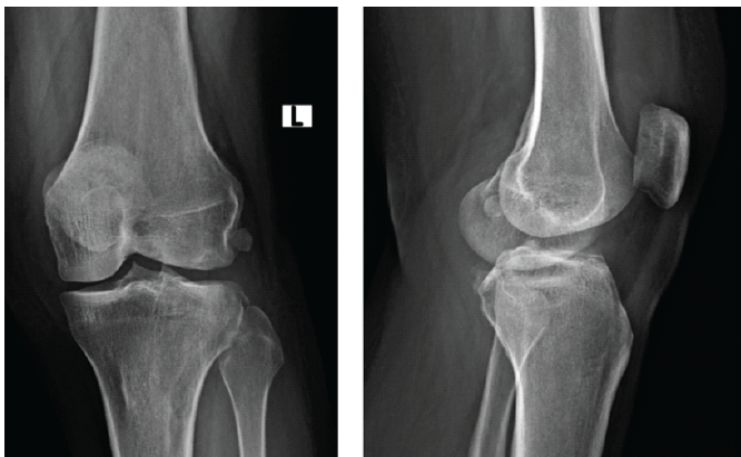
Figure 1: X-ray of left knee suggestive of posterior cruciate ligament avulsion fracture
Magnetic resonance imaging (MRI) of the left knee demonstrated: Complete anterior cruciate ligament (ACL) tear, PCL tibial avulsion fracture, Lateral meniscus posterior root tear, posterior horn medial meniscus tear and significant joint effusion (Fig. 2).
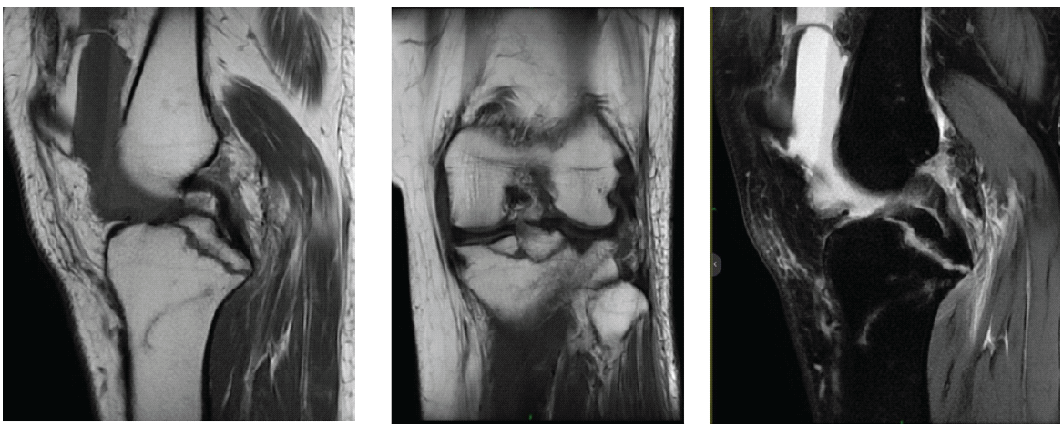
Figure 2: Magnetic resonance imaging of the left knee, suggestive of a complete anterior cruciate ligament tear, posterior cruciate ligament tibial avulsion fracture, lateral meniscus posterior root tear and Posterior horn medial meniscus tear.
Surgery performed
The patient underwent surgery approximately 48 h after injury (second post-injury day) following routine pre-operative evaluation, informed consent, and pre-anesthetic assessment by the consultant anesthesiologist, who declared the patient fit for spinal anesthesia. The decision to proceed with early surgery was not an emergency intervention. Still, it was based on accepted principles for selected multiligament knee injuries associated with repairable meniscal tears, PCL avulsion fracture, and significant knee instability. Early anatomical restoration was planned to optimize functional outcome and reduce the risk of secondary intra-articular damage. There were no neurological or systemic contraindications to surgery, and the intraoperative course under spinal anesthesia was uneventful.
After routine pre-operative evaluation and informed consent, the patient underwent:
- Diagnostic arthroscopy of the left knee
- Arthroscopic hematoma evacuation
- Arthroscopic medial meniscus repair using a suture anchor
- Arthroscopic ACL reconstruction using hamstring autograft
- Arthroscopic lateral meniscus root repair using a transtibial pull-out technique
- Arthroscopic fixation of the PCL avulsion fracture using a transtibial pull-out technique and fixation using a suture disc.
The procedure was completed uneventfully under spinal anesthesia.
Post-operative course
In the immediate post-operative period, the patient developed:
- Drowsiness
- Reduced consciousness
- Dense left-sided weakness.
An urgent neurological evaluation was obtained.
Investigations
Non-contrast computed tomography (CT) of the brain revealed a large acute infarction involving the right cerebral hemisphere and left frontal region (Fig. 3).
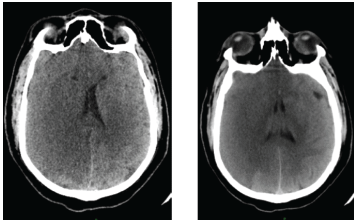
Figure 3: Non-contrast computed tomography brain suggestive of a large acute infarction involving the right cerebral hemisphere and left frontal region.
MRI of the brain confirmed large acute infarcts in the right cerebral hemisphere (Fig. 4)
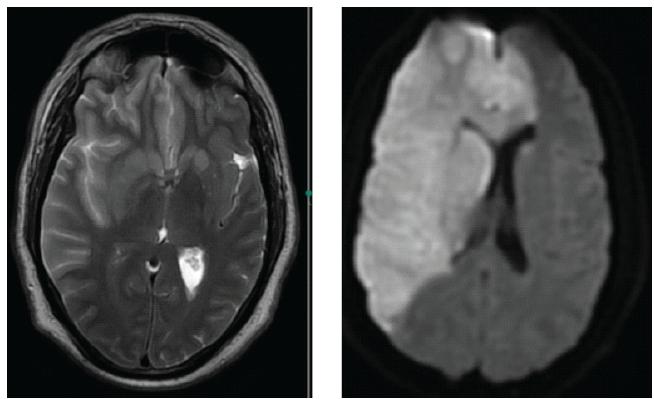
Figure 4: Magnetic resonance imaging of the brain suggestive of large acute infarcts in the right cerebral hemisphere.
CT angiography of the brain and neck (Fig. 5)
- Complete occlusion of the right internal carotid artery (ICA)
- Presence of a dissection flap in the proximal ICA
- No contrast opacification distal to the lesion
- Absent of flow in the right middle cerebral artery (MCA) territory.
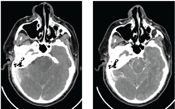
Figure 5: Computed tomography angiography of the brain and neck, suggestive of complete occlusion of the right internal carotid artery (ICA), dissection flap in the proximal ICA with absent flow in the right middle cerebral artery territory.
Magnetic resonance angiography-confirmed complete luminal obstruction of the right ICA and absent flow in the right MCA distribution (Fig. 6).
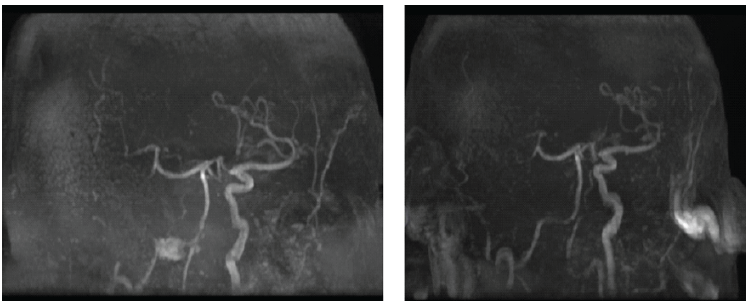
Figure 6: Magnetic resonance angiography suggestive of complete luminal obstruction of the right internal carotid artery and absent flow in the right middle cerebral artery distribution.
Post-operative management
Following neurological deterioration, the patient was shifted to the intensive care unit and managed with endotracheal intubation, mechanical ventilation, inotropic support, and close neurological monitoring.
Medical management included antiedema measures with intravenous mannitol, anticonvulsant therapy with levetiracetam (Levipil), and anticoagulation with low-molecular-weight heparin (Clexane) followed by unfractionated heparin as per neurological advice.
Despite aggressive medical management, the patient’s neurological status continued to deteriorate.
Neurosurgical consultation recommended urgent decompressive craniotomy. However, the patient’s relatives declined further surgical intervention and requested discharge against medical advice.
Knee arthroscopy is generally associated with a very low incidence of major complications. Most reported complications involve local surgical issues rather than systemic neurological events [1].
Carotid artery dissection accounts for approximately 2–5% of ischemic strokes in younger adults and is frequently associated with trauma. Clinical manifestations may be delayed, resulting in a missed diagnosis during the initial assessment [3,4].
In the present case, the patient sustained a scooter-related injury. Although the primary complaint involved the knee, it is likely that an occult cervical vascular injury occurred simultaneously. The perioperative period may have contributed to the progression of thrombosis or embolization from the dissected carotid artery, culminating in a massive ischemic stroke [2,3,5].
An important consideration in this case is the timing of surgery. The patient underwent reconstruction approximately 48 h after injury, after a normal neurological examination and appropriate pre-operative evaluation. Current evidence suggests that neurological manifestations of traumatic carotid artery dissection commonly occur during the subacute phase (1–7 days after injury), with the majority of ischemic events developing during this period. Therefore, postponing surgery by an additional 24 h would not necessarily have prevented the subsequent cerebrovascular event. Moreover, delayed reconstruction of complex multiligament knee injuries may increase the risk of progressive meniscal damage, ligament scarring, persistent instability, and technically more demanding surgery with inferior functional outcomes. Thus, the decision to proceed with early surgery was based on established orthopedic principles rather than emergency necessity. The catastrophic neurological event occurred postoperatively and appeared unrelated to any identifiable pre-operative neurological abnormality.
Current guidelines recommend urgent neuroimaging, vascular assessment, and multidisciplinary management in patients presenting with acute ischemic stroke [6].
The management of cervical carotid artery dissection may include antithrombotic therapy, including antiplatelet or anticoagulant treatment, although the optimal strategy remains controversial [7,8].
Post-operative stroke due to carotid artery pathology has been reported in the literature, emphasizing the importance of early recognition and timely intervention [9].
However, only a few isolated case reports describing catastrophic ischemic stroke following arthroscopic knee surgery have been reported, limiting meaningful comparison with previously reported cases [9].
Although the neurological deterioration occurred in the immediate post-operative period, a direct causal relationship between arthroscopic surgery and ischemic stroke cannot be established. The close temporal proximity between the initial trauma and subsequent surgery makes it difficult to determine whether the cerebrovascular event resulted from an occult traumatic carotid injury, perioperative progression of vascular pathology, or another unrelated mechanism. Therefore, the temporal association observed in this case should not be interpreted as evidence of causation [10].
Limitations
- This report describes a single patient, limiting the generalizability of the findings.
- A definitive causal relationship between arthroscopic knee surgery and the post-operative ischemic stroke cannot be established.
- Despite extensive investigations, the exact etiology of the ICA occlusion remained uncertain.
- Baseline pre-injury vascular imaging was unavailable, making it impossible to determine whether the carotid pathology was pre-existing or trauma-related.
- Screening CT angiography of the neck was not performed immediately after the initial trauma, as there were no clinical features suggestive of blunt cerebrovascular injury; therefore, an occult carotid injury could not be excluded before surgery.
- The close temporal proximity between the road traffic accident and surgery made it difficult to distinguish trauma-related vascular injury from perioperative progression.
- A comprehensive thrombophilia and hypercoagulable workup could not be completed, and other potential embolic or occult vascular etiologies could not be completely excluded.
- The diagnosis of ICA dissection was based on characteristic imaging findings, without surgical or histopathological confirmation.
- The patient was discharged against medical advice after the family declined a decompressive craniectomy, preventing assessment of the outcome following the recommended neurosurgical intervention.
- Long-term neurological and functional follow-up was unavailable, precluding assessment of recovery, disability, or survival.
- As a descriptive single-case report, statistical analysis, estimation of incidence, and assessment of relative risk were not possible.
- Therefore, this report should be interpreted as a description of a rare post-operative neurological complication rather than evidence that arthroscopic knee surgery increases the risk of ischemic stroke. Larger multicenter registries and observational studies are required to better define the incidence, risk factors, and optimal management of such rare events.
Acute ischemic stroke occurring in the immediate post-operative period after arthroscopic knee surgery is an extremely rare and devastating event. Although the exact etiology could not be definitively established in our patient, clinicians should maintain a high index of suspicion whenever unexpected neurological deterioration occurs following an otherwise uneventful procedure.
This report should not be interpreted as evidence that arthroscopic knee surgery independently increases the risk of ischemic stroke; rather, it highlights the importance of prompt recognition and multidisciplinary management of unexpected post-operative neurological deterioration.
Prompt neurovascular evaluation and multidisciplinary management are essential for timely diagnosis and optimal patient care.
Broader recommendations regarding routine vascular screening or changes in perioperative management cannot be derived from a single case. Larger multicenter registries and well-designed observational studies are needed to better define the incidence, risk factors, and optimal management of these exceptionally rare events.
Not all post-operative neurological symptoms are related to anesthesia or routine post-operative recovery. Sudden onset of drowsiness, altered consciousness, or focal neurological deficits after arthroscopic surgery should prompt urgent neuroimaging to rule out catastrophic cerebrovascular events. Early diagnosis may be lifesaving.
References
- 1. Neagoe RM, Bancu S, Muresan M, Sala D. Major vascular injuries complicating knee arthroscopy. Videosurgery Miniinv 2015;10:266-74. [Google Scholar] [PubMed]
- 2. Janczak D, Ziomek A, Lesniak M, Malinowski M, Pormanczuk K, Dorobisz T, et al. The endovascular emergency treatment of an acute carotid artery dissection after Krav Maga training-a case report. Phlebology 2018;33:140-4. [Google Scholar] [PubMed]
- 3. Debette S, Leys D. Cervical-artery dissections: Predisposing factors, diagnosis, and outcome. Lancet Neurol 2009;8:668-78. [Google Scholar] [PubMed]
- 4. Schievink WI. Spontaneous dissection of the carotid and vertebral arteries. N Engl J Med 2001;344:898-906. [Google Scholar] [PubMed]
- 5. Marnane M, Duggan CA, Sheehan OC, Curtin D, Harris D, Williams D,et al. Stroke caused by internal carotid artery dissection: Clinical and radiological characteristics. Cerebrovasc Dis 2011;31:456-63. [Google Scholar] [PubMed]
- 6. Powers WJ, Rabinstein AA, Ackerson T, Adeoye OM, Bambakidis NC, Becker K, et al. Guidelines for the early management of patients with acute ischemic stroke: 2019 Update to the 2018 guidelines for the early management of acute ischemic stroke: A guideline for healthcare professionals from the American heart association/American stroke association. Stroke 2019;50:e344-418. [Google Scholar] [PubMed]
- 7. Menon R, Kerry S, Norris JW, Markus HS. Treatment of cervical artery dissection: A systematic review and meta-analysis. J Neurol Neurosurg Psychiatry 2008;79:1122-7. [Google Scholar] [PubMed]
- 8. Engelter ST, Traenka C, Lyrer PA. Dissection of cervical and cerebral arteries. Curr Neurol Neurosci Rep. 2017;17:59. [Google Scholar] [PubMed]
- 9. Nemoto H, Uchida K, Minami T, Yasuda S, Cho T, Masuda M. A massive postoperative stroke caused by a carotid thrombus that occurred during the surgical repair of an aortic dissection. SAGE Open Med Case Rep 2021;9. [Google Scholar] [PubMed]
- 10. Mokin M, Primiani CT, Ren Z, Kan P, Duckworth E, Linfante I, et al. Endovascular treatment of stroke caused by carotid artery dissection. Brain Sci 2020;10:800. [Google Scholar] [PubMed]





Related Articles in Journal of Orthopaedic Case Reports
 February 1, 2026 A Novel Technique of Rerouting Semitendinosus Graft for Medial Collateral Ligament and Medial Patellofemoral Ligament Reconstruction – In a Polytrauma Patient with Multiligament Injury: Kakran et al. Technique
 October 1, 2026 Posterior Hip Dislocation with Ipsilateral Subtrochanteric Femur Fracture: A Rare Case Report
 October 1, 2026 Staged Bilateral Total Elbow Arthroplasty in Advanced Rheumatoid Elbow Disease: A Case Report
 September 1, 2026 Iliopsoas Abscess as the Initial Manifestation of Periprosthetic Hip Joint Infection after Total Hip Arthroplasty: A Case Report and Literature Review