


Introduction
Traumatic hip dislocation is considered an orthopedic emergency and requires urgent reduction to reduce the risk of avascular necrosis of the femoral head and long-term disability. Posterior hip dislocations account for nearly 85–90% of all traumatic hip dislocations and are commonly caused by dashboard injuries during road traffic accidents [1,2]. They are often associated with acetabular fractures, femoral head fractures, or femoral shaft fractures. However, association with an ipsilateral subtrochanteric femur fracture is extremely uncommon [3,4].
When a subtrochanteric fracture is present, management becomes more difficult because the continuity of the proximal femur is disrupted. This makes closed reduction of the dislocated hip difficult or impossible, and open reduction is often necessary [5]. Delay in reduction beyond 6 h is known to increase the risk of avascular necrosis and poorer long-term outcomes [6].
Only a few such cases have been reported in the literature, and most involve femoral shaft or intertrochanteric fractures rather than true subtrochanteric fractures [7,8,9,10]. We report one such rare case managed with open reduction and proximal femoral nail (PFN).
Case Report
A 35-year-old male was brought to the emergency department after a high-velocity road traffic accident involving a motorcycle collision. He complained of severe pain in the left hip and thigh and was unable to stand or bear weight on the affected limb. There was a history of head injury; however, there was no evidence of chest trauma, abdominal injury, or other major associated injuries.
On examination, the patient was hemodynamically unstable and required initial resuscitation and monitoring by the trauma team. Examination of the left lower limb showed shortening, flexion, adduction, and internal rotation, which suggested posterior dislocation of the hip. There was swelling, deformity, and marked tenderness over the proximal thigh. Distal pulses were palpable, and there was no sensory or motor deficit suggestive of sciatic nerve involvement.
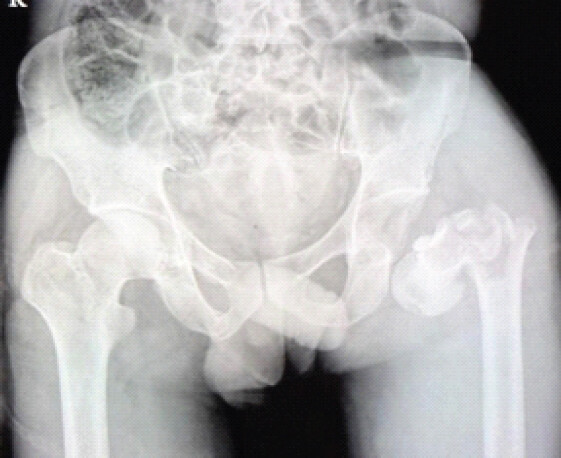
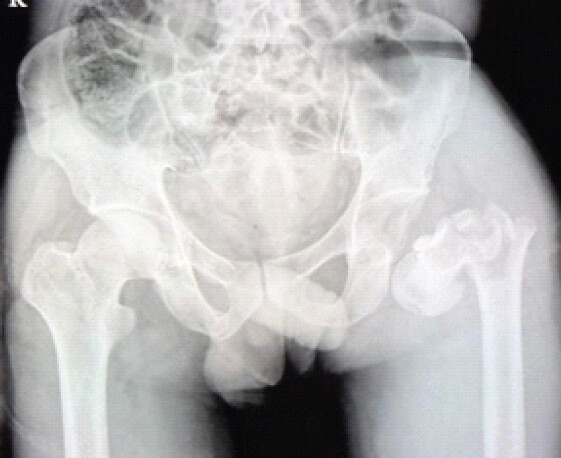
Plain radiographs of the pelvis and left femur showed posterior dislocation of the left hip with an ipsilateral subtrochanteric fracture of the femur. No associated acetabular fracture was seen on X-ray (Fig. 1). Computed tomography of the pelvis and left femur confirmed posterior dislocation of the left femoral head with an ipsilateral subtrochanteric femoral fracture. The femoral head and acetabulum were intact, with no associated acetabular fracture or intra-articular osseous fragment.
Although urgent reduction of the hip dislocation is generally recommended, definitive surgical intervention was initially deferred because the patient was not medically fit for anesthesia and surgery at presentation. The associated head injury necessitated further neurological evaluation and delayed anesthetic clearance. Following adequate resuscitation, stabilization, and optimization of the patient’s general condition, definitive surgical management was undertaken within 72 h of the trauma.
Surgical technique
The patient was taken to the operating room after anesthesia clearance. Under spinal anesthesia, he was placed in the lateral decubitus position on a radiolucent table. All pressure points were padded properly, and the entire limb was prepared and draped.
Because of the associated subtrochanteric fracture, standard closed reduction of the hip was not possible due to loss of the proximal femoral lever arm. Initially, we attempted to stabilize the subtrochanteric fracture first so that the dislocated femoral head could be reduced later in the usual manner after restoration of femoral continuity. However, because of the unstable fracture pattern and poor control of the proximal fragment, this could not be achieved.
A decision was then made to reduce the hip first through an open posterior approach. A standard Kocher–Langenbeck approach was used. After skin incision and fascial exposure, the gluteus maximus was split in line with its fibers to expose the posterior aspect of the hip (Fig. 2).
The femoral head was found displaced posteriorly outside the acetabulum. Since the hip had remained dislocated for some time, the acetabulum was filled with hematoma and clots. These were thoroughly evacuated before reduction. This was important not only for achieving proper concentric reduction but also for reducing the chances of post-operative infection.
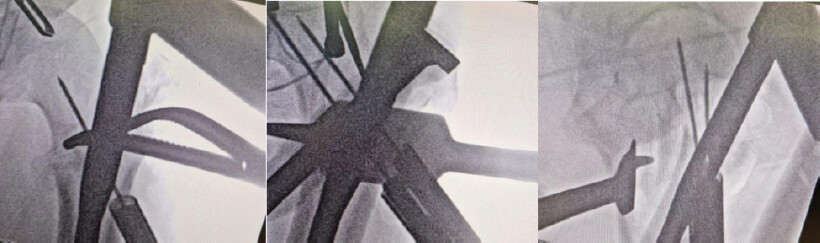
Because direct manipulation through the fractured femur was difficult, a Steinmann pin was inserted into the proximal fragment and used as a joystick. Gentle traction and controlled rotation helped guide the femoral head back into the acetabulum under direct vision. Reduction was confirmed clinically and under fluoroscopy (Fig. 3).
After reduction of the hip, fixation of the subtrochanteric fracture was carried out in the same lateral position, although the original plan had been to shift to the supine position after hip reduction. The entry point for the PFN was made through the piriformis fossa under direct vision.
During entry preparation, it was noted that the greater trochanter was also fractured and became further comminuted. The fragments were reconstructed and tied together using strong Ethibond sutures to restore continuity and maintain the abductor mechanism.
Because the fracture was highly unstable, sequential reaming was done carefully to avoid further displacement. A PFN was then inserted under fluoroscopic guidance. Proximal and distal locking was completed, and alignment, limb length, and rotational stability were checked.
Final fluoroscopic images confirmed satisfactory reduction of the hip joint, proper fracture alignment, and good implant position. The wound was washed thoroughly and closed in layers over a suction drain.
Post-operative rehabilitation and follow-up
After surgery, the patient was kept non-weight-bearing with walker support. Ankle pump exercises, quadriceps strengthening, and bedside hip and knee range-of-motion exercises were started as tolerated. Strict non-weight-bearing walking was advised for the first 6 weeks.
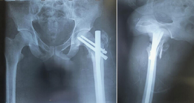
Post-operative radiographs showed maintained reduction of the hip joint, good alignment of the subtrochanteric fracture, and satisfactory implant position without any early implant-related problems (Fig. 4).
At 15 days follow-up, the surgical wound was healthy, and sutures were removed. The patient had mild pain, which was gradually improving, and was mobilizing comfortably with walker support. There was no wound infection, neurovascular deficit, implant failure, redislocation, or any other early post-operative complication.
As the follow-up period is still short, assessment of fracture union, avascular necrosis, post-traumatic arthritis, and long-term functional outcome is ongoing. The patient remains under regular follow-up.
Discussion
Posterior hip dislocation with an ipsilateral subtrochanteric femur fracture is an uncommon injury caused by high-energy trauma. The usual mechanism is force transmission through a flexed hip and knee, which pushes the femoral head posteriorly out of the acetabulum while also producing a fracture in the proximal femur [1,3].
In isolated posterior hip dislocation, closed reduction is usually possible. However, when a subtrochanteric fracture is present, the proximal femoral continuity is lost, and the normal lever arm required for reduction is absent. This makes closed reduction difficult and often requires open reduction [5]. In our case, a Steinmann pin was used as a joystick to control the proximal fragment and help reduce the femoral head safely.
Timing of reduction is important because delayed reduction increases the risk of avascular necrosis. Ideally, reduction should be done within 6 h [6]. In our patient, surgery was delayed because of hemodynamic instability, but it was completed within 72 h after proper stabilization.
The order of treatment in such injuries is still debated. Some authors recommend reducing the hip first, while others prefer fracture fixation first to restore femoral continuity [7,8]. In our case, fracture fixation was attempted first, but because of instability, this was not possible. Therefore, the hip was reduced first, and fracture fixation was completed afterward in the same lateral position.
PFN was chosen because it provides good biomechanical stability in subtrochanteric fractures. It acts as a load-sharing implant, requires less soft tissue dissection, preserves fracture biology, and allows earlier rehabilitation compared to plate fixation [11]. Greater trochanteric comminution can make nail entry difficult, as seen in our case, and Ethibond reconstruction helped maintain stability.
Another important step was evacuation of the hematoma from the acetabulum. A retained hematoma can interfere with proper reduction and may also increase the risk of post-operative infection. Careful post-operative wound care is therefore necessary.
To the best of our knowledge, very few cases of true posterior hip dislocation associated specifically with ipsilateral subtrochanteric femur fracture have been reported. Most available reports involve femoral shaft, femoral neck, or intertrochanteric fractures rather than true subtrochanteric fractures [8,9,10,12]. This makes the present case rare and technically challenging. Previously reported similar cases are summarized in Table 1.
Previously reported similar cases
| Author | Year | Injury pattern | Reduction type | Timing of surgery | Definitive management | Final outcome |
|---|---|---|---|---|---|---|
| Iftekhar et al. | 2020 | Posterior hip dislocation with ipsilateral femoral shaft fracture | Closed reduction | Early intervention | Closed reduction+interlocking intramedullary nailing | Good functional recovery |
| Su et al. | 2020 | Posterior hip dislocation with femoral neck and intertrochanteric fracture | Open reduction | Early intervention | Open reduction+internal fixation | Satisfactory fracture union |
| Nohmi S et al. | 2022 | Posterior hip fracture-dislocation with acetabular posterior wall fracture and trochanteric fracture | Open reduction | Early intervention | ORIF of acetabulum+proximal femur fixation | Good radiological and clinical outcome |
| Zarei et al. | 2023 | Posterior hip dislocation with posterior wall acetabular fracture and femoral shaft fracture | Open reduction | Early intervention | Open reduction+fracture fixation | Good post-operative recovery |
| Wuhib et al. | 2024 | Obturator (anterior) hip dislocation with ipsilateral subtrochanteric femur fracture | Open reduction | Early intervention | Open reduction+antegrade intramedullary nailing | Good clinical outcome |
| Present case | 2026 | Posterior hip dislocation with ipsilateral subtrochanteric femur fracture | Open reduction | Within 72 h of trauma | Open reduction using Steinmann pin joystick+PFNN fixation | Early post-operative recovery satisfactory |
ORIF: Open reduction internal fixation, PFN: Proximal femoral nail
Limitation
The main limitation of this report is the short follow-up period of only 15 days. Because of this, long-term complications such as avascular necrosis of the femoral head, post-traumatic arthritis, fracture healing, and final functional outcome cannot yet be assessed. Continued follow-up is necessary.
Conclusion
Posterior hip dislocation with ipsilateral subtrochanteric femur fracture is a rare and difficult injury that requires early recognition and proper surgical planning. Closed reduction is often not possible because of the disrupted femoral lever arm. Open reduction using a Steinmann pin as a joystick, careful evacuation of acetabular hematoma, and stable fixation with PFN can provide good early results. Long-term follow-up remains important to assess final outcome and complications.
Clinical Message
Timely reduction is crucial; Steinmann-pin joystick-assisted open reduction facilitates hip reduction, followed by stable PFN fixation for early mobilization in complex ipsilateral subtrochanteric fractures.
Conflict of Interest:
Nil
Source of Support:
Nil
Consent
The authors confirm that informed consent was obtained from the patient for publication of this article
How to Cite this Article
Goyal A, Lakhera VK, Agarwal S. Posterior Hip Dislocation with Ipilateral Subtrochanteric Femur Fracture: A Rare Case Report. Journal of Orthopaedic Case Reports 2026 October;16(10): 124-128.
References
- Epstein HC. Traumatic dislocations of the hip. Clin Orthop Relat Res 1973;92:116-42. [Google Scholar] | [PubMed]
- Brav EA. Traumatic dislocation of the hip: Army experience and results over a twelve-year period. J Bone Joint Surg Am 1962;44:1115-34. [Google Scholar] | [PubMed]
- Thompson VP, Epstein HC. Traumatic dislocation of the hip; a survey of two hundred and four cases covering a period of twenty-one years. J Bone Joint Surg Am 1951;33:746-78. [Google Scholar] | [PubMed]
- Stewart MJ, Milford LW. Fracture-dislocation of the hip; an end-result study. J Bone Joint Surg Am 1954;36:315-42. [Google Scholar] | [PubMed]
- Tornetta P. Hip dislocation associated with femoral shaft fracture: Current concepts. J Am Acad Orthop Surg 1997;5:27-36. [Google Scholar] | [PubMed]
- Hougaard K, Thomsen PB. Traumatic posterior dislocation of the hip--prognostic factors influencing the incidence of avascular necrosis of the femoral head. Arch Orthop Trauma Surg (1978) 1986;106:32-5. [Google Scholar] | [PubMed]
- Iftekhar N, Rasool T, Zhitny VP, Rasool S. Ipsilateral posterior hip dislocation and femur shaft fracture - a rare presentation. J Orthop Case Rep 2020;10:3-5. [Google Scholar] | [PubMed]
- Su C, Liu Y, Wu P, Yuan J, Lang J, Wu C. Ipsilateral femoral neck and intertrochanteric fractures with posterior dislocation of the hip: A report of two cases. SAGE Open Med Case Rep 2020;8:1-6. [Google Scholar] | [PubMed]
- Nohmi S, Oishi H, Sakamoto Y. Posterior hip fracture-dislocation associated with posterior wall fracture of the acetabulum and ipsilateral comminuted trochanteric fracture of the femur: A case report. Int J Surg Case Rep 2022;94:107075. [Google Scholar] | [PubMed]
- Zarei R, Tavassoli M, Baroutkoub M, Afzal S. Concurrent ipsilateral fractures of acetabular posterior wall and femoral shaft associated with posterior hip dislocation: A case report and literature review of a rare injury. Int J Surg Case Rep 2023;113:109035. [Google Scholar] | [PubMed]
- Wiss DA, Brien WW. Subtrochanteric fractures of the femur. Results of treatment by interlocking nailing. Clin Orthop Relat Res 1992;283:231-6. [Google Scholar] | [PubMed]
- Wuhib G, Wuhib M, Mekonnen L, Assefa M. Traumatic obturator dislocation of the hip associated with ipsilateral subtrochanteric femur fracture in a young adult: A case report. J Med Case Rep 2024;18:607. [Google Scholar] | [PubMed]
© 2026 Journal of Orthopaedic Case Reports - Published by Indian Orthopaedic Research Group
About the Authors
-

Amrit Goyal
-

Vishal Kumar Lakhera
-

Sudhanshu Agarwal
Related Articles in Journal of Orthopaedic Case Reports
 April 1, 2026 Management of Subtrochanteric Femur Fracture with Pre-existing Grade IV Psoriatic Hip Arthritis Using Primary Total Hip Arthroplasty with a Long Distal-Loading Stem: A Case Report
 October 1, 2026 A Prospective Study of Functional Outcome of Intertrochanteric Fracture of Proximal Femur in Adults Treated with Proximal Femoral Nail
 October 1, 2026 Posterior Fibular Entrapment in a Bosworth Fracture-Dislocation: A Case Report and Review of Diagnostic and Operative Considerations
 October 1, 2026 Recurrent Ventral Scapular Osteochondroma Presenting with Pseudo-winging in an Adolescent: A Case Report and Follow-up of a Previously Published Case