


Introduction
Chronic osteomyelitis is a vexing condition that was traditionally treated using lengthy antibiotic regimes and repeated surgical debridement [1]. It represents a major economic burden for every healthcare system and substantially affects the quality of life of affected patients and their families [1]. Defect management following eradication of infection is the foremost challenge, for which a multitude of options, ranging from vascularized or non-vascularized bone grafting to bone transport, are available, each having its own benefits and demerits. The aim of this study was to put forth an innovative new way of managing large peri-articular cavitary defects, using a modified Masquelet technique, combining the structural durability of a non-vascularized fibula with the superior osteoinductive properties of cancellous bone graft, along with the addition of a surgical polypropylene mesh. We have termed this innovative method the “bell-tent technique.”
Case Report
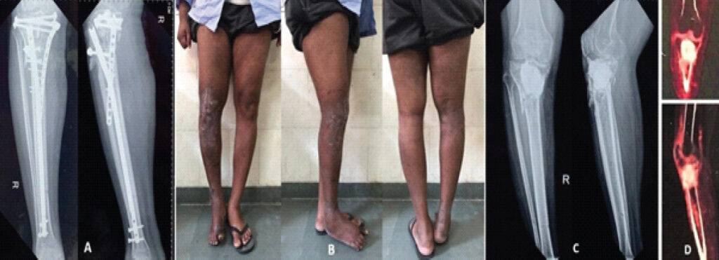
A 34-year-old male soldier, a non-smoker, suffered a compound fracture of the proximal tibia during a road accident, for which he underwent wound debridement and internal fixation of the fracture using a nail-plate construct (Fig. 1a). He subsequently developed surgical site infection, for which multiple debridements along with a cement spacer insertion were performed. A few months later, he presented to our center with pain and an inability to bear weight on the affected limb. Clinical examination revealed a stiff knee with only a jog of movement, a discharging sinus, and a varus deformity of the proximal tibia, along some abnormal mobility at the fracture site (Fig. 1b). Radiographs showed a non-union fracture of the proximal tibia with a cement spacer in situ (Fig. 1c). An 18-flourodeoxyglucose positron emission tomography (FDG-PET) scan was performed to delineate the anatomical extent of infection within the bone, which revealed tracer concentration in the proximal tibial metaphyseal region (Fig. 1d). Quantitative C-reactive protein (Q-CRP) values were recorded for serial monitoring. The patient was given an “antibiotic-free holiday” of 2 weeks before contemplating any surgical intervention.
Using the previous midline scar and a medial parapatellar arthrotomy (Fig. 2a), the knee joint was exposed after clearing all intra- and extra-articular adhesions. The cement spacer was removed carefully without damaging the existing bone stock. After obtaining five specimens for culture from the medullary canal, posterior cortex, subchondral bone, surrounding soft tissue, and joint fluid, an extensive debridement was performed until satisfactory clearance of all inviable bone and soft tissues was obtained. The posterior cortex of the proximal tibia was intact, and on making multiple cortical drill holes, fresh punctate bleeding was observed. The entire anterior cortex, comprising approximately 35–40% of the circumference, was absent, exposing a large, 6 × 8 cm cavitary defect in the proximal tibia, extending up to the posterior cortex (Fig. 2b). The articular surface was intact, with around 5 mm of subchondral bone centrally. The meta-diaphyseal junction was in a state of fibrous union.
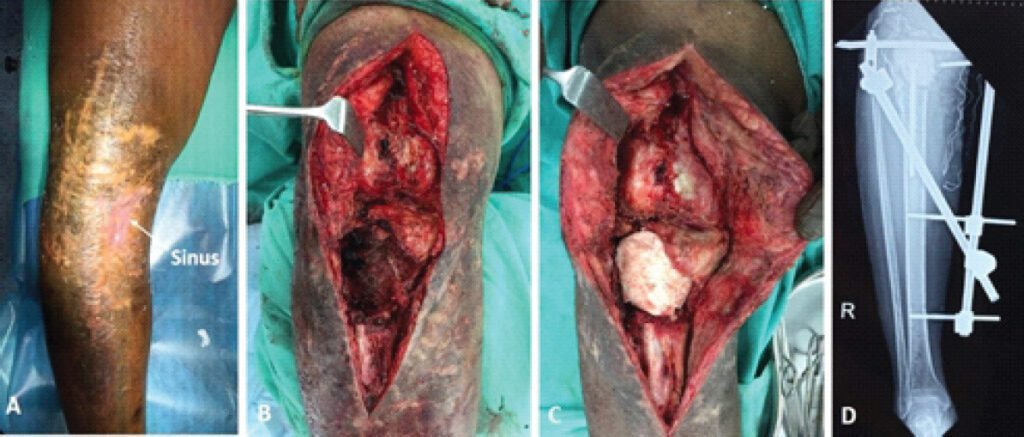
Next, the medullary canal was reamed with flexible reamers and irrigated using pulsatile jet lavage. An antibiotic-impregnated cement spacer mounted on an Ilizarov rod (as a stem) was prepared, to which 4 g of Vancomycin and 4 g of Meropenem were added. The spacer was carefully inserted into the defect, followed by application of a temporary external fixator (Fig. 2c and d). Postoperatively, 4/5 cultures were positive for Klebsiella pneumoniae, sensitive to Meropenem and Tigecycline; the patient was started on 2 weeks of injectable culture-specific antibiotics, followed by oral antibiotics for 6 weeks. At the end of 2 months, the wound condition improved, and serial Q-CRP monitoring showed a declining trend, which prompted us to plan a definitive reconstructive procedure.
Surgical technique
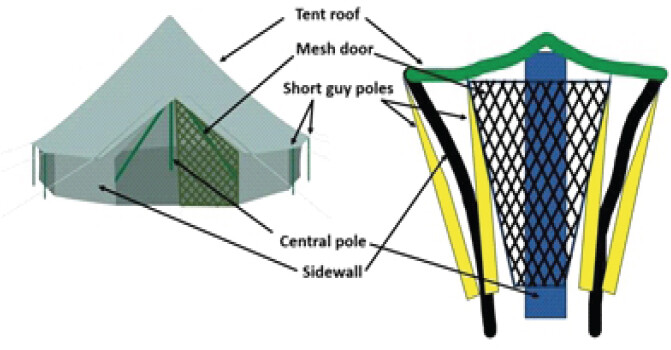
Through the previous surgical approach, the knee joint was exposed, the cement spacer removed, and a thorough debridement with pulsatile lavage was performed. As before, intra-operative samples were sent from different anatomical sites, which later came out negative for any growth. For reconstruction of the cavitary defect, the patient’s ipsilateral fibula, along with iliac crest bone graft, was harvested and combined in a unique way, which we have termed the “bell-tent technique,” akin to the design of a camping tent (Fig. 3). Details of this technique are as follows:
-
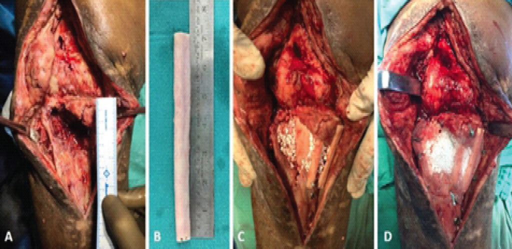
1. To provide a robust central pivot, the length of the fibula required was estimated to be at least twice the maximum length of the defect. Hence for a 6 cm-long cavity (Fig. 4a), a graft measuring 18 cm was harvested, sparing the upper and lower fourths of the fibula (Fig. 4b). The graft was cut into two portions, one of 12 cm, which became the “central pole;” the other 6 cm segment was cut into three longitudinal struts of cortical bone, much like “short-guy poles” of a bell-tent that support the roof circumferentially (Fig. 3 and 4c). The posterior tibial cortex was in a state of fibrous union, which facilitated prying open of the anterior cortical defect to impact the fibular strut in the canal. The canal itself was widened beforehand using appropriately sized K-nails and flexible reamers, up to the required depth, which enabled unhindered passage of the fibula. The strut was inserted antegrade and was then tapped in a retrograde fashion using a bone holding forceps, until its subchondral position was confirmed by direct vision and by fluoroscopy. Once secured, the strut was held in position by two cortical screws, while K-wire bits were used to affix the short-guy poles.
-
2. Next, a mixture of morselized iliac crest bone graft and synthetic bone graft (Allogran R, Biocomposites, USA) in the ratio of 3:1, was prepared and packed into the void between the central pole, side walls, and roof (Fig. 4c). The anterior cortical defect was closed using a surgical polypropylene mesh (Ethicon, J&J, USA), analogous to a “mesh door,” to contain the graft within the cavity (Fig. 3 and 4d).
-
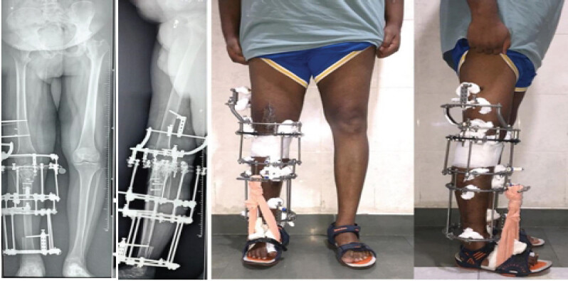
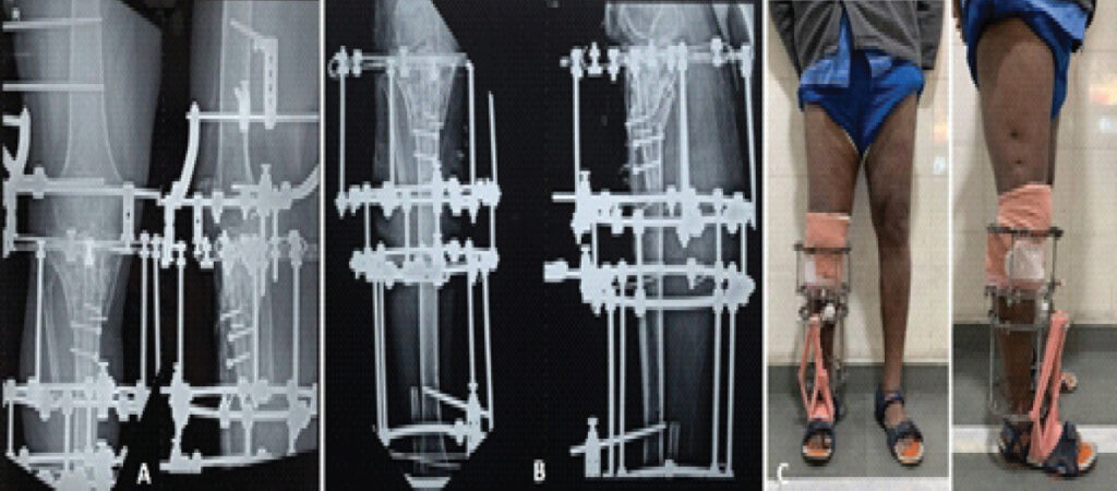
3. Following wound closure, a pre-constructed, knee-spanning Ilizarov frame was applied, using multiple subchondral “raft wires,” to support the articular surface, affixed to a radiolucent carbon-fiber ring, that enabled clear visualization of the joint and status of graft integration (Fig. 5). The varus malunion was corrected acutely at this stage; post-operative long leg films showed good limb alignment (Fig. 5). Weight bearing was permitted as per tolerance, using a walking aid. In about 4 months, the graft looked sufficiently consolidated to remove the upper (knee-spanning) part of the frame and commence range of motion (ROM) exercises (Fig. 6a, b, c). Active quadriceps strengthening exercises and full weight bearing were permitted with the help of two crutches. The knee ROM achieved was 0–30° with a 10° extensor lag.
-
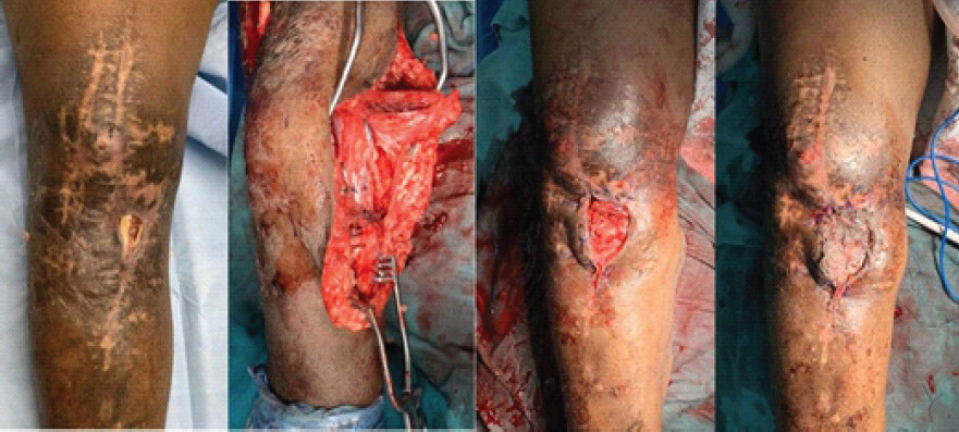
4. Clinico-radiological union was seen at 9 months post-operative, and the frame was removed. By this time, the fragile skin over the anteromedial aspect of the proximal tibia had dehisced, exposing the underlying bone, for which a medial gastrocnemius flap, along with patellofemoral adhesiolysis, was performed (Fig. 7). The previously inserted mesh was found to be adherent to the overlying redundant skin and had to be excised in totality, warranting the need for flap cover. Within the erstwhile cavity, the graft was completely incorporated, hence the screws and wire-bits were removed, leaving behind no metallic implants. Continuous passive motion as well as strengthening exercises were commenced.
Follow-up and Results
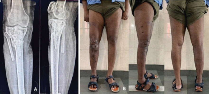
At final follow-up of 37 months, he demonstrated a clinically stable knee with ROM of 0–70° (5° extensor lag persisted), which enabled him to use stairs and public transport, and radiographs showed a consolidated proximal tibia in good alignment (Fig. 8). Table 1 shows a comparative analysis of his radiological deformity parameters before and following the intervention. No major complications apart from superficial pintract infections were encountered.
Comparison of pre- and post-operative radiographic parameters
| Parameter | Pre-operative | Post-operative |
|---|---|---|
| MPTA | 81° | 86° |
| PPTA | 87° | 84° |
| MAD | Not done | 9 mm |
| LLD | 28 mm | 10 mm |
MPTA: Medial proximal tibial angle, PPTA: Posterior proximal tibial angle, MAD: Mechanical axis deviation, LLD: Limb length discrepancy
Discussion
Chronic osteomyelitis is a progressive, low-grade inflammation of bone caused by pathogens, resulting in destruction and sequestrum formation [2]. The symptoms include chronic pain, swelling, persistent sinus or drainage, impaired wound healing, and their duration may vary considerably, with intermittent waxing and waning, giving the false impression of a remission. Imaging modalities include plain radiography, magnetic resonance imaging, and FDG-PET, which has the highest sensitivity and specificity in detecting inflammatory activity in remote locations such as the medullary cavity [3]. The most widely used classification system of chronic osteomyelitis in adults is the Cierny-Mader classification [4]. We have limited our discussion to the management of Type 3 (localized) osteomyelitis, with full-thickness cortical involvement, requiring complex dead-space management.
Adequate surgical debridement remains the cornerstone of treatment, and soft-tissue reconstruction is an obligatory part of the procedure. In the tibia, the anteromedial cortex has the least muscle cover and blood supply, while the posterior cortex has a more vascular muscle bed and a better blood supply; hence, it was not disturbed. Antibiotic delivery and duration remain a topic of contention; however, there is enough evidence to support polymethylmethacrylate (PMMA) as an efficient vehicle for local antibiotic delivery in high concentrations [5]. A number of alternatives with antimicrobial and osteoconductive properties, such as calcium sulfate-hydroxyapatite composite (Cerament G/V, Bonesupport, Lund, Sweden), calcium sulfate (Stimulan, Biocomposities, USA), and Bioactive glass S53P4 (BonAlive, Turku, Finland), have been used with reasonable success for infection control as well as defect management [6,7]. Bone regeneration using the time-tested Ilizarov method has shown wonderful results, but it is interminable, and active cooperation of the patient is essential [8]. Isolated uni-cortical defects have been successfully treated with hemi-corticotomy, leaving the posterior cortex intact, and gradual segmental transport using the Ilizarov technique [9]. The proponents of this method of semi-focal bone transport have reported shorter external fixator indices and fewer complications as compared to traditional Ilizarov bone transport [10].
The induced membrane technique of Masquelet has been widely used for defect management with encouraging results [11]. Cancellous bone grafting can be combined with a non-vascularized fibular autograft, as in our described technique, to provide structural support and reduce the quantity of cancellous graft required [12]. We believe this results in earlier weight bearing and prevents a late collapse. Vascularized bone grafts, by virtue of their inherent vascularity, unite more rapidly with the host bone and are more resistant to infection; however, their application in the management of long bone defects remains limited, due to inaccessibility to microsurgery at many centers [13]. Our described technique uses a polypropylene mesh to substitute the deficient anterior wall and contain a large volume of bone graft within the cavity. Kalantar et al. described a modified Masquelet technique using polypropylene mesh and PMMA together for the treatment of large traumatic bone defects, without any major complications [14]. It is prudent to state that polypropylene invokes severe tissue reactions and carries a substantial risk of catching an infection; hence, it should be used with caution.
Conclusion
Chronic cavitary bone defects involving the periarticular region of long bones can be successfully salvaged with the described “bell-tent technique,” in combination with stable external fixation. The method offers the unique advantage of early weight bearing and joint mobilization, while minimizing the drawbacks associated with prolonged duration of external fixation. Further studies are required to establish its reproducibility and long-term outcomes.
Clinical Message
Soft-tissue reconstruction should be planned in conjunction with bone reconstruction and preferably done in the same sitting, to reduce the chances of wound dehiscence and shorten the duration of treatment.
Conflict of Interest:
Nil
Source of Support:
Nil
Consent:
The authors confirm that informed consent was obtained from the patient for publication of this article
References
- Panteli M, Giannoudis PV. Chronic osteomyelitis: What the surgeon needs to know. EFORT Open Rev 2016;1:128-35. Google Scholar | PubMed
- Lew DP, Waldvogel FA. Osteomyelitis. Lancet 2004;364:369-79. Google Scholar | PubMed
- enter V, Albert NL, Brendel M, Fendler WP, Cyran CC, Bartenstein P. FDG PET accurately differentiates infected and non-infected non-unions after fracture fixation. Eur J Nucl Med Mol Imaging 2017;44:432-40. Google Scholar | PubMed
- Cierny G 3rd, Mader JT, Penninck JJ. A clinical staging system for adult osteomyelitis. Clin Orthop Relat Res 2003;414:7-24. Google Scholar | PubMed
- Hake ME, Young H, Hak DJ, Stahel PF, Hammerberg EM, Mauffrey C. Local antibiotic therapy strategies in orthopaedic trauma: Practical tips and tricks and review of the literature. Injury 2015;46:1447-56. Google Scholar | PubMed
- Niemann M, Graef F, Ahmad SS, Braun KF, Stöckle U, Trampuz A. Outcome analysis of the use of Cerament® in patients with chronic osteomyelitis and corticomedullary defects. Diagnostics 2022;12:1207. Google Scholar | PubMed
- Van Vugt TA, Heidotting J, Arts JJ, Ploegmakers JJ, Jutte PC, Geurts JA. Mid-term clinical results of chronic cavitary long bone osteomyelitis treatment using S53P4 bioactive glass: A multi-center study. J Bone Jt Infect 2021;6:413-21. Google Scholar | PubMed
- Paley D, Maar DC. Ilizarov bone transport treatment for tibial defects. J Orthop Trauma 2000;14:76-85. Google Scholar | PubMed
- Emara KM. Hemi-corticotomy in the management of chronic osteomyelitis of the tibia. Int Orthop 2002;26:310-3. Google Scholar | PubMed
- Wang Q, Ma T, Li Z, Zhang K, Huang Q. Semi-focal bone transport versus traditional bone transport technique for the management of large tibial bone defects after trauma. Sci Rep 2024;14:7982. Google Scholar | PubMed
- Masquelet AC, Begue T. The concept of induced membrane for reconstruction of long bone defects. Orthop Clin North Am 2010;41:27-37. Google Scholar | PubMed
- annamani S, Rachakonda KR, Tellakula Y, Takkalapally H, Maryada VR, Reddy AG. Combining non-vascularized fibula and cancellous graft in the masquelet technique: A promising approach to distal femur compound fracture management with large defects. Injury 2024;55:111233. Google Scholar | PubMed
- Hertel R, Pisan M, Jakob RP. Use of the ipsilateral vascularised fibula for tibial reconstruction. J Bone Joint Surg Br 1995;77:914-9. Google Scholar | PubMed
- Kalantar SH, Saffar H, Hoveidaei AH. Bone reconstruction with modified Masquelet technique in open distal femoral fractures: A case series. BMC Musculoskelet Disord 2024;25:26. Google Scholar | PubMed
© 2026 Journal of Orthopaedic Case Reports - Published by Indian Orthopaedic Research Group
About the Authors
-

Rajiv Kaul
-

Zafar Israil
-

Manish Prasad
-

Neha Akhoon
Related Articles in Journal of Orthopaedic Case Reports
 June 1, 2026 Atypical Bilateral Cystic Foot Swellings with Osteomyelitis-Like Features: A Diagnostic Dilemma
 November 1, 2025 Primary Bone Lymphoma of the Tibia Mimicking Chronic Osteomyelitis: A Case Report
 March 1, 2025 Creation of Single-Bone Forearm in Case of Diffuse Chronic Osteomyelitis of Diaphysis of Radius in an Adolescent Treated with Two Staged Procedure: A Case Report
 November 1, 2024 Unusual Sites of Brodie’s Abscess and the use of Calcium Sulfate Beads: A Case Series