


Introduction
Patella dislocation is one of the leading causes of traumatic knee lesions at 3%, with the majority of patients under 20 years old [1]. This injury is often encountered by athletic trainers, emergency medicine providers, and orthopedic surgeons. In comparison with other joint dislocations, patellar dislocations are often considered lower complexity and can be managed by non-orthopedic providers [2]. However, the elusive irreducible patella does exist, and it is important to understand so that healthcare providers know when to seek specialist care for these injuries. Similarly, atypical presentations of patellar dislocations, such as the knee stuck in extension instead of flexion, should alert first responders to engage specialist care early [3]. With the aging population, it is particularly important to be aware of irreducible patella dislocations, as degenerative changes to the knee can prevent simple closed reduction maneuvers.
Case Report
An 81-year-old woman presented to the emergency department for a left lateral patellar dislocation after tripping and falling while crossing the street. While she had a remote history of contralateral patellar dislocation at 16 years old, she denied any history of left knee pain or prior dislocation of the left patella. After failed closed reduction attempts by the emergency medicine team involving analgesia without sedation and repeat attempts of controlled knee flexion with lateral to medial pressure along the lateral patella border, the orthopedic surgery team was consulted.
On examination, the patient’s left leg was locked in extension, secondary to pain, with the patella dislocated to the superolateral aspect of the knee. There was an abrasion over the tibia tubercle, but the skin was otherwise intact. She was unable to straight leg raise, presumably secondary to pain. Alternatively, the extensor mechanism may have been due to over-tensioning due to the dislocation. She was neurovascularly intact.
X-rays demonstrated a superolaterally displaced patella with no acute fracture. The tibiofemoral joint space was preserved with moderate tricompartmental osteoarthritis (Fig. 1, 2, 3).
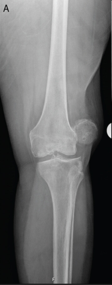
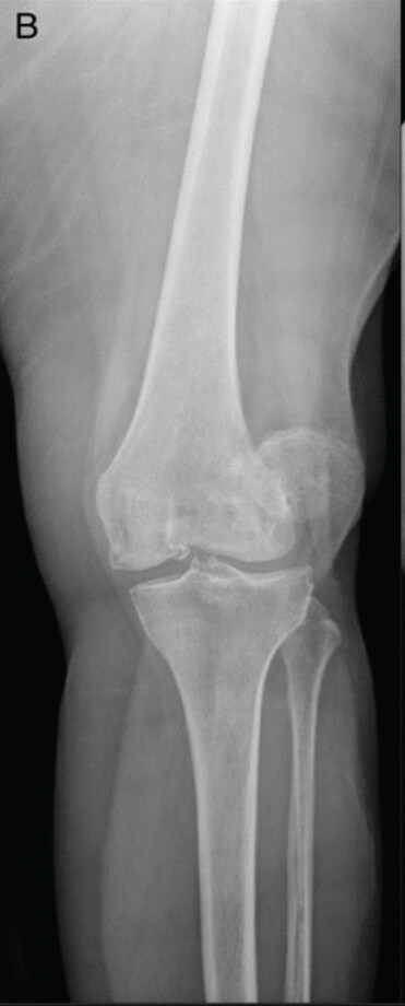
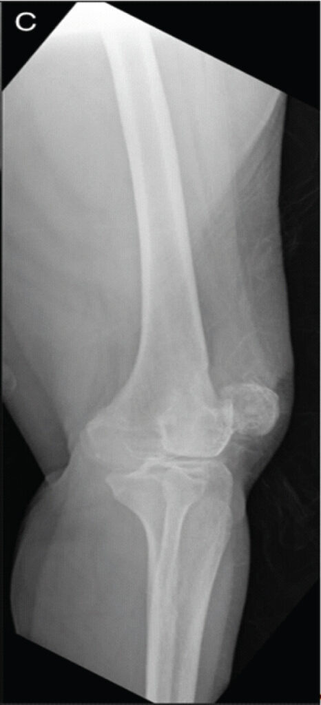
Multiple failed attempts at closed reductions were attempted by the orthopedics team. These attempts included fluoroscopy and conscious sedation. A variety of maneuvers were attempted, including pressure along the lateral patella to guide back into track with controlled flexion of the knee, hyperextension of the knee, varus stress, valgus stress, internal and external hip rotation, hyperflexion of the hip to relax the quadriceps, and combinations of the above.
With every attempt, the patella remained displaced. It was noted that the patella could be shifted medially, but never to the midline of the knee. Once at the lateral edge of the trochlea, the patella would not translate further medially, but instead flip internally on its axial axis, stopping with the lateral patella border facing anteriorly. The articular surfaces were unable to be reduced. Due to the multiple failed attempts at closed reduction, the patient was admitted for further care, including pain control, magnetic resonance imaging (MRI) of the knee, and planned open reduction.
The MRI demonstrated full-thickness tearing of the medial retinaculum, a moderate to high-grade sprain of the lateral retinaculum, a large knee joint effusion, and osseous contusion deformity at the anterior tibial tubercle with marrow edema. The medial border of the patella was impacted into an osseous defect of the lateral femoral condyle (Fig. 4, 5, 6, 7). Additional findings include quadriceps and patellar tendinosis, degeneration of the lateral meniscus, a lateral collateral ligament sprain, maceration of the medial meniscus, and a medial collateral ligament sprain.
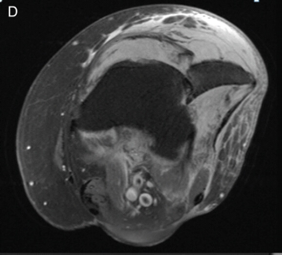
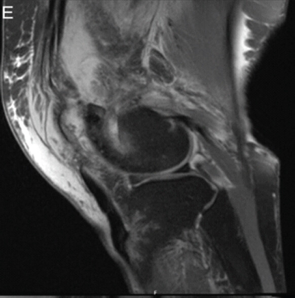
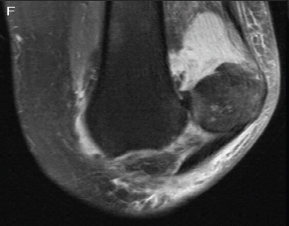
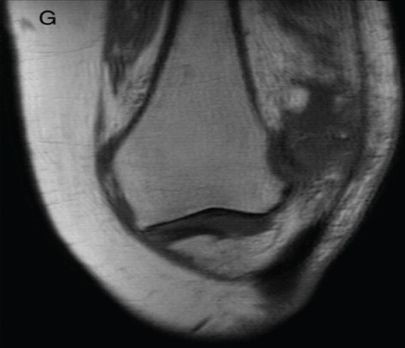
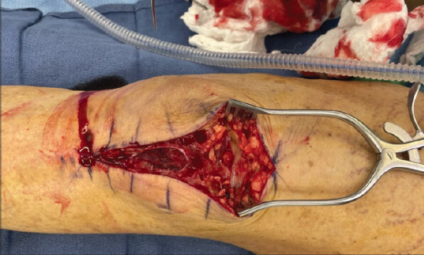
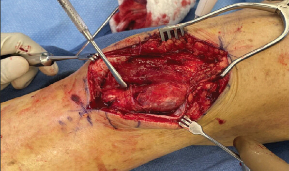
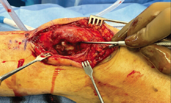
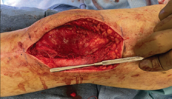
The patient was subsequently taken to the operating room for an open reduction. A standard midline incision with sharp dissection was performed. There was significant soft-tissue trauma from the injury (Fig. 8). The patella was identified with its lateral border tilted anterolaterally and buttonholed through the lateral retinaculum and soft tissue (Fig. 9 and 10). The medial border of the patella was engaged on the lateral femur, which was noted to have a reciprocal impaction fracture. The patella was disengaged from the soft tissue and manually reduced, retaining reduction throughout the full knee range of motion. Identified, small loose bodies were removed. No additional osteophyte excision was performed. The lateral retinaculum was advanced from its retracted medial position to the lateral remanent edge, and its tears were repaired with 0-vicryl. The proximal medial retinaculum was found to be significantly attenuated, but not torn, prompting its imbrication with 0-vicryl. Finally, the wound was closed with 2-0 vicryl and 3-0 nylon sutures (Fig. 11).
Immediately postoperatively, the patient was made touch-down weight-bearing in a knee immobilizer to best protect the repaired medial retinaculum. At 2 weeks post-operative, she demonstrated full knee extension with intact straight leg raise without lag, with a well-healing incision. The patella remained reduced without any deviation through the arc of knee flexion, albeit knee stiffness. Sutures were removed, the knee immobilizer was discontinued, and she was advanced to weight-bearing as tolerated at that time. As the patient was visiting the region, she elected to continue follow-up with a local orthopedics provider and physical therapist for strengthening and range of motion. One year post-operative, she reports full extension and knee flexion to 95°. She has returned to ambulatory status without an assistive device and denies any knee pain.
Discussion
Patellar dislocations make up roughly 2–3% of knee injuries [3,4]. They require reduction that is noted to be simple techniques, requiring minimal force and manipulation, not requiring much experience or specialist care. Most commonly, the patella dislocates laterally, and the knee is stuck in flexion [2]. The reduction manipulation involves applying lateral-to-medial pressure along the lateral aspect of the patella to guide it back into the trochlea as the knee is brought into extension. Patella dislocation reductions are often performed by basic life support providers pre-hospital, and by emergency medicine providers if within the Emergency Department [2]. Very rarely are patellar dislocations “irreducible.”
Few cases of irreducible patella dislocations have been reported, particularly in the adult population. Benjamin and Percy described an irreducible patella requiring open reduction and soft-tissue reconstruction in their 1984 publication [5]. Then, in 2012, a case reported a 50-year-old gentleman with an irreducible patella dislocation due to a medial retinaculum tear and rotation of the patella with the articular surface facing outward [6]. In 2013, Yerimah et al. described a 21-year-old male with an irreducible patella due to impaction against the femoral condyle [7]. Similar to this case, this caused the patella to “key into” the femoral condyle, preventing closed reduction [7]. However, unlike this case, the patient fit the usual demographic and direct, high-energy mechanism of injury for patellar dislocations. A 2016 case report by Delagrammaticas and Cordes noted fracture incarcerations as the cause of an irreducible patella [8]. A rare cause of irreducible patella was introduced by Duarte-Silva et al. in 2019 and referred to as “the notched patella.” While presenting with a femoral condyle impaction preventing the reduction, this patient had a notch in the medial facet of the patella that had keyed into the condyle [9]. This same notch was found on the contralateral, uninjured knee, highlighting a novel variance of bony anatomy [9]. These types of case reports are sporadic and underline the rarity of the irreducible patella dislocation, but also describe patterns in what prevents closed reduction.
More recently, in 2022, Tanos et al. reported a middle-aged woman with an irreducible patella dislocation that was treated arthroscopically [4]. Their literature review noted common trends in irreducible patella dislocations, notably femoral condyle impaction and button-holing defects [4]. They noted that the mean age of irreducible patella dislocations was 29 years old, and that the oldest reported case was 66 years old [4]. Including their case, six patients were managed with closed reduction, twenty required open arthrotomy, two cases underwent arthroscopic management, and one underwent acute total knee arthroplasty to manage the dislocation along with diffuse articular cartilage thinning and a degenerative medial meniscal tear [4].
Kwan et al. published in 2024 a case of a 60 year-old man with an irreducible patella [10]. It was deemed that the degenerative changes of the knee hindered closed reduction attempts [10]. They introduced a novel technique to reduce patella dislocations caught on osteophytes [10]. This method involves hip flexion to 90° with hyperflexion of the knee to disengage the impacted patella with relaxation of the quadriceps. Lateral to medial force is then applied while extending the knee, reducing the patella. Their discussion, along with this current case report, underlines the need for healthcare providers to be aware of osteophytes’ effects on patella dislocations. The typical young adult requiring a simple closed maneuver to treat this injury may be more prevalent, but the aging population requires acknowledgement that degenerative knees are presenting with more complex patella dislocations. Comparatively, herein is presented a patient well outside the typical demographics for this injury, with multiple contributing factors, including bone architecture (i.e. osteophytes) and likely composition (i.e. osteoporosis), having both femoral impaction and a button-holing defect requiring open reduction.
This case report is unique in its complexity. The patient does not meet the usual demographics for patellar dislocations, and as such, had a pathophysiology preventing the usual closed treatment. In addition, she suffered not one, but two common causes preventing reduction: femoral condyle impact and a button-holing retinacular tear.
Conclusion
While the majority of patella dislocations may be treated with closed reduction, when encountering the irreducible patella, multiple etiologies should be considered, with additional consideration given to the patient’s age. Awareness that the patella may be lodged on an impaction fracture, soft-tissue tear, or osteophyte, is important for first care providers to ensure orthopedics consultation is timely manner. Similarly, osteoporotic bone likely increases an elderly patient’s susceptibility to impaction deformities, complicating treatment. Early decision to obtain advanced imaging is vital in these cases, as closed reduction may be impossible, and additional attempts can impart unnecessary pain to the patient and injury to the surrounding and involved tissues. We recommend obtaining advanced imaging after three failed closed reduction attempts with sufficient analgesia and relaxation. This case report and literature review add to the understanding of the causes of the rare irreducible patella dislocation, with a focus on older patients’ bone quality and architecture complicating simple closed reductions.
Clinical Message
While most patella dislocations are managed with closed reduction, the irreducible patella presents on occasion, at which point bone architecture and composition should be taken into consideration, particularly in elderly patients.
Conflict of Interest:
Nil
Source of Support:
Nil
Consent:
The authors confirm that informed consent was obtained from the patient for publication of this article
References
- Duthon VB. Acute traumatic patellar dislocation. Orthop Traumatol Surg Res 2015;101 Suppl 1:S59–67. [Google Scholar] | [PubMed]
- Lord S, Brodell J, Lenhardt H, Dailey M, Cushman J. Implementation of a prehospital patella dislocation reduction protocol. Prehosp Emerg Care 2020;24:800–3. [Google Scholar] | [PubMed]
- Hayat Z, El Bitar Y, Case JL. Patella Dislocation. In: StatPearls. Treasure Island, FL: StatPearls Publishing; 2026. Available from: https://www.ncbi.nlm.nih.gov/books/nbk538288[1.1]. [Google Scholar] | [PubMed]
- Tanos P, Farook MZ, Volpin A. Arthroscopically reduced, irreducible patella dislocation. BMJ Case Rep 2022;15:e248398. [Google Scholar] | [PubMed]
- Benjamin J, Percy EC. Irreducible lateral dislocation of the patella: A case report. Orthopedics 1984;7:1845–7. [Google Scholar] | [PubMed]
- Lowe M, Meta M, Tetsworth K. Irreducible lateral dislocation of patella with rotation. J Surg Case Rep 2012;2012:10. [Google Scholar] | [PubMed]
- Yerimah G, Eisenstein N, Turner R. Irreducible lateral dislocation of patella. BMJ Case Rep 2013;2013:bcr2013009832. [Google Scholar] | [PubMed]
- Delagrammaticas DE, Cordes SD. A rare case of an irreducible patella dislocation. Case Rep Med 2016;2016:3728425. [Google Scholar] | [PubMed]
- Duarte-Silva M, Rodeia J, Gomes TM, Guerra-Pinto F. Irreducible acute patellar dislocation due to a new anatomical variant - the notched patella. Rev Bras Ortop (Sao Paulo) 2019;54:90–4. [Google Scholar] | [PubMed]
- Kwan B, Flaig B, Shafer D, Ragsdale M. Novel technique for closed reduction of an irreducible patella dislocation in a degenerative knee. J Am Acad Orthop Surg Glob Res Rev 2024;8:e24.00156. [Google Scholar] | [PubMed]
© 2026 Journal of Orthopaedic Case Reports - Published by Indian Orthopaedic Research Group
About the Authors
-

Olivia Jagiella-Lodise
-

Weston Smith
-

Mitchell Hallman
-

Lorraine Boakye
Related Articles in Journal of Orthopaedic Case Reports
 February 1, 2026 Irreducible Periprosthetic Hip Dislocation Due to Muscular Entrapment with Concomitant Sciatic Nerve Involvement
 October 1, 2025 Lax Medial Patellofemoral Ligament Syndrome as a Cause of Recurrent Patellar Instability: A Radiologic Case Series
 November 10, 2023 Patellofemoral Arthroplasty for Irreducible Chronic Bilateral Patella Dislocation-A Case Report
 August 10, 2023 Irreducible Dislocation of the Great Toe Interphalangeal Joint, Dorsal, and Plantar. Management and Long-term Evolution