


Introduction
Aneurysmal bone cysts (ABCs) of the cervical spine in children are uncommon, but when present, they pose a dual challenge of tumor control and preservation of spinal stability [1,2].
ABCs often affect the posterior elements of the spine. Their rapid expansion and proximity to neural structures force surgeons to balance total intralesional excision with the need to maintain spinal stability. Management protocols for these pediatric cases lack a clear consensus [3,4].
Atlantoaxial rotatory subluxation (AARS) usually occurs after trauma or infection in children [5]. Finding AARS together with a primary spinal tumor is rare. The pathophysiology of AARS that persists or returns after tumor removal is not well defined. Such recurrence indicates that standard stabilization methods might fail in these combined pathologies.
This report follows an 8-year-old child with a cervical spine ABC and AARS. Initial treatment utilized extended curettage and halo-vest immobilization. The lesion showed osseous healing, but mechanical instability returned. The patient required subsequent atlantoaxial fusion. Following a limited unilateral posterior exposure, the patient developed unexpected subaxial cervical ankylosis. This specific outcome appears infrequently in pediatric spine literature.
Successful eradication of the primary lesion does not ensure long-term mechanical stability. These cases require extended surveillance. Even limited surgical interventions can result in permanent biomechanical changes within the pediatric spine.
Case Report
An 8-year-old boy presented to our outpatient clinic with neck pain and torticollis that grew worse over time. He had no history of trauma, fever, or respiratory infections. His parents noted that he often supported his head with his hands to ease the discomfort.
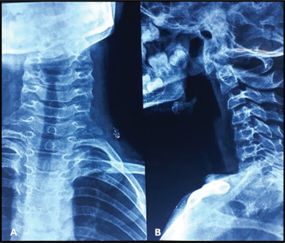
During the physical examination, the boy was afebrile but kept his neck tilted to the right. He guarded the area closely. Every direction of cervical movement was painfully restricted. A neurological examination showed that he had normal motor and sensory function. X-rays demonstrated loss of cervical lordosis and a persistent head tilt (Fig. 1). Blood tests, including white blood cell count and inflammatory markers such as C-reactive protein, were all normal.
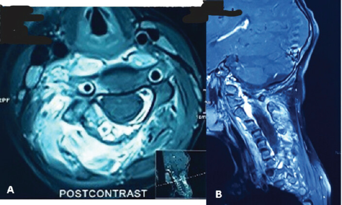
Since there were no urgent signs such as fever or nerve deficits, conservative management with a soft collar and pain medication was attempted first. Due to worsening of symptoms over the next 3 months, contrast-enhanced magnetic resonance imaging was done. The scan revealed an expansile lesion involving the right lamina of the C4 vertebra, characterized by multiseptate fluid–fluid levels and surrounding soft-tissue enhancement, suggestive of an ABC (Fig. 2). A computed tomography scan confirmed that the lesion had broken through the bone cortex and showed some new bone growth.
The patient’s torticollis indicated possible instability in his neck. However, we focused on the bone lesion first and did not perform specific imaging for AARS at the start.
Given the characteristic imaging features and progressive symptoms, a decision was made to proceed with definitive surgical management, with histopathological confirmation.
Access to the C4 lamina was achieved through a unilateral posterior midline subperiosteal approach. Extended intralesional curettage was performed using a high-speed burr. Pre-operative embolization was omitted, and intraoperative hemostasis was maintained. An associated AARS was identified during the procedure. A closed reduction was performed under general anesthesia. Alignment was secured with a halo vest. Histopathology confirmed the diagnosis of ABC. The sample contained focal giant cell-rich areas but lacked the architectural features of a primary giant cell tumor.
Osseous consolidation progressed, and the volume of the lesion decreased. The child remained asymptomatic and maintained a neutral head position for 3 months. The halo vest was removed at that time. Severe neck pain and torticollis recurred 2 weeks after removal.
Radiographs confirmed recurrent AARS, classified as Fielding and Hawkins Type I. This recurrence, despite apparent healing of the primary lesion, suggested persistent ligamentous instability. Given the failure of prior stabilization, definitive surgical management was undertaken.
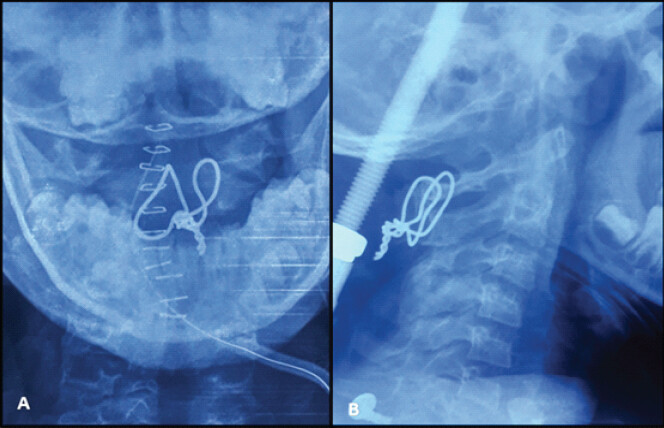
A posterior C1–C2 fusion using the Gallie technique was performed, supplemented with halo vest immobilization (Fig. 3). Autologous iliac crest bone graft was used to achieve arthrodesis. Over the next 3 months, solid fusion was achieved, and the halo was removed. The child became asymptomatic with preserved horizontal gaze and was able to perform activities of daily living without restriction.
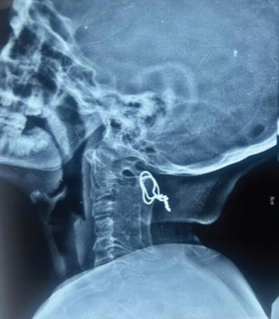
At the latest follow-up of 7 years, there was no evidence of recurrence of the ABC, and the atlantoaxial fusion remained stable. However, ankylosis involving the subaxial cervical spine (C3–C6) was evident (Fig. 4). Despite this, the child maintained a functional range of vision, normal daily activity levels, and adapted well without significant disability (Fig. 5).
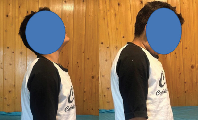
To the best of our knowledge, this is a rare reported case of a cervical spine ABC associated with AARS that recurred after apparent tumor healing and required subsequent fusion, which was further complicated by long-term subaxial cervical ankylosis. This case highlights the difficulty in handling spinal instability and tumors in the pediatric cervical spine.
Discussion
Pediatric cervical ABCs present difficult therapeutic challenges because of aggressive growth near major neurovascular structures and the risk of compromising spinal stability [6,7]. While spinal involvement occurs in a minority of cases, cervical lesions are less frequent and often involve the posterior elements [4,8]. Intralesional curettage with high-speed burring is a common treatment with acceptable control rates, though recurrence exists among younger patients [3,9,10].
This case showed no tumor recurrence at a 7-year follow-up, which supports the use of extended curettage in the cervical spine. AARS associated with spinal ABC is rare [11,12], and few documented cases describe the recurrence of AARS after the lesion has healed. Factors beyond the primary tumor, such as persistent ligamentous laxity or changed cervical biomechanics, likely play a primary role.
Halo immobilization often works for reducible AARS, but the early recurrence in this case showed the limitations of this method when underlying instability exists. This outcome supports the need for definitive stabilization [5]. Posterior C1–C2 fusion followed established principles for recurrent AARS, despite the risk of motion restriction in children. Other methods, such as selective arterial embolization, exist for vascular or complex lesions [9], but were unnecessary here.
A further unusual finding was the development of subaxial cervical ankylosis after a limited unilateral posterior approach. This phenomenon is rare and appears related to the high osteogenic potential of the pediatric spine, prolonged immobilization, and changed load distribution after fusion. Despite this outcome, the child reached an excellent functional state at the 7-year follow-up. This result shows the adaptability of the pediatric cervical spine.
Distinguishing between tumor resolution and biomechanical stability is necessary for guiding the timing of intervention. Management of pediatric cervical ABCs must include an assessment of persistent instability and long-term biomechanical effects.
Conclusion
ABCs in the pediatric cervical spine often involve instability that persists after tumor treatment. Osseous healing of the lesion does not ensure spinal stability. Recurrent AARS sometimes requires posterior fusion. Limited surgical exposure can lead to unintended subaxial ankylosis in children. Management involves tumor eradication and stabilization combined with extended clinical follow-up to monitor biomechanical changes.
Clinical Message
Successful treatment of cervical spine ABCs does not guarantee restoration of stability – persistent or recurrent AARS may require early definitive fusion, and even limited surgery in children can lead to long-term ankylosis, warranting careful follow-up.
Conflict of Interest:
Nil
Source of Support:
Nil
Consent:
The authors confirm that informed consent was obtained from the patient for publication of this article
References
- Sebaaly A, Ghostine B, Kreichati G, Mallet JF, Glorion C, Moussa R. Aneurysmal bone cyst of the cervical spine in children: A review and a focus on available treatment options. J Pediatr Orthop 2015;35:693-702. [Google Scholar] | [PubMed]
- Protas M, Jones LW, Sardi JP, Fisahn C, Iwanaga J, Oskouian RJ. Cervical spine aneurysmal bone cysts in the pediatric population: A systematic review of the literature. Pediatr Neurosurg 2017;52:219-24. [Google Scholar] | [PubMed]
- Kieser DC, Mazas S, Cawley DT, Fujishiro T, Tavolaro C, Boissiere L. Bisphosphonate therapy for spinal aneurysmal bone cysts. Eur Spine J 2018;27:851-8. [Google Scholar] | [PubMed]
- Zileli M, Isik HS, Ogut FE, Is M, Cagli S, Calli C. Aneurysmal bone cysts of the spine. Eur Spine J 2013;22:593-601. [Google Scholar] | [PubMed]
- Umebayashi D, Hara M, Nishimura Y, Wakabayashi T. A morphologically atypical case of atlantoaxial rotatory subluxation. J Korean Neurosurg Soc 2014;55:284-8. [Google Scholar] | [PubMed]
- Mascard E, Gomez-Brouchet A, Lambot K. Bone cysts: Unicameral and aneurysmal bone cyst. Orthop Traumatol Surg Res 2015;101 1 Suppl:S119-27. [Google Scholar] | [PubMed]
- Stevens KJ, Stevens JA. Aneurysmal Bone Cysts. In: StatPearls. Treasure Island, FL: StatPearls Publishing; 2026. Available from: https://www.ncbi.nlm.nih.gov/books/nbk546654. [Last accessed on 2023 Aug 14]. [Google Scholar] | [PubMed]
- Sayago LR, Remondino RG, Tello CA, Piantoni L, Francheri Wilson IA, Galaretto E. Aneurysmal bone cysts of the spine in children: A review of 18 cases. Global Spine J 2020;10:875-80. [Google Scholar] | [PubMed]
- Amendola L, Simonetti L, Simoes CE, Bandiera S, De Iure F, Boriani S. Aneurysmal bone cyst of the mobile spine: The therapeutic role of embolization. Eur Spine J 2013;22:533-41. [Google Scholar] | [PubMed]
- Doyle A, Field A, Graydon A. Recurrent aneurysmal bone cyst of the cervical spine in childhood treated with doxycycline injection. Skeletal Radiol 2015;44:609-12. [Google Scholar] | [PubMed]
- Lu VM, Daniels DJ. Giant cervical aneurysmal bone cyst and its multimodal management. World Neurosurg 2019;131:207-8. [Google Scholar] | [PubMed]
- Ehlers LD, McMordie J, Lookian P, Surdell D, Puccioni M. Cervical spine aneurysmal bone cyst in a pediatric patient: Embolization Considerations and potential pitfalls. World Neurosurg 2020;139:163-8. [Google Scholar] | [PubMed]
© 2026 Journal of Orthopaedic Case Reports - Published by Indian Orthopaedic Research Group
About the Authors
-

Reyaz Ahmed
-

Abhishek Raina
-

Nasir Shah
-

Sumit Kumar
-

Amber Agarwal
-

Anirudh Dwajan
Related Articles in Journal of Orthopaedic Case Reports
 September 1, 2026 Combined Ultrasound- and Fluoroscopy-guided Percutaneous Cementoplasty and Ablation for a C2 Pathologic Fracture: A Case Report
 August 1, 2026 O-arm Navigation-assisted Excision of Cervical Osteoid Osteoma Adjacent to the Vertebral Artery: Technical Note and Case Report
 July 1, 2026 Aneurysmal Bone Cyst of Calcaneum in a Young Adult: A Rare Case Report
 July 1, 2026 Aneurysmal Bone Cyst of the Proximal Humerus Managed with En Bloc Resection, Fibular Strut Grafting, and PHILOS Fixation: A Case Report