


Introduction
Posterior cruciate ligament (PCL) is less commonly seen injuries compared to anterior cruciate ligament (ACL) injuries [1]. PCL avulsion fractures are a rare subset of PCL injuries contributing to significant knee instability [2]. Accurate reduction and healing are essential to avoid complications. They are generally associated with other ligamentous injuries and intra-articular meniscal injuries, and therefore proper workup has to be done to avoid missing out of injuries [3]. Lateral collateral ligament (LCL) injuries are commonly seen with ACL tears and uncommon with PCL injuries [4]. LCL and biceps femoris complex are rare and serious injuries too, which are typically seen in high-energy trauma leading to gross instability of the knee and have to intervened promptly. This work highlights on rarity of the complex injury at the knee involving PCL avulsion fracture, LCL – biceps femoris complex injury in the presence of ipsilateral diaphyseal tibia fracture. This case report also signifies on how proper diagnosis, meticulous approach and proper surgical planning will yield high satisfactory outcome even in such a complex rare injury.
Case Report
A 25-year-old farmer presented to our casualty with swelling over the right knee and deformity over the right mid-leg following a fall from a two-wheeler. Patient fell with direct impact on the proximal tibia, with the knee stretched, causing knee hyperextension and with varus force at the knee. On examination, the patient was conscious, oriented and hemodynamically stable. On local clinical examination, right leg showed swelling and deformity over mid leg with abnormal movement and crepitus over both bone leg. There was a small punctured wound of size 1 × 1 cm over the anterior aspect of mid-leg near to fracture site. Right knee examination showed swelling with effusion, tenderness and significant posterior drawer of the proximal tibia. Furthermore, there was marked varus instability at the knee. Distal pulses were present with no distal neurological deficits.
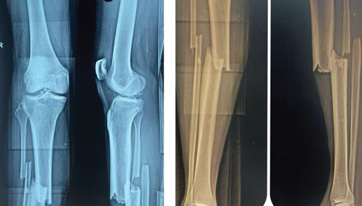
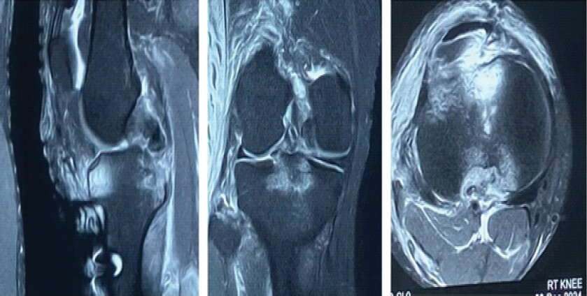
Plain radiography showed both bone mid-shaft displaced fracture on the right leg. Knee radiography showed a PCL avulsion fracture with minimal comminution of fragments (Fig. 1). Magnetic resonance imaging (MRI) of the knee joint post tibia intramedullary nailing was performed confirmed PCL avulsion fracture with comminution at fracture site and LCL avulsion at fibular head, along with biceps femoris tendon avulsion (Fig. 2). Significant hemarthrosis was noted inside the knee joint.
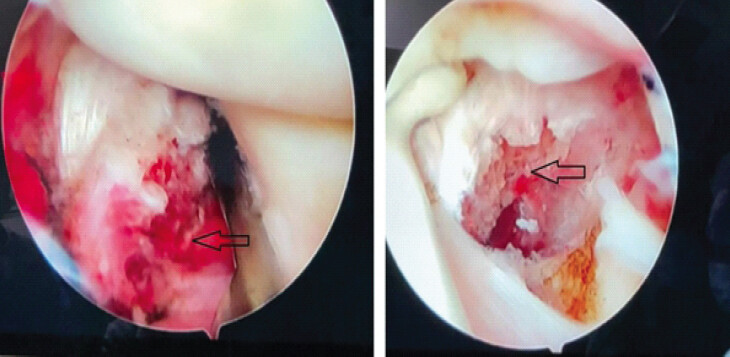
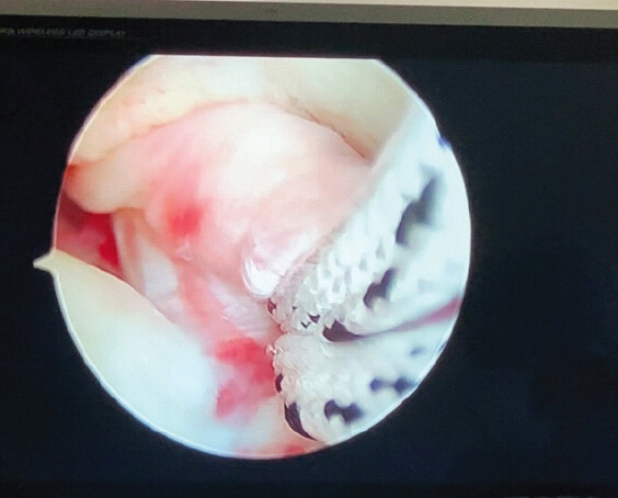
We performed urgent wound debridement and tibial nailing for mid-shaft tibia fracture on an emergency basis in view of open fracture. The next day, following MRI evaluation of the knee joint, it was planned for elective arthroscopic PCL avulsion fracture fixation with hematoma evacuation and open fixation of LCL with biceps femoris complex with suture anchors. After 48 h of primary surgery, a second surgery was performed. Under spinal anesthesia and under a tourniquet, the knee was kept in 90° flexion to relax posterior neurovascular structures. Initial anterolateral (AL) and anteromedial (AM) portals were made. With 30° arthroscope from the AL portal and a shaver instrument from the AM portal, thorough debridement with hematoma evacuation done. Findings were noted, including pseudolaxity of ACL. With a blunt trocar passed from the AM portal between the medial femoral condyle (MFC) and PCL into posteromedial (PM) compartment of the knee, the arthroscope was railroaded. With arthroscope in the PM compartment, 2 PM portals high and low were made under direct visualization, with initial percutaneous entry made from 2 spinal needles. 8 mm cannulas were passed into both PM portals. Now arthroscope was switched into the low PM portal, and the high PM portal was used as a working portal. With the probe, the avulsed PCL fracture fragment was probed, and the fracture pattern was noted (Fig. 3). With shaver passed from the high PM portal, blood clot evacuation, debridement, and loose bony fragments were removed. Freshening of the fracture was done with removal of interposed soft tissue. Care was taken about not to splinter the fracture fragment further and avoiding injury to posterior neurovascular structures. Scorpion suture passer loaded with No. 2 fibre wire was introduced from the high PM portal, and a cinch suture was placed at the PCL-bone junction (Fig. 4). To ascertain firm fixation, another suture was placed with No. 2 fibre wire. Under direct visualization, the PCL guide passed into the space between PCL and MFC at center fracture bed. On the outer side of the tibia, beath pin entry was made slightly posterior to the second locking screw of the tibia nail. At this point, it was made sure that our drill site was not abutting any tibial locking screw or nail. With PCL protection guide passed from high PM portal at the tip of beath pin, overdrilling was done with 4 mm cannulated drill bit. With Ethilon No.1 passed at the eyelet of drillbit passed from the tibial tunnel, a grasper was used to retrieve it from the high PM portal. 4 limbs of No. 2 fibre wire looped through Ethilon were retrieved back onto the anterior surface of the tibia through the tibial tunnel. Fracture reduction was assisted with a blunt trocar passed from the high PM portal and under direct vision with arthroscope in low PM portal; fiber wires were secured onto tibial nail locking screws after gently withdrawing them with knee in complete extension (Fig. 5). With this, we did not need any accessory fixation device like endobutton or suture disk for PCL avulsion fixation on tibial surface. Stability of fracture checked with probe from high PM portal. Confirmation of fixation with satisfactory reduction was done with fluoroscopy. Focus was now shifted to torn.
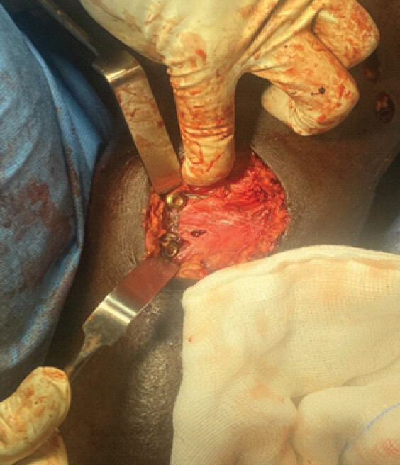
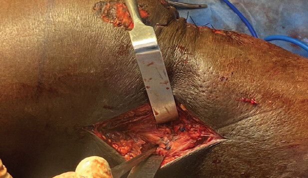
Lateral structures, namely. LCL and biceps femoris tendon complex. With the limb in 30° flexion and a small sand bag under the pelvis to tilt the limb, a 5 cm curvilinear incision was made on the fibular head. Dissection of AL fascia was made, exposing the torn end of LCL. Care was taken not to damage the peroneal nerve. Just posterior to LCL attachment at fibular head, biceps femoris tendon tear was noted and isolated. Two titanium double-loaded anchors were passed at the insertion site of LCL and biceps femoris at the fibular head from AL to the posteromedial direction. Each anchor was used repair the LCL and biceps femoris separately, with locking stiches and tying of knots were done with varus stress applied at knee (Fig. 6). Both posterior stability and varus stability were checked and seemed stable. Post-operative radiograph showed satisfactory reduction of PCL fracture fragment. Patient was advised on a knee brace for 6 weeks with isometric quadriceps exercises started on day 1. Knee range of movements (ROM) up to 90° was started at 3 weeks, with complete ROM at 6 weeks. Partial weight bearing was started at 3 weeks with full weight bearing at 6 weeks.
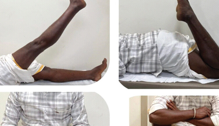
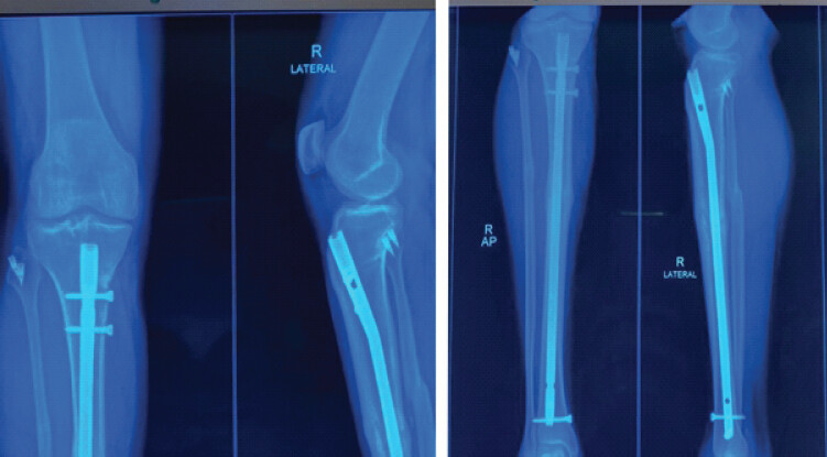
Patient was followed up for 18 months postoperatively, and at his latest follow-up he was back to his regular activities of farming. He had an excellent functional score (Lysholm Knee Score -96) (Fig. 7). Plain radiography of the knee and leg showed complete radiological union of PCL avulsion fracture with complete union of tibial diaphyseal fracture (Fig. 8).
Discussion
To the best of our knowledge and after extensive search in the literature, we found one case report with a similar injury where it was coined as Triple Treat Trauma [5]. Houass et al. reported a PCL avulsion fracture in a diaphyseal tibia fracture with LCL avulsion at the femoral side. However, no mention was made on biceps femoris injury. In their case report, they managed the PCL avulsion fracture with open reduction and fixation with cannulated screws. Another study showed proximal tibial fracture managed with plating and fixation of PCL avulsion with open screw fixation [6]. However, no LCL injury was found.
The mechanism of such injuries is similar to posterolateral corner injuries of the knee which involves direct blow to upper tibia with knee in complete stretched position or hyperextension with varus [7]. Dashboard injuries are also classically seen in isolated PCL avulsion fractures. In our case, it was probably a direct fall on the knee with the knee in hyperextension with varus force involved causing significant injury.
Treatment options for PCL avulsion fractures include both operative and non-operative management. Conservative management is generally considered if displacement of fracture is <5 mm [8]. However, a considered while making a decision, namely, size of the fragment, comminution, posterior displacement tibia on stress views, and presence of other concomitant injuries [9]. Timely intervention avoids complications like persistent instability or fragment non-union, which can lead to exaggerated arthritis of the knee joint.
When indicated, both arthroscopic and open procedures can be opted for fixation of PCL avulsion fracture, though no significant differences have been noted among both techniques [10]. Though arthroscopic techniques have the advantage of addressing other injuries at the same time apart from being minimally invasive, it is a technically challenging procedure with potential danger to neurovascular structures nearby. Difficult scenarios like the presence of an intramedullary nail, as in our case, the procedure becomes even more difficult since tunnel position has to be precise. We decided for slightly posterior tunnel placement than usual due to the presence of an intramedullary nail. Fixation modalities are several in arthroscopic procedures. It mainly depends on the size of the fragment. If larger fragment, screw fixation is ideal, and in cases of small comminuted fractures, suture-based fixation is better [11]. Chen et al., in their case series, described a modified way of fixing PCL avulsion, particularly in small and comminuted fragments, using stainless steel wires and high-strength sutures with a three-tibial-tunnel technique in an inverted triangular fashion and observed excellent fixation allowing early knee ROMs [12]. In our patient, we decided for novel way of fixing onto the tibial surface without additional fixation devices like endobutton or suture disk. We gently withdrew two locking proximal screws of the tibial nail and tied onto them achieving stable fixation. However, it was made sure that the tunnel was placed slightly posterior onto the tibia, avoiding any abutment to the nail or locking screws.
It is equally important to achieve balance between fixation and early mobilization to avoid stiffness in the knee joint, which is the most common complication following PCL avulsion fixation, particularly in arthroscopic techniques than open procedure [13]. Therefore, fixation can be particularly challenging in small comminuted fractures.
LCL – biceps femoris complex is again a rare injury and has been reported scarcely [14]. However, though rare injuries, they can lead to significant instability at the knee and lead to long-term complications if not treated immediately. Therefore, early diagnosis and primary repair are essential to achieve maximum outcome than delayed intervention, as it restores anatomy and allows superior healing [15]. Primary repair can be augmented with allografts like semitendinosus to strengthen the repair [16].
Conclusion
PCL avulsion fractures and LCL-biceps femoris complex avulsion injuries are serious injuries compromising knee stability significantly. Added with, ipsilateral tibia fracture makes the surgical decision very challenging. Therefore, it needs careful planning and a meticulous approach to attain a complete functional outcome and to avoid complications.
Clinical Message
PCL avulsion fractures and LCL-biceps femoris complex avulsion injuries are relatively uncommon injuries, and associated diaphyseal tibia fracture further is rarest combination. It is challenging to diagnose such complex injuries, and missed injuries particularly at knee can lead to significant instability, increasing patients’ morbidity. It is always important to assess neighboring joints while addressing diaphyseal fractures. Arthroscopic fixation of PCL avulsion fracture can be a safe and minimally invasive technique even in the presence intramedullary nail, which needs careful placement of the tibial tunnel, and fixation at the tibial side does not need any additional fixation device. This case report mainly highlights the rarity of complex injury at the knee involving PCL avulsion fracture, LCL -biceps femoris avulsion at fibular side in diaphyseal tibia fracture and how careful planning and management would yield high satisfactory results.
Conflict of Interest:
Nil
Source of Support:
Nil
Consent:
The authors confirm that informed consent was obtained from the patient for publication of this article
References
- Allen CR, Kaplan LD, Fluhme DJ, Harner CD. Posterior cruciate ligament injuries. Curr Opin Rheumatol 2002;14:142-9. Google Scholar | PubMed
- Katsman A, Strauss EJ, Campbell KA, Alaia MJ. Posterior cruciate ligament avulsion fractures. Curr Rev Musculoskelet Med 2018;11:503-9. Google Scholar | PubMed
- Pardiwala DN, Agrawal D, Patil V, Saini U, Dhawal P. Paper 133: Comparison of open versus arthroscopic fixation for isolated PCL tibial bony avulsions: A prospective randomized study with minimum 2 year follow up. Arthroscopy 2012;28:e413-4. Google Scholar | PubMed
- Ramos LA, Zogbi T, De Andrade EF, De Oliveira GT, Nicolini AP, Krob JJ. Treatment and outcomes of lateral collateral ligament injury associated with anterior and posterior cruciate ligament injury at 2-year follow-up. J. Orthop 2019;16:489-92. Google Scholar | PubMed
- Houass E, Elgaliou MR, Haouzi MA, Mekkaoui MJ, Boufettal M, Bassir RA. Triple threat ttrauma: A rare case report of avulsion fracture of the posterior cruciate ligament, lateral collateral ligament, and diaphyseal tibia fracture. Trauma Case Rep 2023;47:100910. Google Scholar | PubMed
- Goyal A, Vimal Y, Mittal V, Gupta M, Kapoor R, Rohela R. A rare case report: Managing PCL avulsion fracture with a fracture of the proximal tibial shaft. J Orthop Case Rep 2025;15:177-82. Google Scholar | PubMed
- Shon OJ, Park JW, Kim BJ. Current concepts of posterolateral corner injuries of the knee. Knee Surg Relat Res 2017;29:256-68. Google Scholar | PubMed
- Zhao J, He Y, Wang J. Arthroscopic treatment of acute tibial avulsion fracture of the posterior cruciate ligament with suture fixation technique through Y-shaped bone tunnels. Arthroscopy 2006;22:172-81. Google Scholar | PubMed
- White EA, Patel DB, Matcuk GR, Forrester DM, Lundquist RB, Hatch GF3rd. Cruciate ligament avulsion fractures: Anatomy, biomechanics, injury patterns, and approach to management. Emerg Radiol 2013;20:429-40. Google Scholar | PubMed
- Ling HM, Wang CJ, Tu YK, Yeh WL. Arthroscopy in avulsion fracture of posterior cruciate ligament. Chang Gung Med J 2001;24:313-7. Google Scholar | PubMed
- Kim SJ, Shin SJ, Cho SK, Kim HK. Arthroscopic suture fixation for bony avulsion of the posterior cruciate ligament. Arthroscopy 2001;17:776-80. Google Scholar | PubMed
- Chen X, Cao Y, Shi Z, An L, Zhao J, Li X. Arthroscopic Treatment of Avulsion Fracture of the Posterior Cruciate Ligament from the Tibia: A Modified Surgical Technique and Case Series, Journal of ISAKOS https://doi.org/10.1016/ j.jisako.2026.101086. Google Scholar | PubMed
- Hooper PO 3rd, Silko C, Malcolm TL, Farrow LD. Management of posterior cruciate ligament tibial avulsion injuries: A systematic review. Am J Sports Med 2018;46:734-42. Google Scholar | PubMed
- Szukics P, Lerebours F, Robaina J, Yagnik G, Ross J. Lateral collateral ligament repair with reconstruction augmentation. Video J Sports Med 2025;5. (3) doi.org/10.1177/2635025424129158.. Google Scholar | PubMed | CrossRef
- McCarthy M, Ridley TJ, Bollier M, Cook S, Wolf B, Amendola A. Posterolateral knee reconstruction versus repair. Iowa Orthop J 2015;35:20-5. Google Scholar | PubMed
- Dabis J, Wilson A. Repair and augmentation with internal brace in the multiligament injured knee. Clin Sports Med 2019;38:275-83. Google Scholar | PubMed
© 2026 Journal of Orthopaedic Case Reports - Published by Indian Orthopaedic Research Group
About the Authors
-

Raghavendra Kembhavi
-

Jojin Jose Chitten
-

Siddharath S Parameshwar
Related Articles in Journal of Orthopaedic Case Reports
 May 1, 2026 A Novel Failure Interface in Constrained Total Hip Arthroplasty: Dissociation at the Constrained Liner Insert-Bipolar Head Junction Following Periprosthetic Joint Infection
 April 1, 2026 A Rare Case of Chronic Pan-Diaphyseal Osteomyelitis of the Fibula in a Child Treated by Near Total Fibulectomy: A Case Report
 March 1, 2026 Vascularized fibular flap for gunshot-induced composite foot defects: Two-case report and comprehensive literature review
 December 1, 2025 Engagement of the Radial Head in Posterior Capitellum Fracture: Defining the Critical Point