


Introduction
Giant cell tumor (GCT) of bone is one of the most common benign bone tumors encountered in orthopedic practice [1]. It accounts for approximately 22% of benign bone tumors and 6.6% of all skeletal tumors [2]. Unlike many other bone tumors, GCT of bone shows a slight female predominance (56%) [2]. It most commonly affects young adults between 20 and 40 years of age, with peak incidence in the third decade of life [2, 3, 4, 5]. It is uncommon in children and adolescents, with the reported incidence in the immature skeleton ranging from 1.8% to 10.6% [4, 6]. Because of its rarity and overlapping radiological features with other benign lesions, diagnosis in this age group can be challenging. Only a limited number of isolated case reports and small case series have described GCT involving the proximal tibia in skeletally immature patients [1, 3, 4, 5, 6, 7, 8, 9, 10, 11].
We report a histopathologically confirmed case of GCT of the proximal tibia in a 7-year-old girl presenting with a pathological fracture. The unusual age at presentation, metadiaphyseal location with epiphyseal sparing, and clinicoradiological overlap with other benign lesions posed a diagnostic challenge. A review of the available literature was performed to better understand the epidemiology and diagnostic considerations of GCT in skeletally immature patients. This case highlights the importance of considering GCT even in very young patients and emphasizes the role of histopathological confirmation in such scenarios. The course of disease and treatment principles in this group largely mirrors those in adults [1].
Case Report
A 7-year-old girl presented to the emergency department with pain, swelling, and deformity of the left upper leg following trivial trauma while playing. Clinical examination revealed diffuse swelling and tenderness over the proximal leg with intact distal neurovascular status.
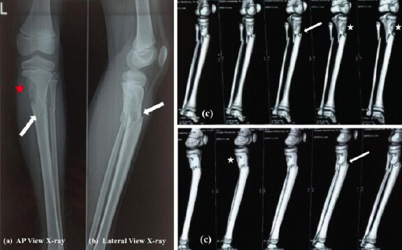
Anteroposterior and lateral radiographs demonstrated an eccentric osteolytic lesion in the proximal tibial metaphysis with cortical destruction and an associated mildly displaced fracture involving the proximal tibia and fibula (Fig. 1a and b). A non-contrast computed tomography (CT) scan showed an ill-defined, lobulated, expansile lytic lesion measuring approximately 29 × 14 × 15 mm in the metadiaphyseal region of the medial proximal tibia, with thin and sclerotic cortical margins and a fracture line extending through the lesion (Fig. 1c). Based on age and imaging features, provisional differential diagnoses included aneurysmal bone cyst, non-ossifying fibroma, and simple bone cyst.
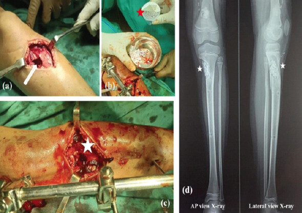
After routine pre-operative evaluation and parental consent, surgery was performed under spinal anesthesia. Through an anteromedial approach, a cortical window was created and thorough intralesional curettage was performed. The cavity was irrigated and filled with autologous cancellous bone graft harvested from the ipsilateral distal femur, supplemented with 5.0 cc chronOS granules (1.4–2.8 mm), a synthetic β-tricalcium phosphate (β-TCP) (chronOS, Johnson and Johnson Medical Pty Ltd t/a DePuy Synthes). The fracture was stabilized using a spanning external fixator (Fig. 2).
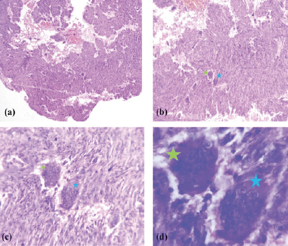
Histopathological examination revealed a moderately cellular lesion composed of sheets of round to ovoid mononuclear stromal cells admixed with numerous osteoclast-like multinucleated giant cells, without anaplasia, necrosis, or increased mitotic activity, confirming the diagnosis of GCT of bone (Fig. 3).
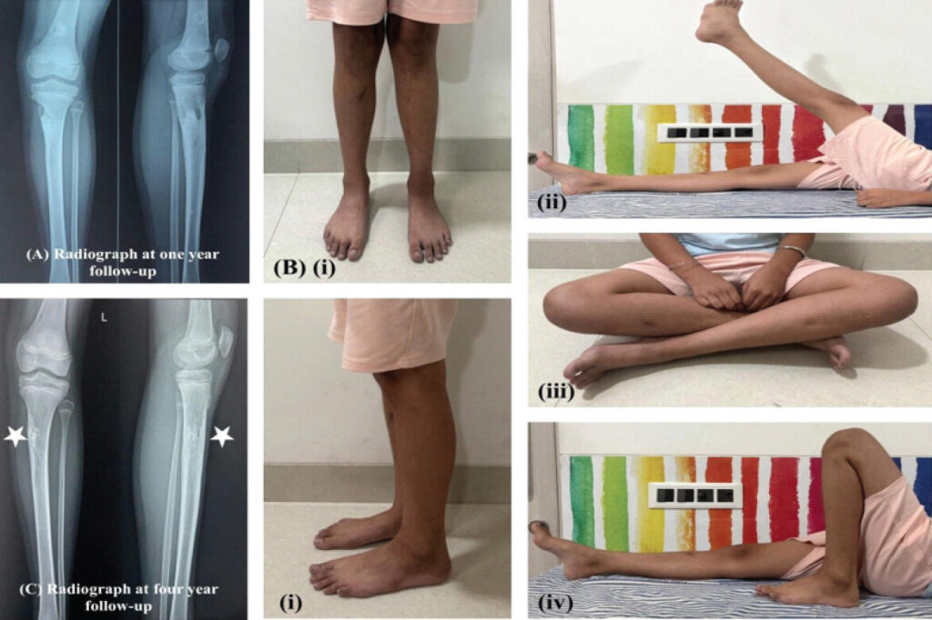
The external fixator was removed at 6 weeks, followed by application of a patellar tendon-bearing cast for another 6 weeks and gradual weight-bearing. Serial radiographs demonstrated progressive healing. At 1 year, the patient was pain-free with full knee range of motion. At 4-year follow-up, she remained asymptomatic with no limb-length discrepancy, angular deformity, or radiological recurrence (Fig. 4). Informed consent for the publication of this case report was obtained from the patient’s parents.
Discussion
Giant cell tumor of bone predominantly affects skeletally mature young adults, with approximately 85% of patients older than 19 years [2]. GCT of the proximal tibia in children is rarely reported in the literature [1, 3, 4, 5, 6, 7, 8, 9, 10, 11].
While most pediatric GCTs are reported in older adolescents, only a few cases have been reported in younger children. Strøm et al. and Zhang et al. described GCT in a 9-year-old girl, Kransdorf et al. reported a case in a 7-year-old girl, and Rajesh et al. described GCT in a 4-year-old boy, the youngest among reported patients [3, 8, 9, 10]. Our case describing GCT in a 7-year-old girl adds to this limited literature, highlighting the presentation at an unusually young age with a pathological fracture.
In contrast to many bone tumors that show male predominance, several series of pediatric GCT have demonstrated a female preponderance, with reported female predominance ranging from 63 to 82% [1,3,4,12]. Our case aligns with this observation, although a few authors have reported male predominance [5, 8, 9, 11].
Approximately 90% of pediatric GCT lesions occur in long tubular bones [3, 11]. Similar to adults, it shows a predilection for the ends of long bones around the knee joint (46–50%), with the distal femur being the most frequent location, followed by the proximal tibia and distal radius [1, 2, 3, 12]. However, Kransdorf, Schütte, and Strøm et al. reported the proximal tibia as the most common site of GCT in the immature skeleton [3, 4, 9].
GCT in adults typically involves the metaphysio-epiphyseal region of long bones; however, there has been debate regarding the site of origin in the immature skeleton [3, 4, 8, 10]. Kransdorf et al., citing observations by Picci et al., suggested that most pediatric GCTs originate in the metaphysis with subsequent epiphyseal extension at or after physeal closure [9]. Schütte and Strøm et al. also supported a metaphyseal origin in children, with increasing epiphyseal involvement with age [3,4]. In our case, the lesion was located in the metaphyseo-diaphyseal region with epiphyseal sparing, supporting the concept of metaphyseal origin in younger patients.
Moderate pain and swelling around the joint are usually the initial symptoms, while restriction of joint movements is seen in advanced stages [3, 12]. Pathological fractures occur in approximately 10–18% of cases, as seen in our patient [3, 4, 10, 12].
Radiographs, CT, and magnetic resonance imaging (MRI) are commonly used imaging modalities in the evaluation of GCT, while histopathological examination confirms the diagnosis [7, 10]. The classic histological appearance includes uniformly distributed characteristic osteoclast-like multinucleated giant cells in a background of round-to-oval and spindle-shaped mononuclear stromal cells [4]. Although MRI may provide additional information regarding soft-tissue extension and physeal involvement, CT adequately characterized the osseous lesion in our patient, whereas the definitive diagnosis was established by histopathological examination.
Management of GCT is primarily surgical and aims to achieve local control while preserving joint function [1, 3, 5, 12]. Extended intralesional curettage with local adjuvants such as phenol, hydrogen peroxide, liquid nitrogen, or polymethylmethacrylate is commonly employed [5, 7, 12]. The defect created after curettage is reconstituted with autograft, allograft, synthetic bone substitutes like calcium phosphate, cement, or a combination [1, 7]. While Strøm et al., Sharma et al., and Vijay et al. used cement, Sharma et al. used maternal iliac crest-based allograft for filling the defect [3, 5, 7, 8]. We performed intralesional curettage, autologous cancellous bone grafting, and β-TCP supplementation with external stabilization for fracture, following which the lesion healed uneventfully.
Earlier studies reported recurrence rates of up to 50% following curettage, whereas with modern techniques and use of adjuvants, recurrence rates have been reduced to approximately 14–25% and even between 8% and 20% in younger patients [2, 3, 4, 7, 8]. However, adequate tumor removal remains the key prognostic factor [1].
The diagnosis of GCT in skeletally immature patients is challenging due to overlapping clinical and radiological features with other eccentric osteolytic lesions of the metadiaphyseal region such as aneurysmal bone cyst, non-ossifying fibroma, and simple bone cyst. Aneurysmal bone cyst is often considered the primary differential diagnosis of expansile lytic metaphyseal lesions in young children presenting with pathological fracture. In our case, the patient’s age, metadiaphyseal location, and fracture presentation initially favored these diagnoses, thus highlighting the importance of histopathological examination for definitive diagnosis. Other differential diagnoses include giant-cell–rich lesions of bone, namely metaphyseal fibrous cortical defect, giant cell reparative granuloma, brown tumor of hyperparathyroidism, benign chondroblastoma, and giant cell-rich osteosarcoma [2, 4].
Epiphyseal invasion and destruction by the tumor are considered signs of aggressive growth that may lead to angular deformities and growth disturbances [1, 4, 5, 6]. Since these features were absent in our patient, no such complications were observed. Although most recurrences typically occur within the first 2 years [1], continued long-term surveillance is advisable. At 4-year follow-up, our patient remains asymptomatic with no clinical or radiological evidence of recurrence.
This case highlights that GCT, although rare in early childhood, can present with atypical clinicoradiological features and should remain an important differential diagnosis in pediatric osteolytic lesions of long bones.
Conclusion
Although GCT is primarily a tumor of skeletally mature individuals, it can rarely occur in young children and may demonstrate a metaphyseal location with female predominance. The diagnosis in young children can be challenging because of overlapping imaging features with other giant cell–rich lesions of long bones. An eccentric osteolytic lesion in the metaphyseal or metadiaphyseal region of long bones in the immature skeleton should raise suspicion for GCT, and histopathological confirmation remains essential. Adequate intralesional curettage with reconstruction provides favorable functional outcomes with a low risk of recurrence. GCT should be considered in the differential diagnosis of metaphyseal lytic lesions with pathological fracture in young children, as appropriate surgical management can achieve excellent functional outcomes.
Clinical Message
Giant cell tumor of bone, though rare in skeletally immature patients, should be considered in the differential diagnosis of metadiaphyseal osteolytic lesions, as it may mimic conditions such as aneurysmal bone cyst or non-ossifying fibroma. Histopathological examination remains essential for definitive diagnosis. Intralesional curettage with appropriate reconstruction provides good functional outcomes in young children, with long-term follow-up required to monitor recurrence.
Conflict of Interest:
Nil
Source of Support:
Nil
Consent:
The authors confirm that informed consent was obtained from the parent for publication of this article
References
- Puri A, Agarwal MG, Shah M, Jambhekar NA, Anchan C, Behle S. Giant cell tumor of bone in children and adolescents. J Pediatr Orthop 2007;27:635-9. [Google Scholar] | [PubMed]
- Unni KK, Inwards YC. Giant cell tumor (osteoclastoma). In: Dahlin's Bone Tumors: General Aspects and Data on 10,165 Cases. 6th ed.. Philadelphia, PA:Wolters Kluwer/Lippincott Williams & Wilkins Health2010225-38. [Google Scholar] | [PubMed]
- Strøm TM, Skeie AT, Lobmaier IK, Zaikova O. Giant cell tumor: A rare condition in the immature skeleton - A retrospective study of symptoms, treatment, and outcome in 16 children. Sarcoma 2016;2016:3079835. [Google Scholar] | [PubMed]
- Schütte HE, Taconis WK. Giant cell tumor in children and adolescents. Skeletal Radiol 1993;22:173-6. [Google Scholar] | [PubMed]
- Sharma V, Sharma S, Mistry KA, Awasthi B, Verma L, Singh U. Giant cell tumor of bone in skeletally immature patients - A clinical perspective. J Orthop Case Rep 2015;5:57-60. [Google Scholar] | [PubMed]
- Picci P, Manfrini M, Zucchi V, Gherlinzoni F, Rock M, Bertoni F. Giant-cell tumor of bone in skeletally immature patients. J Bone Joint Surg Am 1983;65:486-90. [Google Scholar] | [PubMed]
- Vijay C, Nekkanti S, RaviShankar R, Shashikiran R, Sunila R. An unusual presentation of giant cell tumor of the tibia in a skeletally immature patient. J Orthop Case Rep 2018;8:10-3. [Google Scholar] | [PubMed]
- Sharma R, Meena RP. Management of giant cell tumor (GCT) of proximal tibia in a skeletally immature patient: A rare case report. Int J Innov Sci Res Technol 2018;3:181-3. [Google Scholar] | [PubMed]
- Kransdorf MJ, Sweet DE, Buetow PC, Giudici MA, Moser RPJr. Giant cell tumor in skeletally immature patients. Radiology 1992;184:233-7. [Google Scholar] | [PubMed]
- Zhang XP, Lu XC, Wang LL, Wei JQ, Yan J, Shao XN. Giant cell tumors of bone in patients aged 18 years old or younger: Imaging features and tumor characteristics. Int J Gen Med 2021;14:8389-97. [Google Scholar] | [PubMed]
- Kim WJ, Kim S, Choi DW, Lim GH, Jung ST. Characteristics of giant cell tumor of the bone in pediatric patients: Our 18-year, single-center experience. Children 2021;8:1157. [Google Scholar] | [PubMed]
- Campanacci M. Giant cell tumors of bone. Curr Orthop 1993;7:26-35. [Google Scholar] | [PubMed]
© 2026 Journal of Orthopaedic Case Reports - Published by Indian Orthopaedic Research Group
About the Authors
-

Nadeem Akhtar Qureshi
-

Ravi Prakash Futela
Related Articles in Journal of Orthopaedic Case Reports
 October 10, 2022 Giant Cell Tumor of Talus – A Case Report
 October 1, 2026 Giant Cell Tumor of the Distal Phalanx of the Great Toe: Case Report
 October 1, 2026 Giant Cell Tumor of the Lateral Malleolus Treated by Tumor Excision and Reconstruction using A 180° Rotated Proximal Fibular Autograft – A Rare Case Report
 September 1, 2026 “A Giant Shoulder”: Proximal Humerus Giant Cell Tumor Managed With Extended Curettage and Proximal Humerus Internal Locking System Plating with Fibular Strut Graft – A Case Report