


Introduction
The proximal humerus represents the fourth most common site for primary malignant bone tumors and is the most frequent location in the upper limb [1]. The most common malignant tumors at this site include chondrosarcoma, osteosarcoma, and Ewing sarcoma [2].
Limb-sparing surgery of the shoulder girdle is technically demanding and must strictly respect oncological principles. Functional outcomes depend largely on the preservation of the glenoid, rotator cuff, deltoid muscle, and axillary nerve [3]. We report a case of proximal humerus Ewing sarcoma managed with conservative resection and biological reconstruction, highlighting technical considerations and reconstructive strategies.
Case Report
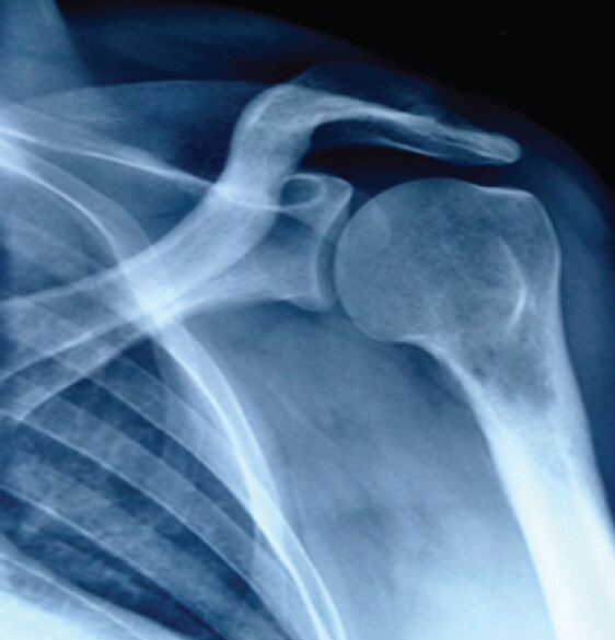
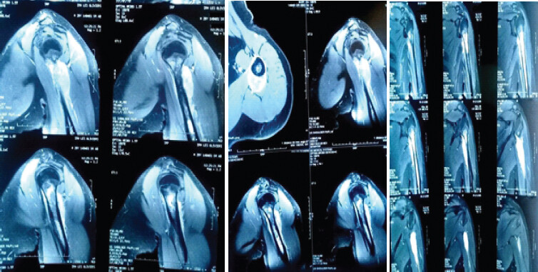
We present the case of a 29-year-old, right-handed, manual worker, with no significant medical history, who presented with mechanical left shoulder pain. Active and passive range of motion of the glenohumeral joint was preserved. No local inflammatory signs or satellite lymphadenopathy were noted. Plain radiography of the left shoulder revealed an osteolytic lesion of the left proximal humerus (Fig. 1). Magnetic resonance imaging (MRI) demonstrated an aggressive soft-tissue mass arising from the proximal humerus measuring 4 × 2 cm with cortical breakthrough and focal soft-tissue invasion (triceps long head). There was no evidence of involvement of the deltoid, rotator cuff, glenoid, or lymph nodes (Fig. 2). A surgical biopsy confirmed the diagnosis of Ewing sarcoma. Staging evaluation revealed a pulmonary metastasis but no other distant localizations.
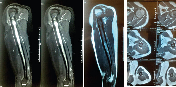
Three cycles of neoadjuvant chemotherapy were initiated. A follow-up thoraco-abdomino-pelvic computed tomography (CT) scan showed complete resolution of pulmonary nodules. A follow-up MRI demonstrated the complete regression of the soft-tissue extension (Fig. 3).
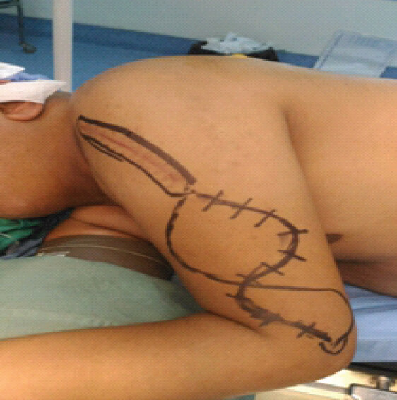
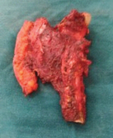
A standard deltopectoral approach was used (Fig. 4). The patient underwent diaphyso-metaphyseal tumor resection, preserving the proximal two-thirds of the humeral head, which was not involved by the tumor. The resected specimen was sent for histopathological examination (Fig. 5). Reconstruction was performed using a non-vascularized fibular autograft stabilized with an anatomical plate and an intramedullary pin. Post-operative radiographs confirmed satisfactory implant positioning (Fig. 6). Macroscopic surgical margins appeared clear. However, microscopic margins were determined to be R0 at the epiphysis but R1 at the diaphysis. The patient received three additional cycles of adjuvant chemotherapy.
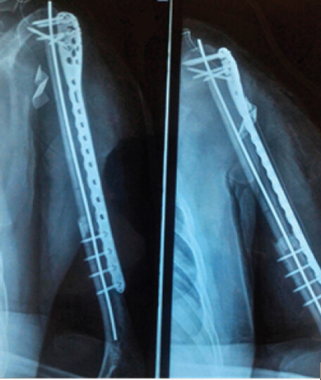
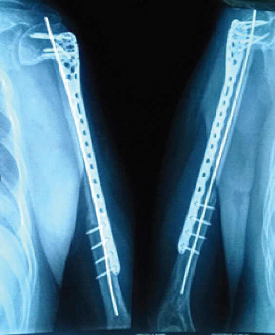
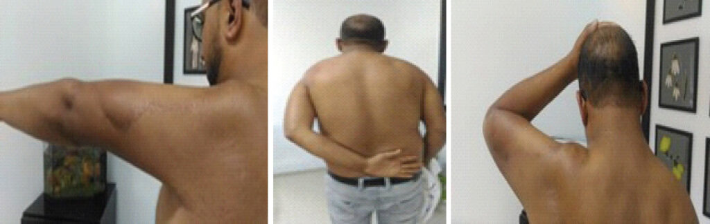
At the last follow-up of 30 months, clinical and radiological results were good with no evidence of tumor recurrence (Figs. 7 and 8).
Discussion
Conservative treatment for malignant tumors of the proximal humerus is gaining importance, largely due to advancements in chemotherapy. Indications for amputation and shoulder disarticulation have decreased [4,5,6]. Resection of these tumors must be oncologically sound and preoperatively planned. Accurate radiological evaluation before surgery is mandatory [7].
Limb-sparing surgery for proximal humerus tumors is often technically challenging as it involves a complex joint with intimate muscular, tendinous, and neurovascular relationships, rendering oncologic resection difficult. The essential risk of conservative treatment remains locoregional recurrence: a 10% recurrence rate at 5 years in O’Connor’s meta-analysis [8] and 34.5% in the Cochin series.
The method of reconstruction following proximal humerus tumor resection depends on several factors.
-
If the glenoid, rotator cuff, deltoid, and axillary nerve are intact, a tumor prosthesis is indicated in the case of humeral head involvement [3].
-
If the humeral head is intact, reconstruction can be performed using an intramedullary nail or an anatomical plate combined with a bone graft.
-
If the deltoid, glenoid, or axillary nerve is involved, glenohumeral arthrodesis is the treatment of choice [3]. Arthrodesis provides a stable, pain-free construct, allowing mobility through the scapulothoracic articulation, but it often yields less satisfactory functional outcomes [8,9].
In our case, we opted for reconstruction using a non-vascularized fibular autograft fixed with an anatomical plate and an intramedullary pin, given the preserved humeral head, glenoid, deltoid, rotator cuff, and axillary nerve.
Literature review indicates that allografts carry disadvantages including bone resorption and fatigue fractures [10]. Indeed, allografts possess osteoconductive but not osteoinductive properties. O’Connor recommends combining an allograft with a vascularized fibular autograft, which provides primary stability via the allograft and secondary strength through the hypertrophy of the vascularized fibula [8].
Conclusion
Conservative treatment of proximal humeral tumors faces several challenges: achieving oncological resection due to the complex musculotendinous and neurovascular relationships and the complexity of the shoulder joint, and the subsequent challenge of reconstructing the bone stock. Indications and functional outcomes depend heavily on the involvement of the deltoid, axillary nerve, glenoid, and rotator cuff.
Clinical Message
Preservation of the deltoid muscle, rotator cuff, glenoid, and axillary nerve allows effective biological reconstruction after proximal humerus tumor resection, providing durable function and oncological safety.
Conflict of Interest:
Source of Support:
Nil
Consent:
The authors confirm that informed consent was obtained from the patient for publication of this article
References
- Raza AA, Whaley MJ, Shakir M.Management and novel treatment of degloving soft tissue injuries: A case report. Cureus 2023;15:e49999. Google Scholar | PubMed
- Giotis D, Kotsias C, Plakoutsis S, Malahias MA, Konstantinidis C.Management of heel pad degloving injury after severe foot crush injury: A case report study. Cureus 2021;13:e14191. Google Scholar | PubMed
- Luze H, Nischwitz SP, Smolle C, Zrim R, Kamolz LP.The use of Acellular fish skin grafts in burn wound management-a systematic review. Medicina (Kaunas) 2022;58:912. Google Scholar | PubMed
- Lekuya HM, Alenyo R, Kajja I, Bangirana A, Mbiine R, Deng AN.Degloving injuries with versus without underlying fracture in a Sub-Saharan African tertiary hospital: A prospective observational study. J Orthop Surg Res 2018;13:2. Google Scholar | PubMed
- Zhou F, Zhang X, Zhang Y, Xiang G, Luo P, Hu W.Comprehensive management of degloving soft tissue injuries of the extremity: A 12-year retrospective study. Injury 2024;55:111939. Google Scholar | PubMed
- Cantrell WA, Lawrenz JM, Vallier HA.A salvage strategy for heel pad degloving injury: A case report. OTA Int 2018;1:e007. Google Scholar | PubMed
- Jørgensen U, Bojsen-Møller F.Shock absorbency of factors in the shoe/heel interaction--with special focus on role of the heel pad. Foot Ankle 1989;9:294-9. Google Scholar | PubMed
- Krishna D, Chaturvedi G, Khan MM, Cheruvu VP, Laitonjam M, Minz R.Reconstruction of heel soft tissue defects: An algorithm based on our experience. World J Plast Surg 2021;10:63-72. Google Scholar | PubMed
- De Francesco F, Zingaretti N, Parodi PC, Riccio M.The evolution of current concept of the reconstructive ladder in plastic surgery: The emerging role of translational medicine. Cells 2023;12:2567. Google Scholar | PubMed
- Kudsk KA, Sheldon GF, Walton RL.Degloving injuries of the extremities and torso. J Trauma 1981;21:835-9. Google Scholar | PubMed
- Waikakul S.Revascularization of degloving injuries of the limbs. Injury 1997;28:271-4. Google Scholar | PubMed
- Sakai G, Suzuki T, Hishikawa T, Shirai Y, Kurozumi T, Shindo M.Primary reattachment of avulsed skin flaps with negative pressure wound therapy in degloving injuries of the lower extremity. Injury 2017;48:137-41. Google Scholar | PubMed
- Pilancı O, Aköz Saydam F, Başaran K, Datlı A, Güven E.Management of soft tissue extremity degloving injuries with full-thickness grafts obtained from the avulsed flap. Ulus Travma Acil Cerrahi Derg 2013;19:516-20. Google Scholar | PubMed
- Simman R, Abbas FT.Foot wounds and the reconstructive ladder. Plast Reconstr Surg Glob Open 2021;9:e3989. Google Scholar | PubMed
- Posner KM, Bakus C, Sodha S.Rapid healing of necrotizing fasciitis using the kerecis fish skin xenograft: A clinical case report. Cureus 2024;16:e73060. Google Scholar | PubMed
- Ivana J, Sanjaya IG.Evaluation of the efficacy of fish skin grafts as wound dressings: A systematic review. Eur Burn J 2025;6:50. Google Scholar | PubMed
- Tan AP, Chng JK.Fish skin acellular dermal matrix combined with negative pressure wound therapy for diabetic foot ulcers. Cureus 2025;17:e80488. Google Scholar | PubMed
- El-Shazly M, Yassin O, Kamal A, Makboul M, Gherardini G.Soft tissue defects of the heel: A surgical reconstruction algorithm based on a retrospective cohort study. J Foot Ankle Surg 2008;47:145-52. Google Scholar | PubMed
- Herold J, Kamin K, Bota O, Dragu A, Rammelt S.Complete avulsion of the heel pad with talar and calcaneal fracture: Salvage with multiple K-wire anchorage, internal fixation and free ALT flap. Arch Orthop Trauma Surg 2023;143:2429-35. Google Scholar | PubMed
- Howard JL, Buckley R, McCormack R, Pate G, Leighton R, Petrie D.Complications following management of displaced intra-articular calcaneal fractures: A prospective randomized trial comparing open reduction internal fixation with nonoperative management. J Orthop Trauma 2003;17:241-9. Google Scholar | PubMed
- Lutnick E, LaNicca M, Montero J, Ritter C, Mutty C.Heel pad degloving injuries: A case series. J Am Acad Orthop Surg 2026;34:e1915-26. Google Scholar | PubMed
- Esmaeili A, Biazar E, Ebrahimi M, Heidari Keshel S, Kheilnezhad B, Saeedi Landi F.Acellular fish skin for wound healing. Int Wound J 2023;20:2924-41. Google Scholar | PubMed
- Kotronoulas A, Jónasdóttir HS, Sigurðardóttir RS, Halldórsson S, Haraldsson GG, Rolfsson Ó.Wound healing grafts: Omega-3 fatty acid lipid content differentiates the lipid profiles of acellular Atlantic cod skin from traditional dermal substitutes. J Tissue Eng Regen Med 2020;14:441-51. Google Scholar | PubMed
- Magnusson S, Baldursson BT, Kjartansson H, Rolfsson O, Sigurjonsson GF.Regenerative and antibacterial properties of acellular fish skin grafts and human amnion/chorion membrane: Implications for tissue preservation in combat casualty care. Mil Med 2017;182:383-8. Google Scholar | PubMed
- Cerceo JR, Malkoc A, Nguyen A, Daoud A, Wong DT, Woodward B.Management of large full-thickness burns using kerecis™ acellular fish skin graft and recell™ autologous skin cell suspension: A case report of two patients with large surface area burns. Cureus 2024;16:e71101. Google Scholar | PubMed
- Heitzmann W, Enzmann J, Von Kohout M, Mattern MM, Akkan J, Fuchs PC.Accelerated wound healing of enzymatically debrided deep dermal burn wounds after the use of fish skin (Kerecis Omega3 Wound®) in comparison to Suprathel®. Burns 2025;51:107471. Google Scholar | PubMed
- Dardari D, Piaggesi A, Potier L, Sultan A, Diener H, Francois M.Intact fish skin graft to treat deep diabetic foot ulcers. NEJM Evid 2024;3:EVIDoa2400171. Google Scholar | PubMed
- Yoon J, Yoon D, Lee H, Lee J, Jo S, Kym D.Wound healing ability of acellular fish skin and bovine collagen grafts for split-thickness donor sites in burn patients: Characterization of acellular grafts and clinical application. Int J Biol Macromol 2022;205:452-61. Google Scholar | PubMed
- Gonzalez SR, Wolter KG, Yuen JC.Infectious complications associated with the use of integra: A systematic review of the literature. Plast Reconstr Surg Glob Open 2020;8:e2869. Google Scholar | PubMed
- Struble SL, Patel NK, Graham EM, Tipps JA, Vaile JR, Leeflang EJ.Outcomes of biodegradable temporizing matrix for soft tissue reconstruction of the hand and extremities. Plast Reconstr Surg Glob Open 2024;12:e5956. Google Scholar | PubMed
© 2026 Journal of Orthopaedic Case Reports - Published by Indian Orthopaedic Research Group
About the Authors
-
Dr. Rami Ben Mhenni
-

Dr. Sadok Chtai
-

Dr. Mohamed Ali Khlifa
-
Dr. Adnene Siala
-
Dr. Mahmoud Ben Maitigue
-

Dr. Karim Bouattour
Related Articles in Journal of Orthopaedic Case Reports
 September 1, 2026 Limb Salvage for Entire Tibial Ewing Sarcoma in a 4-year-Old Child Using Extracorporeal Irradiation and Free Flap Reconstruction: A Case Report and Surgical Technique
 September 1, 2026 “A Giant Shoulder”: Proximal Humerus Giant Cell Tumor Managed With Extended Curettage and Proximal Humerus Internal Locking System Plating with Fibular Strut Graft – A Case Report
 September 1, 2026 Proximal Midfoot Amputation as a Functional Alternative to Major Amputation in Advanced Diabetic Foot Infection: A Case Report
 September 1, 2026 Successful Limb Salvage Using Staged Applications of a Fish-Skin Xenograft (Kerecis) in a Heel Pad Degloving Injury: A Case Report