


Introduction
Giant cell tumors of the bone (GCTB) are challenging neoplasms in orthopedic practice. Although benign, GCTB is locally aggressive, has a high recurrence rate, and can metastasize [1]. GCTB affects individuals in their third and fourth decades, representing 5% of primary bone tumors, with a 2:1 female prevalence after epiphyseal closure [2,3]. GCTB commonly affects the metaphyseal-epiphyseal regions of long bones, mainly the distal femur, proximal tibia, and distal radius. The proximal humerus, the fourth most common site, presents challenges because of its shoulder anatomy, biomechanics, and proximity to vital structures. The proximal humerus location creates difficulties owing to shoulder joint motion, muscular stability, and proximity to the axillary nerve and rotator cuff [4,5].
The pathophysiology of GCTB involves three cell populations: neoplastic giant cell tumor stromal cells, mononuclear histiocytic cells, and multinucleated giant cells (MNGC). These interact through the RANK/RANKL/Osteoprotegerin pathway, which has advanced the understanding of GCTB and led to new therapies [6]. Proximal humerus GCTB presents with deep and constant shoulder pain, affecting daily activities. Patients may develop swelling over the lateral proximal arm, with reduced shoulder motion. Mechanical symptoms include cortical destruction and glenohumeral joint involvement [7].
Diagnosis requires clinical assessment, imaging, and histopathological examination. Radiographs show a “soap bubble” appearance with bone destruction, and magnetic resonance imaging (MRI) assesses tumor spread, cortical integrity, and joint extension for surgery [8]. Managing proximal humerus GCTB requires examination of tumor characteristics, patient factors, and surgical experience. Treatment options include prolonged excision with supplemental treatment, extensive removal with repair, and surgical removal in uncontrollable cases. Extended curettage with additional therapy is preferred for bone preservation with reasonable recurrence rates [9]. This case report examined proximal humeral GCTB surgery and documented the outcomes to inform future treatment.
Case Report
A 46-year-old male presented with 3 months of increasing pain and limited shoulder mobility, affecting his activities and sleep. The patient had no history of trauma, fever, weight loss, or comparable swellings. Clinical examination revealed shoulder swelling, pain over the proximal humerus, and impaired mobility with unimpaired neurovascular function. General examination was normal, with plain radiography showing a multiloculated, expansile osteolytic lesion peripherally in the proximal humerus with cortical thinning and breach areas, without periosteal reaction. Computed tomography (CT) and MRI confirmed an expansile lytic lesion in the metaphyseal–epiphyseal region, without skip lesions or metastases.
Laboratory investigations, including hemogram, leukocyte count, renal function, and coagulation profile, were normal. A CT-guided core biopsy revealed gray-white and gray-brown tissue fragments. Histopathological examination revealed a neoplasm with osteoclast-type MNGC among mononuclear stromal cells with eosinophilic cytoplasm and pleomorphic vesicular nuclei. The giant cell nuclei resembled those of mononuclear stromal cells. The stroma showed fibrocollagenous tissue with inflammatory infiltrates, congested vessels, and hemorrhage. These findings confirmed a Campanacci Grade III giant cell tumor of the proximal humerus. Pre-operative range of movements with abduction of about 50°, flexion of about 60°, and external rotation of 20°, with further movements being painful, were restricted with a disability of the arm, shoulder, and hand (DASH) score of 42, which signified a moderate-to-high level of disability.
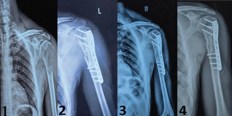
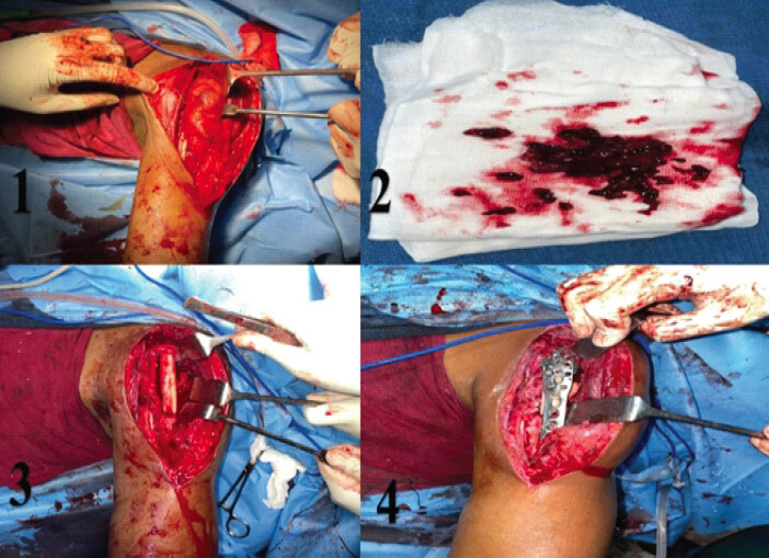
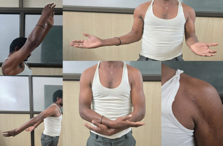
In the absence of metastasis, surgery focused on local control and functional preservation. Extended intralesional curettage was performed using the deltopectoral approach on January 24, 2024. Thorough tumor removal was performed using high-speed burrs, followed by cryotherapy. Reconstruction was performed using a 12-cm fibular strut graft to bridge the humeral defect. Fixation was performed using a proximal humerus internal locking system (PHILOS) plate with screws. The radiological images and intra operative images are shown in Fig. 1 and 2 respectively. The patient received three doses of zoledronic acid, administered at 3 weeks apart. The recovery was uneventful, with primary wound healing observed. Rehabilitation progressed from immobilization to mobilization. He was discharged on day 12 with instructions regarding medications, wound care, physiotherapy, and follow-up. At 24 months, the patient had regained near-complete shoulder mobility with a near-normal range of movements as shown in Table 1 and Fig. 3. (Abduction: 150°, Flexion: 170°, External rotation: 70°). The DASH score was 20.8, indicating excellent function. Imaging (MRI and CT of the shoulder at 24 months postoperatively) revealed complete graft incorporation without any recurrence. There were no signs of donor-site morbidity related to fibular graft harvesting and no implant-related complications such as screw loosening, plate irritation, graft fracture, or hardware failure during follow-up.
Range of movements comparison.
| Time point | Flexion | Abduction | External rotation | Internal rotation |
|---|---|---|---|---|
| Pre-operative | 60° | 50° | 20° | 30° |
| 6 months | 90° | 80° | 40° | 50° |
| 1 year | 130° | 120° | 60° | 70° |
| 2 years | 170° | 150° | 70° | 90° |
Discussion
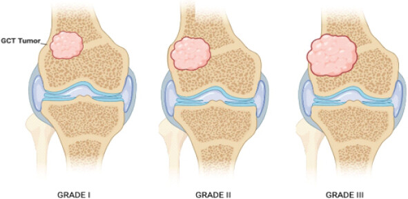
This proximal humerus giant cell tumor case managed with extended curettage, PHILOS plating, and fibular strut grafting represents a comprehensive approach to treatment. Compared to the literature, key aspects emerge regarding the treatment outcomes. The selection of extended curettage over wide resection aligns with current treatment algorithms. Studies have shown that while wide resection offers lower recurrence rates (7.7% vs. 28.4%), extended curettage provides better functional outcomes with superior DASH scores. The DASH score of 20.8 at the 2-year follow-up in this case (Fig. 4) compared favorably with reported outcomes versus typical post-resection scores of 13–30 points [9,10,11]. The literature supports extended curettage for Campanacci Grade II-III (Fig. 5) lesions when adequate bone stock remains. A multicentre study of 82 patients with Grade III giant cell tumors reported zero recurrence when combining extended curettage with pseudocapsule excision. Liquid nitrogen cryotherapy protocols show 25–50% recurrence rates with mechanical curettage [12,13,14,15].

PHILOS plating with fibular strut grafting provides stability and reconstruction of the bone. The angular stability of the PHILOS system enables superior metaphyseal fixation and early mobilization [16]. Fibular strut grafting enables biological reconstruction that cement cannot provide, resulting in better long-term outcomes than cemented reconstruction. A proximal femur study reported successful bone healing at the 40-month follow-up [17]. While cement reconstruction achieves 89.2% musculoskeletal tumor society scores, it lacks biological reconstruction potential [18]. The combined use of cryotherapy, fibular grafting, PHILOS plating, and zoledronic acid may offer a cost-effective treatment strategy with efficient resource utilization. Post-operative zoledronic acid represents an adjuvant strategy with mixed results in the literature. A meta-analysis showed that zoledronic acid reduces recurrence rates (P = 0.007) through osteoclast apoptosis and differentiation. Studies have shown that zoledronic acid and denosumab have equivalent outcomes, with zoledronic acid being more cost-effective than denosumab. The three-dose protocol follows the established recurrence-prevention regimens [19].
Proximal humerus giant cell tumors present challenges because of their anatomy and neurovascular proximity. The deltopectoral approach provides tumor access while preserving critical structures. The 2-year follow-up requires interpretation within the natural history of the tumor, as recurrences occur within 2 years. Current follow-up shows encouraging results, although surveillance is ongoing. Reduced mechanical stress may contribute to lower recurrence rates. Long-term outcomes showed sustained improvement, with imaging and DASH scores used for monitoring. This approach balances tumor control and functional preservation.
Conclusion
Extended curettage with adjuvant cryotherapy and reconstruction using a fibular strut graft provides durable structural support and preserves function in patients with proximal humeral giant cell tumors. The addition of zoledronic acid may reduce the risk of recurrence. This multimodal approach yielded excellent functional recovery and tumor control at short-term follow-up.
Clinical Message
Giant cell tumor of the proximal humerus challenges achieving tumor control while preserving shoulder function. Extended intralesional curettage with adjuvant therapy and reconstruction using fibular strut graft and PHILOS plate fixation balances oncological safety with function. The successful graft incorporation and absence of recurrence at 24 months demonstrate that joint-preserving strategies remain effective in selected patients.
Conflict of Interest:
Nil
Source of Support:
Nil
Consent:
The authors confirm that informed consent was obtained from the patient for publication of this article
References
- Gharpinde MR, Pisulkar G, Taywade S, Awasthi AA, Suneja A. Excision of a giant cell tumor with bone grafting and bone cementing of the proximal humerus: A case report. Cureus 2024;16:e59492. Google Scholar | PubMed
- Todorova T, Gramatnikovski N, Angelovska T, Kostadinova-Kunovska S, Manevska N, Foteva M. Autologous vascular proximal fibular graft in the treatment of giant cell tumor of the distal radius-a case report. J Surg Case Rep 2024;2024:rja.e490. Google Scholar | PubMed
- Ahilan S, Harshavardhan JK. Giant cell tumor of distal radius treated with wide local excision and reconstruction with ipsilateral proximal fibular autograft - a follow-up. J Orthop Case Rep 2025;15:41–5. Google Scholar | PubMed
- Carrascal GJ, Pinto JA, Carranza MA, Chaparro AF, Lucero DS. Massive allograft in salvage surgery for giant cell tumor of the proximal humerus. World J Adv Res Rev 2025;26:3786–91. Google Scholar | PubMed
- Maniar MH, Mankar S, Sakhre R. En bloc resection with reconstruction using a customized megaprosthesis in a case of proximal humerus giant cell tumor: A case report. Cureus 2023;15:e34217. Google Scholar | PubMed
- Rekhi B, Dave V. Giant cell tumor of bone: An update, including spectrum of pathological features, pathogenesis, molecular profile and the differential diagnoses. Histol Histopathol 2023;38:139–53. Google Scholar | PubMed
- Wiratnaya IG, Raditya RH. Bone tumor wide excision due to giant cell tumor of proximal humerus: A case report. Int J Health Med Sci 2023;6:201–3. Google Scholar | PubMed
- Alhulaimi Y, AlAbbasi KK, AlShaya OS, Alrawaf TN, Aldosari NH, Zogel B. Giant cell tumor with secondary aneurysmal bone cyst in the left distal humerus: A case report. Cureus 2024;16:e65507. Google Scholar | PubMed
- Van Der Heijden L, Dijkstra PD, Campanacci DA, Gibbons CL, Van De Sande MA. Giant cell tumor with pathologic fracture: should we curette or resect? Clin Orthop Relat Res 2013;471:820–9. Google Scholar | PubMed
- Eldebeis HM, El Masry AM, Elmowafi MA. Management of giant cell tumor of the distal end radius: Extended curettage and adjuvant therapy versus wide resection and reconstruction: A systematic review and meta-analysis. Egypt Orthop J 2024;59:144–53. Google Scholar | PubMed
- Masry AM, Mowafi MA, Hassan Yousef Eldebies HM. Management of giant cell tumor of distal end radius: Extended curettage and adjuvant therapy vs wide resection and reconstruction: A systematic review and meta-analysis. QJM Mon J Assoc Phys 2024;117:671; hcae175.671. Google Scholar | PubMed
- Harshwal RK, Sehrawat M, Saini N, Modi P, Kumawat KK, Meena DS. Reducing recurrence in Campanacci type three giant cell tumor of bone: Outcomes from an 82-case study evaluating psuedocapsule excision with extended curettage. BJKines 2023;15:57–65. Google Scholar | PubMed
- Şirin E, Akgülle AH, Topkar OM, Sofulu Ö, Baykan SE, Erol B. Mid-term results of intralesional extended curettage, cauterization, and polymethylmethacrylate cementation in the treatment of giant cell tumor of bone: A retrospective case series. Acta Orthop Traumatol Turc 2020;54:524–9. Google Scholar | PubMed
- Lackman RD, Crawford EA, King JJ, Ogilvie CM. Conservative treatment of campanacci grade III proximal humerus giant cell tumors. Clin Orthop Relat Res 2009;467:1355–9. Google Scholar | PubMed
- Balke M, Campanacci L, Gebert C, Picci P, Gibbons M, Taylor R. Bisphosphonate treatment of aggressive primary, recurrent and metastatic giant cell tumour of bone. BMC Cancer 2010;10:462. Google Scholar | PubMed
- Doshi C, Sharma GM, Naik LG, Badgire KS, Qureshi F. Treatment of proximal humerus fractures using PHILOS plate. J Clin Diagn Res 2017;11:RC10–3. Google Scholar | PubMed
- Song J, Liu B, Jin K, Yao Q. Giant cell tumor of proximal femur managed by extended curettage with fibular strut allograft using long intramedullary interlocking nail: A case report and literature review. Medicine (Baltimore) 2024;103:e40960. Google Scholar | PubMed
- Sukpanichyingyong S, Salang K, Sangkomkamhang T. Clinical outcomes of extended intralesional curettage with cementation without implant augmentation in non-fracture giant cell tumor of bone around the knee. J Surg Case Rep 2022;2022:rjac197. Google Scholar | PubMed
- Kumar A, Sinha S, Haider Y, Jameel J, Kumar S. Role of zoledronic acid supplementation in reducing post-surgical recurrence of giant cell tumor of bone: A meta-analysis of comparative studies. Cureus 2021;13:e16742. Google Scholar | PubMed
© 2026 Journal of Orthopaedic Case Reports - Published by Indian Orthopaedic Research Group
About the Authors
-

M Antony Vimal Raj
-

Lakshmikanth Manikkam Ettiappan
-

Arvindh Venkatesan
-

Aditya Vezhaventhan
Related Articles in Journal of Orthopaedic Case Reports
 July 1, 2026 Aneurysmal Bone Cyst of the Proximal Humerus Managed with En Bloc Resection, Fibular Strut Grafting, and PHILOS Fixation: A Case Report
 April 1, 2026 A Novel Approach to Proximal Humerus Giant Cell Tumor: Cement Spacer and Screw Interdigitation for Optimal Stability
 June 10, 2023 Giant Cell Tumor of Thumb Proximal Phalanx-A Case Report
 September 1, 2026 Proximal Tibial Giant Cell Tumor in a Skeletally Immature Child: A Diagnostic Dilemma and Review of the Literature