


Introduction
Heel pad degloving injuries frequently result from trauma, notably motor vehicle collisions, crush injuries, and falls [1]. They are characterized by avulsion of skin and subcutaneous tissue from fascia, muscle, or bone, often with soft-tissue damage, contamination, vascular compromise, and osseous injury [2,3]. Reconstruction is challenging when the heel is involved due to its role in weight-bearing and shock absorption, limited tissue coverage, and the unique biomechanical properties of plantar skin [4, 5, 6, 7, 8]. Adjacent tissue does not adequately replicate the properties of native heel skin, making restoration of durable coverage difficult. Management requires progression along the reconstructive ladder, ranging from secondary intention and skin grafting to local rotational flaps and free tissue transfer [9]. Although flap reconstruction remains the standard option for plantar soft-tissue loss, it may not be feasible in the acute setting due to contamination, necrosis, evolving zones of injury, uncertain tissue viability, or patient-specific considerations [10,11,12,13,14]. In these circumstances, staged wound-bed optimization using biologic or synthetic matrices provides an opportunity to preserve future reconstructive options while reducing the risk of wound failure.
Among available biologic matrices, Kerecis is a decellularized fish-skin xenograft derived from Gadus morhua. Its preserved extracellular matrix and omega-3 fatty acids support infiltration, neovascularization, angiogenesis, and modulation of the inflammatory response while maintaining compatibility with negative-pressure wound therapy [15, 16]. Although Kerecis has demonstrated favorable outcomes in chronic wounds, ulcers, burns, and necrotizing soft-tissue infections [15,16,17], its application in lower-extremity trauma, particularly heel pad degloving injuries, remains poorly described.
We present a case of a patient with a heel pad degloving injury who developed plantar necrosis despite serial debridement and fracture stabilization. Given its complexity, options across the reconstructive ladder were considered. Although flap reconstruction is traditionally favored for plantar heel defects, patient-specific considerations supported another approach. Following shared decision-making, the patient chose staged fish-skin xenograft reconstruction to optimize the wound bed before definitive skin grafting. At 1-year follow-up, the patient achieved soft-tissue coverage capable of withstanding the demands of a weight-bearing heel. This case highlights the role of staged fish-skin xenograft reconstruction as a biologic bridge to definitive reconstruction in carefully selected patients with complex heel injuries.
Case Report
A 24-year-old male was riding an all-terrain vehicle (ATV) at a low speed when the ATV rolled, landing on his left foot. Orthopedic surgery was consulted for a large degloving injury with an open fracture of the left foot and ankle, with an intact dorsalis pedis pulse. Radiographs revealed a lateral malleolus fracture, fractures of the fourth and fifth metatarsals, and a fracture of the base of the first proximal phalanx (Fig. 1).
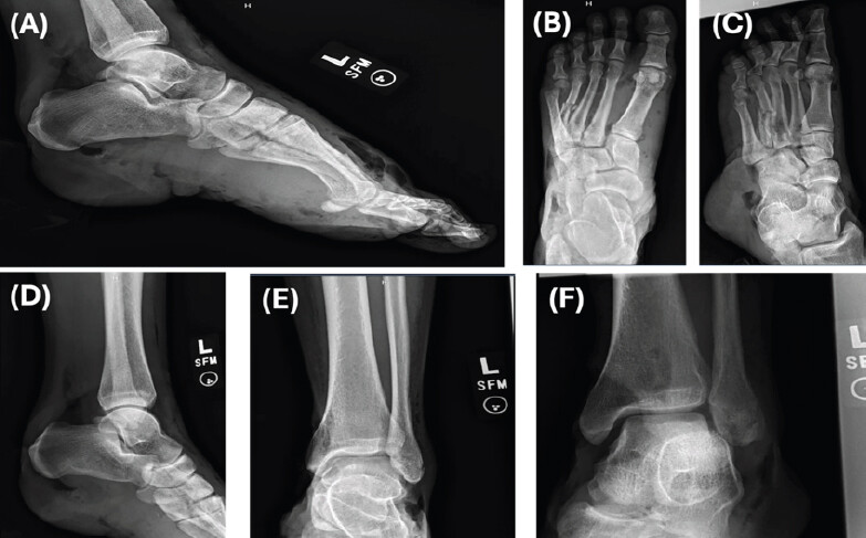
Computed tomography confirmed comminuted, displaced, and intra-articular fractures involving the calcaneus, navicular, cuneiforms, multiple metatarsals and phalanges, as well as extensive soft-tissue injury, including a large skin defect and exposure of the calcaneus. The patient denied tobacco, vaping, and drug use, with rare alcohol intake. He used no chronic medications and reported allergies to morphine and peanuts. The clinical timeline is illustrated in Fig. 2.
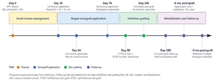
The patient underwent irrigation and debridement, closed reduction with percutaneous pinning of the heel pad and metatarsals, and application of negative-pressure wound therapy on the day of injury. Despite serial debridement and aggressive wound management, progressive plantar heel necrosis developed, resulting in a large full-thickness soft-tissue defect extending to the calcaneus.
Given the severity of soft-tissue loss, reconstructive options spanning the reconstructive ladder were considered, including below-knee amputation, free tissue transfer, and staged biologic reconstruction. Immediate flap reconstruction was considered less favorable due to the evolving zone of injury, ongoing tissue demarcation, and uncertainty regarding the extent of viable soft tissue. Following multidisciplinary discussion and consideration of patient-specific factors, the patient elected to pursue limb salvage using a staged reconstruction with fish-skin xenograft despite its non-traditional application in this setting. The objective was to optimize the wound bed before definitive soft-tissue coverage while preserving future reconstructive options. Kerecis was selected due to its biocompatibility, resistance to infection, and potential to promote granulation tissue in complex wounds [16, 17].
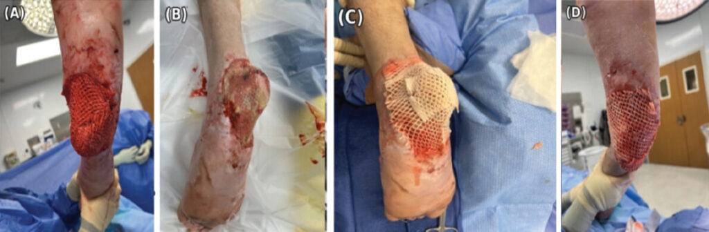
Thirty-nine days following the injury, the patient returned to the operating room for the first application. The wound, measuring 22 × 15 × 0.5 cm and extending to the level of the calcaneus, underwent debridement until bleeding tissue was achieved at the wound bed. Kerecis was placed over the wound bed and secured with staples. Non-adherent dressing and black foam were applied, followed by continuous negative-pressure wound therapy at 125 mmHg. The extremity was immobilized in a well-padded AO splint constructed with posterior and stirrup slabs. Two additional staged xenograft applications were performed on post-injury days 44 (Fig. 3b and c) and 79 (Fig. 3d) following debridement of residual non-viable tissue. Serial applications demonstrated progressive incorporation of the xenograft, healthy granulation tissue, and gradual optimization of the wound bed without infection, graft intolerance, or wound deterioration. At the third application, the wound measured 24 × 14 × 0.5 cm and was reconstructed using a 7 × 20 cm Kerecis sheet.
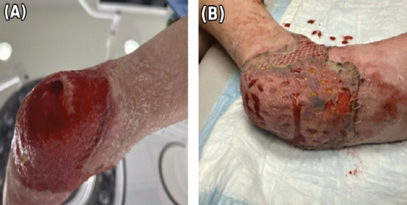
With a well-granulated wound bed achieved using Kerecis (Fig. 4a), the patient underwent definitive soft-tissue reconstruction 3 months later, including a full-thickness skin graft to the heel and split-thickness skin grafts to the anterior ankle and lateral heel (Fig. 4b). The patient received follow-up care, and his graft incorporated well without erythema, drainage, or purulence 3 months following his skin graft (Fig. 5). At 6 months following definitive grafting, the patient developed hypertrophic scarring with contracture involving the grafted region of the left foot and the medial thigh donor site. He was treated with a single session of fractional carbon dioxide laser therapy (120 mJ energy, 3% density, 250 Hz frequency) over a 15 × 35 cm area of the left foot and a 20 × 6 cm area of the medial thigh, followed by topical scar therapy and compression dressings. Reassessment documented improvement in scar erythema and tightening. At 9 months, the patient demonstrated excellent wound healing and graft integration, and he was released to weight-bearing as tolerated.
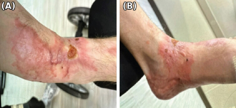
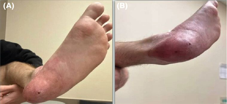
At 1-year follow-up, the heel remained epithelialized without ulceration, drainage, infection, or graft failure (Fig. 6). The patient progressed to full weight-bearing while utilizing a customized offloading ankle-foot orthosis with shoe modifications to redistribute plantar loading. He also completed physical therapy focused on ankle range of motion, strengthening, gait retraining, and scar mobilization, demonstrating functional recovery throughout rehabilitation.
Discussion
Heel pad degloving injuries present a reconstructive challenge because durable coverage must be restored to a weight-bearing surface while minimizing infection risk, wound breakdown, and limb loss [18,19]. Traditional reconstructive options, including local rotational and free tissue flaps, represent the standard of care for plantar heel defects; however, their success depends on adequate vascularity, a stable wound bed, and viable surrounding soft tissue. In acute settings, wound contamination, soft-tissue necrosis, serial debridement, and uncertainty regarding tissue viability may preclude immediate definitive reconstruction. In these situations, staged wound-bed optimization with biologic scaffolds may preserve future reconstructive options while establishing a vascularized wound bed for definitive soft-tissue reconstruction.
This case highlights an approach to a complex reconstructive problem. Following progression of heel pad necrosis despite serial debridement and negative-pressure wound therapy, reconstructive options spanning the reconstructive ladder – including amputation, free tissue transfer, and staged biologic reconstruction – were considered. After multidisciplinary evaluation and shared decision-making, the patient elected to pursue limb salvage using staged fish-skin xenograft reconstruction as a bridge to definitive skin grafting. This allowed continued wound optimization while preserving future reconstructive options.
The primary finding is that staged fish-skin xenograft reconstruction, with negative-pressure wound therapy, prepared a plantar heel defect for definitive reconstruction. Despite an exposed calcaneus and soft-tissue necrosis, sequential xenograft applications generated a vascularized wound bed that supported full- and split-thickness skin grafting. Importantly, the reconstruction remained durable at 1-year follow-up without ulceration, infection, graft failure, or wound breakdown despite the biomechanical demands of the plantar heel [18, 19, 20, 21].
Fish-skin xenografts are biologic matrices that support angiogenesis and granulation beds while maintaining compatibility with negative-pressure wound therapy [22, 23, 24]. Although their use has been reported in chronic wounds, burns, ulcers, and necrotizing soft-tissue infections [3, 15, 25, 26, 27], literature describing their application in traumatic foot and ankle reconstruction remains limited. This case supports the use of fish-skin xenografts as a staged reconstructive adjunct in complex lower-extremity trauma, particularly when immediate definitive reconstruction is infeasible.
Alternative technologies are available for staged wound-bed preparation. Compared with collagen-based dressings, fish-skin xenografts have been associated with accelerated healing and lower infection rates [28]. Dermal regeneration templates such as Integra provide a durable dermal substitute but remain susceptible to infectious complications [29]. Similarly, Biodegradable Temporizing Matrix (BTM) has emerged as an option for complex extremity wounds [30]. In this case, definitive grafting was performed approximately 8 weeks after the initial xenograft application, a timeline comparable to integration periods for BTM. Although conclusions cannot be drawn from a single case, staged fish-skin xenograft reconstruction demonstrated reliable wound-bed preparation while maintaining compatibility with serial debridement and negative-pressure wound therapy.
Another advantage observed was integration with negative-pressure wound therapy. The xenograft tolerated applications beneath wound vacuum dressings without complication, permitting continued wound management while progressive granulation tissue developed. Similar compatibility has been reported in diabetic foot wounds and other complex wounds treated with fish-skin grafts and negative-pressure wound therapy [17, 24].
This case demonstrates the potential for functional reconstruction. The patient progressed to full weight-bearing with customized orthotics and rehabilitation while maintaining soft-tissue coverage without recurrent breakdown at 1-year follow-up. Although long-term surveillance remains warranted because plantar injuries remain susceptible to late ulceration and shear-related complications, this outcome suggests that staged fish-skin xenograft reconstruction can provide durable limb salvage in carefully selected patients.
Ultimately, staged fish-skin xenograft reconstruction facilitated limb salvage in a patient with a heel pad avulsion and associated foot fractures. Successful wound-bed preparation, graft incorporation, and restoration of weight-bearing function support its use as a reconstructive adjunct in complex lower-extremity trauma [18, 19, 21, 22, 23, 24]. Further comparative studies are needed to define its role relative to other wound matrices.
Limitations
As a single-patient report, this case cannot establish efficacy or generalizability. Standardized outcome measures such as the Vancouver Scar Scale or the Patient and Observer Scar Assessment Scale were not used, and no validated lower-extremity functional score was obtained at final follow-up. The 1-year follow-up window, while informative, is insufficient to assess durability under sustained weight-bearing, a particular concern for heel pad reconstructions, where repetitive loading predisposes to breakdown. The absence of a comparison group precludes inference about the relative performance of fish-skin xenograft versus alternative biologic or synthetic matrices. Finally, cost and access considerations were not analyzed and may influence the real-world applicability of this approach.
Patient perspective
Throughout recovery, the patient expressed satisfaction with the reconstructive process and the functional outcome, reporting well-controlled pain and progressive return to daily activities. After participating in shared decision-making regarding reconstructive options, he remained committed to limb salvage despite the anticipated prolonged recovery and multiple staged procedures. The patient provided written informed consent for publication of this case and accompanying clinical photographs with the hope that his experience may help inform future treatment of patients with similarly devastating injuries.
Conclusion
This case demonstrates the successful use of staged fish-skin xenograft reconstruction as a bridge to definitive soft-tissue coverage in a complex heel pad degloving injury. Xenograft applications facilitated wound-bed optimization following progressive plantar necrosis, ultimately allowing successful full- and split-thickness skin grafting and preservation of limb function. At 1-year follow-up, the reconstructed heel remained durable under the demands of weight-bearing without recurrent ulceration, infection, or graft failure. Although free tissue transfer remains the conventional reconstructive option for plantar heel defects, this case illustrates that staged biologic reconstruction may represent a valuable limb-salvage strategy in carefully selected patients when immediate definitive reconstruction is not feasible or when individualized patient factors support an alternative approach. Further comparative studies are warranted to define the role of fish-skin xenografts relative to other biologic and synthetic wound matrices in complex lower-extremity trauma.
Clinical Message
Traumatic degloving injuries, particularly those involving tissues with poor wound-healing properties like the heel, may benefit from sequential applications of fish-skin xenografts in definitive soft-tissue reconstruction.
Conflict of Interest:
Source of Support:
Nil
Consent:
The authors confirm that informed consent was obtained from the patient for publication of this article
References
- Raza AA, Whaley MJ, Shakir M.Management and novel treatment of degloving soft tissue injuries: A case report. Cureus 2023;15:e49999. Google Scholar | PubMed
- Giotis D, Kotsias C, Plakoutsis S, Malahias MA, Konstantinidis C.Management of heel pad degloving injury after severe foot crush injury: A case report study. Cureus 2021;13:e14191. Google Scholar | PubMed
- Luze H, Nischwitz SP, Smolle C, Zrim R, Kamolz LP.The use of Acellular fish skin grafts in burn wound management-a systematic review. Medicina (Kaunas) 2022;58:912. Google Scholar | PubMed
- Lekuya HM, Alenyo R, Kajja I, Bangirana A, Mbiine R, Deng AN.Degloving injuries with versus without underlying fracture in a Sub-Saharan African tertiary hospital: A prospective observational study. J Orthop Surg Res 2018;13:2. Google Scholar | PubMed
- Zhou F, Zhang X, Zhang Y, Xiang G, Luo P, Hu W.Comprehensive management of degloving soft tissue injuries of the extremity: A 12-year retrospective study. Injury 2024;55:111939. Google Scholar | PubMed
- Cantrell WA, Lawrenz JM, Vallier HA.A salvage strategy for heel pad degloving injury: A case report. OTA Int 2018;1:e007. Google Scholar | PubMed
- Jørgensen U, Bojsen-Møller F.Shock absorbency of factors in the shoe/heel interaction--with special focus on role of the heel pad. Foot Ankle 1989;9:294-9. Google Scholar | PubMed
- Krishna D, Chaturvedi G, Khan MM, Cheruvu VP, Laitonjam M, Minz R.Reconstruction of heel soft tissue defects: An algorithm based on our experience. World J Plast Surg 2021;10:63-72. Google Scholar | PubMed
- De Francesco F, Zingaretti N, Parodi PC, Riccio M.The evolution of current concept of the reconstructive ladder in plastic surgery: The emerging role of translational medicine. Cells 2023;12:2567. Google Scholar | PubMed
- Kudsk KA, Sheldon GF, Walton RL.Degloving injuries of the extremities and torso. J Trauma 1981;21:835-9. Google Scholar | PubMed
- Waikakul S.Revascularization of degloving injuries of the limbs. Injury 1997;28:271-4. Google Scholar | PubMed
- Sakai G, Suzuki T, Hishikawa T, Shirai Y, Kurozumi T, Shindo M.Primary reattachment of avulsed skin flaps with negative pressure wound therapy in degloving injuries of the lower extremity. Injury 2017;48:137-41. Google Scholar | PubMed
- Pilancı O, Aköz Saydam F, Başaran K, Datlı A, Güven E.Management of soft tissue extremity degloving injuries with full-thickness grafts obtained from the avulsed flap. Ulus Travma Acil Cerrahi Derg 2013;19:516-20. Google Scholar | PubMed
- Simman R, Abbas FT.Foot wounds and the reconstructive ladder. Plast Reconstr Surg Glob Open 2021;9:e3989. Google Scholar | PubMed
- Posner KM, Bakus C, Sodha S.Rapid healing of necrotizing fasciitis using the kerecis fish skin xenograft: A clinical case report. Cureus 2024;16:e73060. Google Scholar | PubMed
- Ivana J, Sanjaya IG.Evaluation of the efficacy of fish skin grafts as wound dressings: A systematic review. Eur Burn J 2025;6:50. Google Scholar | PubMed
- Tan AP, Chng JK.Fish skin acellular dermal matrix combined with negative pressure wound therapy for diabetic foot ulcers. Cureus 2025;17:e80488. Google Scholar | PubMed
- El-Shazly M, Yassin O, Kamal A, Makboul M, Gherardini G.Soft tissue defects of the heel: A surgical reconstruction algorithm based on a retrospective cohort study. J Foot Ankle Surg 2008;47:145-52. Google Scholar | PubMed
- Herold J, Kamin K, Bota O, Dragu A, Rammelt S.Complete avulsion of the heel pad with talar and calcaneal fracture: Salvage with multiple K-wire anchorage, internal fixation and free ALT flap. Arch Orthop Trauma Surg 2023;143:2429-35. Google Scholar | PubMed
- Howard JL, Buckley R, McCormack R, Pate G, Leighton R, Petrie D.Complications following management of displaced intra-articular calcaneal fractures: A prospective randomized trial comparing open reduction internal fixation with nonoperative management. J Orthop Trauma 2003;17:241-9. Google Scholar | PubMed
- Lutnick E, LaNicca M, Montero J, Ritter C, Mutty C.Heel pad degloving injuries: A case series. J Am Acad Orthop Surg 2026;34:e1915-26. Google Scholar | PubMed
- Esmaeili A, Biazar E, Ebrahimi M, Heidari Keshel S, Kheilnezhad B, Saeedi Landi F.Acellular fish skin for wound healing. Int Wound J 2023;20:2924-41. Google Scholar | PubMed
- Kotronoulas A, Jónasdóttir HS, Sigurðardóttir RS, Halldórsson S, Haraldsson GG, Rolfsson Ó.Wound healing grafts: Omega-3 fatty acid lipid content differentiates the lipid profiles of acellular Atlantic cod skin from traditional dermal substitutes. J Tissue Eng Regen Med 2020;14:441-51. Google Scholar | PubMed
- Magnusson S, Baldursson BT, Kjartansson H, Rolfsson O, Sigurjonsson GF.Regenerative and antibacterial properties of acellular fish skin grafts and human amnion/chorion membrane: Implications for tissue preservation in combat casualty care. Mil Med 2017;182:383-8. Google Scholar | PubMed
- Cerceo JR, Malkoc A, Nguyen A, Daoud A, Wong DT, Woodward B.Management of large full-thickness burns using kerecis™ acellular fish skin graft and recell™ autologous skin cell suspension: A case report of two patients with large surface area burns. Cureus 2024;16:e71101. Google Scholar | PubMed
- Heitzmann W, Enzmann J, Von Kohout M, Mattern MM, Akkan J, Fuchs PC.Accelerated wound healing of enzymatically debrided deep dermal burn wounds after the use of fish skin (Kerecis Omega3 Wound®) in comparison to Suprathel®. Burns 2025;51:107471. Google Scholar | PubMed
- Dardari D, Piaggesi A, Potier L, Sultan A, Diener H, Francois M.Intact fish skin graft to treat deep diabetic foot ulcers. NEJM Evid 2024;3:EVIDoa2400171. Google Scholar | PubMed
- Yoon J, Yoon D, Lee H, Lee J, Jo S, Kym D.Wound healing ability of acellular fish skin and bovine collagen grafts for split-thickness donor sites in burn patients: Characterization of acellular grafts and clinical application. Int J Biol Macromol 2022;205:452-61. Google Scholar | PubMed
- Gonzalez SR, Wolter KG, Yuen JC.Infectious complications associated with the use of integra: A systematic review of the literature. Plast Reconstr Surg Glob Open 2020;8:e2869. Google Scholar | PubMed
- Struble SL, Patel NK, Graham EM, Tipps JA, Vaile JR, Leeflang EJ.Outcomes of biodegradable temporizing matrix for soft tissue reconstruction of the hand and extremities. Plast Reconstr Surg Glob Open 2024;12:e5956. Google Scholar | PubMed
© 2026 Journal of Orthopaedic Case Reports - Published by Indian Orthopaedic Research Group
About the Authors
-

Mr. Dylan Parry
-

Mr. Dylan J. Stephens
-

Miss. Caroline J. Cushman
-

Miss. Emily Uyen Thai
-

Mr. Evan J. Hernandez
-

Dr. Brendan J. MacKay
Related Articles in Journal of Orthopaedic Case Reports
 September 1, 2026 Limb Salvage for Entire Tibial Ewing Sarcoma in a 4-year-Old Child Using Extracorporeal Irradiation and Free Flap Reconstruction: A Case Report and Surgical Technique
 September 1, 2026 Proximal Midfoot Amputation as a Functional Alternative to Major Amputation in Advanced Diabetic Foot Infection: A Case Report
 September 1, 2026 Proximal Humerus Ewing Sarcoma: Technical Challenges and Strategies for Bone Defect Reconstruction
 August 1, 2026 Successful Salvage of Infected Tibial Shaft Non-union After Implant Removal with Ilizarov External Fixation: A Case Report