


Introduction
Chondrosarcoma is the second most common primary malignant tumor of bone accounting for 20–27% of these [1]. Chondrosarcoma is malignant tumor; that produces a chondrosarcoma cartilaginous matrix [2]. Classification of chondrosarcoma is based on histological subtypes, including conventional (grades 1–3), clear cell, mesenchymal, and dedifferentiated 2020 [3].
The World Health Organization classification divides chondrosarcoma into eight subtypes [4]. These are found most commonly, in the bones of the axial skeleton (pelvis, scapula, sternum, and ribs), followed by the proximal femur and proximal humerus [5]. The treatment for proximal femur tumor necessitates a two-part strategy incorporating tumor resection for oncological reasons and bone and soft-tissue reconstruction for mechanical purposes [6]. Perivascular epitheloid cell tumor (PEComa) is a rare mesenchymal neoplasm. These tumors all share a distinctive cell type, the perivascular epitheloid cell (which has no known normal tissue counter-part). These are most commonly found in visceral sites such as gastrointestinal and uterus, in retroperitoneum and abdominal pelvic sites [7], rarely affecting skeletal system.
Case Report
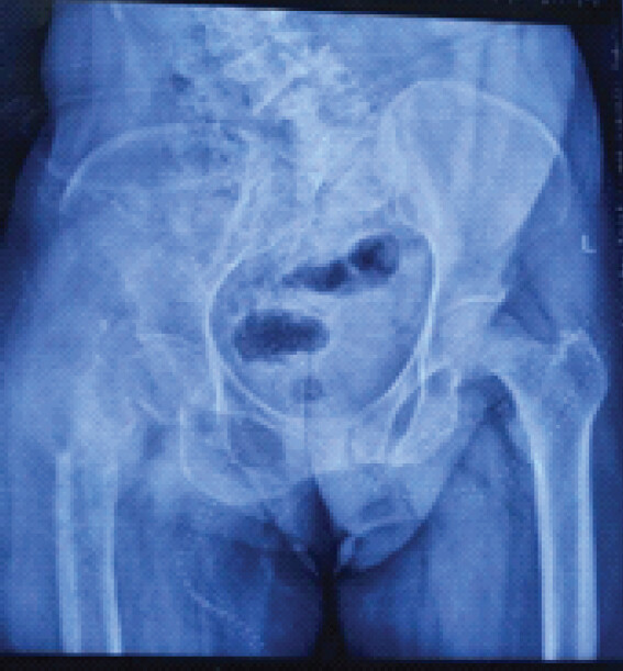
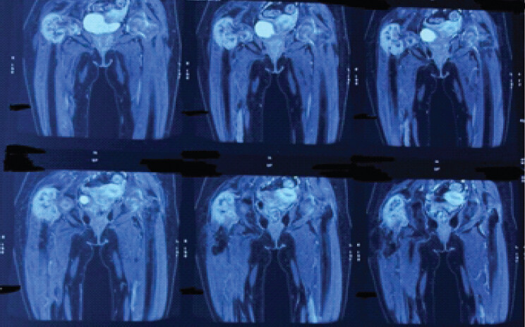
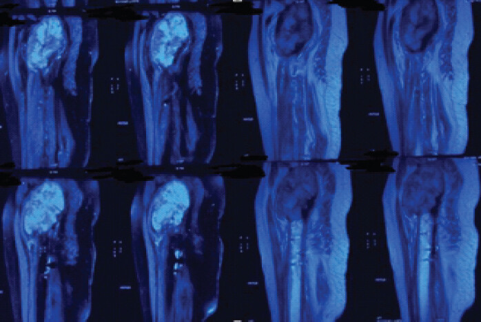
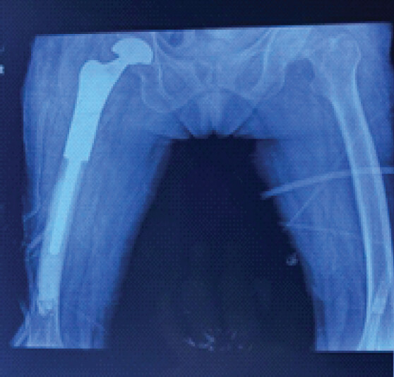
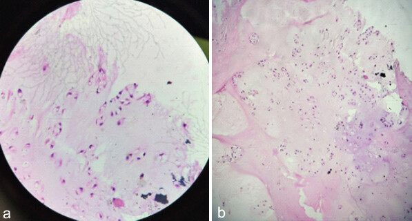
A 45-year-old female was presented in the orthopedic outpatient department with a 2 year history of pain and swelling in her right hip with recent weight-bearing difficulties for 1 week after trivial slip and fall. The patient was unable to bear full weight on affected limb since 1 week. She faced pain at hip joint in squatting and sitting cross legged since 2 year which were gradually increasing in intensity. There was history of weight loss. There was no association with fever, chills, or history of steroid intake. Radiograph (Fig. 1) revealed a lytic lesion in her right femur, involving the entire peri-trochanteric region, whole neck, parts of femur head, and proximal shaft which was compounded by a femur neck fracture. Following initial management with traction and immobilization, further investigations, including magnetic resonance imaging (MRI) and positron emission tomography (PET) scan, were conducted. MRI (Fig. 2 and 3) revealed a T2 hyperintense, and TI hypointense, mass confined to the bone sparing surrounding soft tissues and neurovascular structures. PET scan was negative for metastasis. A core-needle biopsy indicated a perivascular epitheloid tumor. Patient was diagnosed with primary bone PEComa without metastasis in the right proximal femur. Under regional anesthesia, the patient was kept in lateral decubitus position and southern approach was used to expose lesion. The patient underwent wide local excision and reconstruction using a mega-prosthesis. Post-operative radiograph (Fig. 4) showed complete excision of lytic area replaced with mega-prosthesis appropriately. The excised tissue was postoperatively sent for biopsy to confirm the diagnosis. The biopsy result surprisingly came out as low-grade chondrosarcoma showing presence of atypical chondrocytes with hyperchromatic nuclei and calcification (Fig. 5).
Discussion
The case report shows that sometime a common bone tumor can have atypical presentation.
Biopsy of lesion although being gold standard diagnostic tool cannot be 100% reliable due to inadequate tissue sample, wrong tissue sample or maybe due to inexperienced pathologist. PEComas are a family of mesenchymal tumors that rarely arise as a primary bone tumor [8]. Our patient bad involvement of proximal femur which is also a common location for chondrosarcoma. The age of 45 years also favors for chondrosarcoma as it is common in adults of 30–70 years of age. Clinically, most patients present with pain, swelling, and the presence of a mass of variable duration [9]. Radiologically, chondrosarcoma shows lytic lesions (50%) intralesional calcifications: ~70% (ring and arc or popcorn calcification), endosteal scalloping affecting more than two-thirds of the cortical thickness, moth-eaten appearance, or permeative appearance in higher grade tumors, for example, myxoid, dedifferentiated, and mesenchymal chondrosarcoma. Cortical remodeling and periosteal reaction are also seen. MRI shows low-to-intermediate signal intensity lesion on T1 and T2 shows high-signal intensity lesion as was consistent with our patient [10]. Diagnosis is confirmed on biopsy. Macroscopically chondrosarcoma presents as translucent lobular, blue-gray, or white cut surface corresponding to the presence of hyaline cartilage. There may be areas containing myxoid or mucoid material and cystic changes. Microscopically, chondrosarcoma show abundant blue-gray cartilage matrix-production. Irregularly shaped lobules of cartilage varying in size and shape are present. Fibrous bands separate that these lobules or permeate bony trabeculae calcified areas suggesting the presence of a pre-existing enchondroma can often be found. The chondrocytes are atypical, with variable size and shape and contain enlarged hyperchromatic nuclei. Low-grade chondrosarcoma often closely resembles normal cartilage or the benign enchondroma, it is differentiated by presence of “chondrosarcoma permeation pattern,” where the tumor infiltrates through the marrow cavity instead of being confined by the native architecture as present in benign pathology [11]. Low grade or grade 1 chondrosarcoma with no aggressive imaging features can be treated with intralesional curettage and local adjuvants [12]. However, when aggressive biologic behavior is evident on imaging, wide resection following surgical principles of malignant bone tumors seems more appropriate. Our patient underwent wide resection and reconstruction with mega-prosthesis as the pre-operative biopsy diagnosis was PEComa which needs wide surgical excision [13]. However, despite the inconsistency in diagnosis, the patient had optimum final outcome.
Conclusion
This case highlighted an unusual diagnostic discrepancy between pre-operative and post-operative evaluations. Nonetheless, the adopted strategy was effective, allowing the patient to resume daily activities. Surgeon should always send post-operative tumor sample for confirmation of diagnosis and if there is discrepancy in diagnosis do the needful for the well-being of the patient.
Clinical Message
Surgeons should remain aware of the potential for primary bone tumors to masquerade as atypical tumors.
Conflict of Interest:
Source of Support:
Nil
Consent:
The authors confirm that informed consent was obtained from the patient for publication of this article
References
- Murphey MD, Walker EA, Wilson AJ, Kransdorf MJ, Temple HT, Gannon FH.From the archives of the AFIP: Imaging of primary chondrosarcoma: Radiologic-pathologic correlation. Radiographics 2003;23:1245-78. Google Scholar | PubMed
- Marco RA, Gitelis S, Brebach GT, Healey JH.Cartilage tumors: Evaluation and treatment. J Am Acad Orthop Surg 2000;8:292-304. Google Scholar | PubMed
- Aigner T.Towards a new understanding and classification of chondrogenic neoplasias of the skeleton--biochemistry and cell biology of chondrosarcoma and its variants. Virchows Arch 2002;441:219-30. Google Scholar | PubMed
- Choi JH, Ro JY.The 2020 WHO classification of tumors of soft tissue: Selected changes and new entities. Adv Anat Pathol 2021;28:44-58. Google Scholar | PubMed
- Thorkildsen J, Taksdal I, Bjerkehagen B, Haugland HK, Børge Johannesen T, Viset T.Chondrosarcoma in Norway 1990-2013; An epidemiological and prognostic observational study of a complete national cohort. Acta Oncol 2019;58:273-82. Erratum in: Acta Oncol 2019;58:393 Google Scholar | PubMed
- Li B, Yu Y, Bao Y, Song J.Proximal femoral tumor resection followed by joint prosthesis replacement: A systematic review and meta-analysis. BMC Musculoskelet Disord 2023;24:779. Google Scholar | PubMed
- Hornick JL, Fletcher CD.PEComa: What do we know so far?. Histopathology 2006;48:75-82. Google Scholar | PubMed
- Zhong J, Hu Y, Si L, Xing Y, Geng J, Jiao Q.Primary perivascular epithelioid cell tumor (PEComa) in bone: A review of the literature and a case arising in the humerus with multiple metastases. J Bone Oncol 2020;26:100336. Google Scholar | PubMed
- Van Oosterom AT, Dirix LY.Chondrosarcoma and other rare bone sarcomas. Curr Opin Oncol 1990;2:495-9. Google Scholar | PubMed
- Varma DG, Ayala AG, Carrasco CH, Guo SQ, Kumar R, Edeiken J.Chondrosarcoma: MR imaging with pathologic correlation. Radiographics 1992;12:687-704. Google Scholar | PubMed
- Limaiem F, Davis DD, Sticco KL.Chondrosarcoma. In: StatPearls Treasure Island, FL: StatPearls Publishing; 2025 Available from: https://www.ncbi.nlm.nih.gov/books/nbk538132. [Last accessed on 15 Mar 2025]. Google Scholar | PubMed
- Campanacci DA, Scoccianti G, Franchi A, Roselli G, Beltrami G, Ippolito M.Surgical treatment of central grade 1 chondrosarcoma of the appendicular skeleton. J Orthop Traumatol 2013;14:101-7. Google Scholar | PubMed
- Czarnecka AM, Skoczylas J, Bartnik E, Świtaj T, Rutkowski P.Management strategies for adults with locally advanced, unresectable or metastatic malignant perivascular epithelioid cell tumor (PEComa): Challenges and solutions. Cancer Manag Res 2023;15:615-23. Google Scholar | PubMed
© 2026 Journal of Orthopaedic Case Reports - Published by Indian Orthopaedic Research Group
About the Authors
-

Dr. Ananya Mishra
-

Dr. Pulkit Gupta
-

Dr. Manohar
-

Dr. Hoysala Kantharaju
-

Dr. Dhyaan Bhardwaj
-

Dr. Vipul Jhajharia
Related Articles in Journal of Orthopaedic Case Reports
 July 1, 2026 Large Posterior Proximal Femur Osteochondroma Presenting in Adulthood – Diagnostic Challenges and Management Strategy: A Case Report
 July 1, 2026 Combined Mechanical and Biological Salvage of Recalcitrant Subtrochanteric Non-union using Valgus Osteotomy, Blade Plate Fixation, and Endomedullary Fibular Graft
 May 1, 2026 Aneurysmal Bone Cyst of Lateral Cuneiform: Challenge in Diagnosis and Management
 April 1, 2026 A Rare and Interesting Case of Sciatic Nerve Compression Due to Proximal Femoral Osteochondroma in a Young Adult – A Case Report