


Introduction
Reverse total shoulder arthroplasty (RSA) is commonly performed for cuff tear arthropathy (CTA) and irreparable rotator cuff tear (RCT), with favorable clinical outcomes reported.[1] The biomechanical principles of RSA involve humeral distalization and medialization of the glenoid component’s center of rotation. This design enhances deltoid muscle tension, thereby improving functional strength. However, humeral lowering lengthens the upper limb by approximately 15–27 mm,[2] which may lead to neurological complications in 2.0–2.8% of patients postoperatively.[1,3,4] The axillary nerve, which follows a posterior-to-anterior course around the proximal humerus, is particularly susceptible to traction stress following limb lengthening,[4] potentially leading to nerve palsy. Even in the absence of paralysis, patients may experience pain or sensory deficits.
Quadrilateral space (QLS) syndrome is a relatively common condition characterized by lateral shoulder pain and paresthesia due to axillary nerve compression. Although few studies have reported QLS syndrome following RSA,[5] it may be underdiagnosed owing to the difficulty in recognizing its symptoms. However, QLS syndrome following RSA may cause postoperative restricted range of motion (ROM) and affect clinical outcomes.
Hydrorelease (HR) is an ultrasound-guided technique that enables precise injections around target tissues and decreases gliding resistance force between tissues[6,7]. It is used for treating restricted shoulder external rotation via coracohumeral ligament injection and for post-reconstruction knee contracture via infrapatellar fat pad injection.[8,9] HR has also been applied in the management of peripheral nerve entrapment syndromes[10,11] and may be effective for QLS syndrome following RSA; however, no studies have yet evaluated its efficacy.
This study presents three cases of QLS syndrome following RSA, characterized by restricted ROM, in which HR targeting the axillary nerve led to ROM improvement.
Case Reports
Patient selection
Fifteen patients who performed RSA for an CTA (9 patients) or for a massive RCT (6 patients) at our institution between April 2024 and September 2024 were included. Five patients demonstrated anterior elevation (AE) and abduction <100 degrees at 3 months postoperatively, and three of them were diagnosed as QLS syndrome. QLS syndrome was diagnosed based on localized tenderness in the QLS and pain in the posterior to lateral shoulder.
Surgical method
In all cases, RSA was performed under general anesthesia in the beach chair position using a deltopectoral approach. Implant size and humeral osteotomy were determined via preoperative planning. On the scapular side, a small baseplate and a 36 mm sphere was used. On the humeral side, the stem was generally implanted without cement; however, cement was used when primary fixation was inadequate. Humeral retroversion was set at 20 degrees. The insert was selected to achieve sufficient ROM without compromising stability. Target ROM was defined as 150 degrees of AE, 40 degrees of extension, and 60 degrees of external rotation (ER) in the resting position. The subscapularis tendon was detached and reattached to the humerus using three high-strength sutures following implant placement.
Postoperative rehabilitation
A shoulder abduction brace was applied for 2 weeks. During the first postoperative week, shoulder pendulum exercises, elbow and wrist ROM exercises, and periscapular muscle relaxation were initiated. From week 1, passive shoulder exercises commenced; from week 2, active-assisted exercises; and from week 3, active exercises. Rehabilitation emphasized avoidance of arm positions with a high dislocation risk, although ROM was not restricted. Postoperative rehabilitation continued for 6–12 months.
Axillary nerve HR
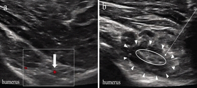
HR of the axillary nerve was conducted in 3 patients who demonstrated AE and abduction <100 degrees at 3 months postoperatively, with a clinical diagnosis of QLS syndrome. The procedure was performed by an orthopedic surgeon with the patient seated and the arm in a resting position. An ultrasound probe was positioned posteriorly along the long axis of the humerus to identify the posterior circumflex humeral artery and accompanying axillary nerve (Figure 1a). A 23-gauge catheterized needle was used to inject 10 mL of saline[12] around the axillary nerve using a parallel technique (Figure 1b).
Clinical outcome
ROM in AE, abduction, ER, and internal rotation (IR) in the resting position was measured before and after axillary nerve HR at 3 months, 6 months, and 1 year postoperatively. Functional outcomes were assessed using the American Shoulder and Elbow Surgeons (ASES) score, Constant score, and Subjective Shoulder Value (SSV) at the same intervals: 3 months (prior to HR), 6 months, and 1 year postoperatively.
Imaging evaluation
Preoperative imaging included Hamada classification[13] on X-ray, and Walch[14] and Favard[15] classifications on computed tomography (CT). Postoperative imaging included assessments of upper limb extension,[16] lateral humeral offset (LHO),[17] baseplate version,[18] baseplate tilt,[19,20] and stem alignment angle.[21] X-ray and CT were obtained 1 week postoperatively. Upper limb extension was measured following the method of Jobin et al.,[16] defined as the distance from the inferolateral acromion to the midpoint of the deltoid tuberosity, calibrated against humeral shaft width. LHO was assessed using the method described by Clinker et al.,[17] as the distance between the lateral edge of the greater tuberosity and the lateral edge of the acromion, with lateralization reported as positive values and medialization as negative values. Inferior baseplate tilt was denoted as positive, and superior tilt as negative.
Case 1
An 82-year-old female underwent RSA for a massive RCT. She was 149.5 cm tall and weighed 48.0 kg. Preoperative ROM was limited to AE: 80°, abduction: 60°, ER: 10°, and IR at the sacral level. Her preoperative ASES, Constant, and SSV scores were 19, 28, and 0 points, respectively. Imaging assessments revealed Hamada classification Type 2, Walch classification Type A1, and Favard classification Type E0.
RSA was performed using the Trabecular Metal Reverse PlusTM system on the glenoid side and the Identity® Shoulder System on the humeral side (Zimmer Biomet, Warsaw, USA). A cementless stem and a minus tray were utilized. Postoperative radiography (Figure 2a) showed an upper limb extension of 14.1 mm and an LHO of 3.1 mm. The baseplate version was 2.9° of anteversion, and baseplate tilt was –4.1°. The stem was inserted at 0.1° of valgus.
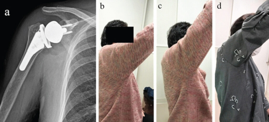
(a) Postoperative anteroposterior X-ray.
(b) At 3 months postoperative, prior to axillary nerve HR.
(c) At 3 months postoperative, immediately after axillary nerve HR.
(d) At 1 year postoperative.
At 3 months postoperatively, ROM improved to AE: 95°, abduction: 90°, ER: 30°, and IR remained at the sacral level. The patient was diagnosed with QLS syndrome and underwent HR of the axillary nerve. ROM at 3 months (after HR)/6 months/1 year following HR was as follows: AE: 135°/135°/135°, abduction: 110°/120°/120°, ER: 30°/30°/30°, and IR: sacral level/sacral/L4. ASES, Constant, and SSV scores at 3 months (before HR)/6 months/1 year were: ASES: 62.5/85/85.2, Constant: 58/72/74, and SSV: 60/80/80. AE images at 3 months (before and after HR) and at 1 year postoperatively are shown in Figure 2b–d.
Case 2
An 82-year-old female underwent RSA for CTA. She was 144.1 cm tall and weighed 44.5 kg. Preoperative ROM included AE: 80°, abduction: 60°, ER: 20°, and IR at the sacral level. Her ASES, Constant, and SSV scores were 37, 35, and 50 points, respectively. Imaging revealed Hamada classification Type 4a, Walch classification Type A1, and Favard classification Type E0.
RSA was performed using the Trabecular Metal Reverse PlusTM system on the glenoid side and the Identity® Shoulder System on the humeral side (Zimmer Biomet, Warsaw, USA). A cemented stem and a minus tray were used. Postoperative radiograph (Figure 3a) showed upper limb extension of 19.5 mm and an LHO of 3.2 mm. The baseplate version was 2.6° anteversion, and tilt was –1.2°. The stem was placed with 4° of varus.
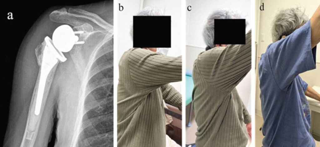
(a) Postoperative anteroposterior X-ray.
(b) At 3 months postoperative, prior to axillary nerve HR.
(c) At 3 months postoperative, immediately after axillary nerve HR.
(d) At 1 year postoperative.
At 3 months postoperatively, ROM included AE: 75°, abduction: 70°, ER: 20°, and IR at the sacral level. Following diagnosis of QLS syndrome, HR of the axillary nerve was performed. ROM at 3 months (after HR)/6 months/1 year after HR was: AE: 115°/120°/120°, abduction: 100°/120°/120°, ER: 20°/20°/30°, and IR: sacral/sacral/L5. ASES, Constant, and SSV scores at 3 months (before HR)/6 months/1 year were: ASES: 46.7/85/85.2, Constant: 48/72/74, and SSV: 60/80/80. AE images at 3 months (before and after HR) and at 1 year postoperatively are presented in Figure 3b–d.
Case 3
An 84-year-old female underwent RSA for CTA. She was 145.8 cm in height and weighed 48.1 kg. Preoperative ROM was AE: 20°, abduction: 20°, ER: 10°, and IR at the buttock level. Her ASES, Constant, and SSV scores were 5, 9, and 5 points, respectively. Imaging showed Hamada classification Type 4a, Walch classification Type A1, and Favard classification Type E0.
RSA was performed using the Equinoxe® platform shoulder system (Exactech, Florida, USA) with a cementless preserve stem. Postoperative imaging (Figure 4a) revealed 14.0 mm of upper limb extension and an LHO of 4.7 mm. The baseplate was positioned at 3.1° of anteversion and –2.3° of tilt. The stem was inserted with 4.1° of varus.
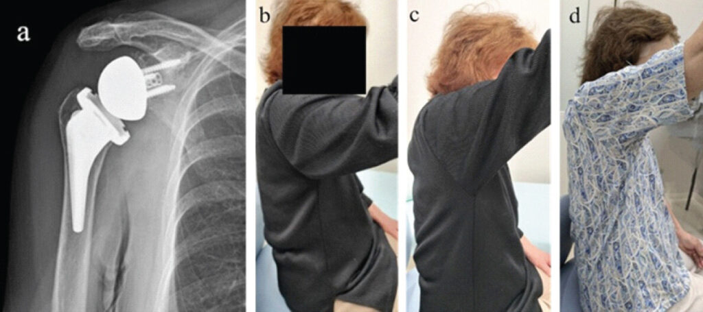
(a) Postoperative anteroposterior X-ray.
(b) At 3 months postoperative, prior to axillary nerve HR.
(c) At 3 months postoperative, immediately after axillary nerve HR.
(d) At 1 year postoperative.
At 3 months, ROM was AE: 65°, abduction: 60°, ER: 30°, and IR at the sacral level. After diagnosis of QLS syndrome, HR of the axillary nerve was conducted. Post-HR ROM at 3 months (after HR)/6 months/1 year was: AE: 110°/110°/110°, abduction: 100°/100°/100°, ER: 30°/40°/50°, and IR: sacral/sacral/L4. ASES, Constant, and SSV scores at 3 months (before HR)/6 months/1 year were: ASES: 33.3/48.3/71.6, Constant: 44/56/72, and SSV: 50/70/70. AE images at 3 months (before and after HR) and at 1 year postoperatively are shown in Figure 4b–d.
Discussion
This study presents three cases of limited AE and abduction at 3 months following RSA, in which HR of the axillary nerve for QLS syndrome led to rapid improvement, sustained at 1 year postoperatively. All three cases involved small female patients, each <150 cm in height. Although upper limb lengthening was modest (14–19 mm), the patients’ small body size may have led to proportionally greater traction on the axillary nerve, potentially contributing to QLS syndrome. The immediate postoperative improvement in ROM following HR suggests a reduction in axillary nerve tension.[6,7] None of the three cases demonstrated issues with implant positioning. Nevertheless, alternative causes of limited ROM after RSA—such as suboptimal implant placement, intraoperative ROM restriction, or reduced scapular mobility—should also be considered alongside QLS syndrome as contributing factors.[20] QLS syndrome following RSA has been underreported and may be underestimated[5]. Some cases may achieve early improvement in ROM with appropriate diagnosis and management of QLS syndrome following RSA.
Rehabilitation is essential for optimizing RSA outcomes. HR of the axillary nerve may facilitate earlier improvements in AE and abduction, potentially enhancing the effectiveness of postoperative rehabilitation as early as three months after RSA. Since patients undergoing RSA are often older adults who may struggle to maintain outpatient rehabilitation, early ROM improvement could help shorten the rehabilitation timeline. Future studies will assess differences in rehabilitation duration by expanding the sample size. Conversely, HR of the axillary nerve did not improve ER or IR. This may be due to unresolved muscle tightness or impingement unrelated to axillary nerve tension.
We have not yet quantified the reduction in axillary nerve stretch tension. Further research, including techniques such as ultrasonic elastography, is required for objective evaluation. Additionally, since follow-up was limited to 1 year, the long-term effects of HR of the axillary nerve warrant further investigation.
Conclusion
This study presents three cases of limited AE and abduction at 3 months after RSA, in which HR of the axillary nerve for QLS syndrome led to rapid improvement that persisted at 1 year postoperatively. HR of the axillary nerve may be effective in alleviating early postoperative limitations in AE and abduction in patients with QLS syndrome following RSA.
Clinical Message
Quadrilateral space syndrome following a reverse total shoulder arthroplasty may be underdiagnosed owing to the difficulty in recognizing its symptoms. Hydrorelease of the axillary nerve may be effective in alleviating early postoperative limitations in anterior elevation and abduction in patients with quadrilateral space syndrome following a reverse total shoulder arthroplasty.
Conflict of Interest:
Nil
Source of Support:
Nil
Consent:
The authors confirm that informed consent was obtained from the patient for publication of this article
References
- Inagaki K, Ochiai N, Hashimoto E, Hattori F, Hiraoka Y, Ise S. Postoperative complications of reverse total shoulder arthroplasty: a multicenter study in Japan. JSES Int 2023;7:642-7. Google Scholar | PubMed
- Lädermann A, Edwards TB, Walch G. Arm lengthening after reverse shoulder arthroplasty: a review. Int Orthop 2014;38:991-1000. Google Scholar | PubMed
- Boileau P, Watkinson D, Hatzidakis AM, Hovorka I, Award N. The Grammont reverse shoulder prosthesis: results in cuff tear arthritis, fracture sequelae, and revision arthroplasty. J Shoulder Elbow Surg 2006 Sep;15(5):527-40. Google Scholar | PubMed
- Lopiz Y, Rodriguez-González A, Martín-Albarrán S, Marcelo H, García-Fernández C, Marco F. Injury to the axillary and suprascapular nerves in rotator cuff arthropathy and after reverse shoulder arthroplasty: a prospective electromyographic analysis. J Shoulder Elbow Surg 2018;27:1275-82. Google Scholar | PubMed
- Harada Y, Iwahori Y, Kajita Y, Morishita Y, Deie M. Short-term outcome of reverse shoulder arthroplasty (in Japanese). Katakansetsu Shoulder Jt 2019;43:900-3. Google Scholar | PubMed
- Shiwaku K, Otsubo H, Suzuki D, Pirri C, Kodesyo T, Kamiya T. Biomechanical effects of fascial hydrorelease: a cadaveric study. BMC Musculoskelet Disord 2025;26(1):306. Google Scholar | PubMed
- Evers S, Thoreson AR, Smith J, Zhao C, Geske JR, Amadio PC. Ultrasound-guided hydrodissection decreases gliding resistance of the median nerve within the carpal tunnel. Muscle Nerve 2018;57(1):25-32. Google Scholar | PubMed
- Kimura H, Suda M, Kobayashi T, Suzuki S, Fukui S, Obata H. Effectiveness of ultrasound-guided fascia hydrorelease on the coracohumeral ligament in patients with global limitation of the shoulder range of motion: a pilot study. Sci Rep 2022;12:19782. Google Scholar | PubMed
- Machida T, Watanabe A, Miyazawa S. A new procedure for ultrasound-guided hydrorelease for the scarring after arthroscopic knee surgery. Cureus [Internet 2020;12:e12405. Google Scholar | PubMed
- Buntragulpoontawee M, Chang KV, Vitoonpong T, Pornjaksawan S, Kitisak K, Saokaew S. The effectiveness and safety of commonly used injectates for ultrasound-guided hydrodissection treatment of peripheral nerve entrapment syndromes: A systematic review. Front Pharmacol 2020;11:621150. Google Scholar | PubMed
- Fukui S, Rokutanda R, Kawai S, Suda M, Iwata F, Okada M. Current evidence and practical knowledge for ultrasound-guided procedures in rheumatology: joint aspiration, injection, and other applications. Best Pract Res Clin Rheumatol 2023;37:101832. Google Scholar | PubMed
- Kobayashi T, Kimura H, Noriyuki O. Effects of interfascial injection of bicarbonated Ringer's solution, physiological saline and local anesthetic under ultrasonography for myofascial pain syndrome -Two prospective, randomized, double-blinded trials-. J Juzen Med Society 2016;125(2):40-9. Google Scholar | PubMed
- Hamada K, Fukuda H, Mikasa M, Kobayashi Y. Roentgenographic findings in massive rotator cuff tears. A long-term observation. Clin Orthop Relat Res 1990(254):92-6. Google Scholar | PubMed
- Walch G, Badet R, Boulahia A, Khoury A. Morphologic study of the glenoid in primary glenohumeral osteoarthritis. J Arthroplasty 1999;14:756-60. Google Scholar | PubMed
- Sirveaux F, Favard L, Oudet D, Huquet D, Walch G, Mole D. Grammont inverted total shoulder arthroplasty in the treatment of glenohumeral osteoarthritis with massive rupture of the cuff. J Bone Jt. Surg Br 2004;86:388-95. Google Scholar | PubMed
- Jobin CM, Brown GD, Bahu MJ, Gardner TR, Bigliani LU, Levine WN. Reverse total shoulder arthroplasty for cuff tear arthropathy: the clinical effect of deltoid lengthening and center of rotation medialization. J Shoulder Elbow Surg 2012;21:1269-77. Google Scholar | PubMed
- Clinker C, Ishikawa H, Presson AP, Zhang C, Joyce C, Chalmers PN. The effect of lateralization and distalization after Grammont-style reverse total shoulder arthroplasty. J Shoulder Elbow Surg 2024;33:2664-70. Google Scholar | PubMed
- Friedman RJ, Hawthorne KB, Genez BM. The use of computerized tomography in the measurement of glenoid version. J Bone Joint Surg Am 1992;74:1032-7. Google Scholar | PubMed
- Boileau P, Gauci M-O, Wagner ER, Clowez G, Chaoui J, Chelli M. The reverse shoulder arthroplasty angle: a new measurement of glenoid inclination for reverse shoulder arthroplasty. J Shoulder Elbow Surg 2019;28:1281-90. Google Scholar | PubMed
- Maksoud A, Steinlechner C, Baldwick C, Tabi W. Associations between radiographic parameters in the reverse shoulder arthroplasty and patient outcomes. Shoulder Elbow 2023;15(3) suppl:19-34. Google Scholar | PubMed
- Abdic S, Athwal GS, Wittmann T, Walch G, Raiss P. Short stem humeral components in reverse shoulder arthroplasty: stem alignment influences the neck-shaft angle. Arch Orthop Trauma Surg 2021;141:183-8. Google Scholar | PubMed
© 2026 Journal of Orthopaedic Case Reports - Published by Indian Orthopaedic Research Group
About the Authors
-

Dr. Kenta Inagaki
-

Dr. Yasuchika Aoki
-

Dr. Seiji Ohtori
Related Articles in Journal of Orthopaedic Case Reports
 April 1, 2026 Management of a Post-traumatic Stiff Elbow: A Case Report
 January 1, 2026 Enhancing Functional Outcomes in Distal End Radius Fracture Rehabilitation
 September 1, 2026 Extensive Post-Traumatic Heterotopic Ossification with Multi-joint Involvement: A Case Report
 August 1, 2026 Reverse Shoulder Arthroplasty with Virtual Implant Positioning System in the Management of Massive Rotator Cuff Tear with Rotator Cuff Arthropathy