


Introduction
The coracoclavicular (CC) ligament consists of two stabilizing bands connecting the clavicle to the coracoid process: The conoid ligament posteromedially and the trapezoid ligament anterolaterally (Fig. 1a and b) [1]. The ligament stabilizes the clavicle by connecting it to the scapular body but still allows some movement [2]. The ossification of the CC ligament is an uncommon finding and has only been described anecdotally [3,4,5,6]. The etiology is unknown, but it seems to be associated with trauma, renal failure or paraplegia. A radiology study by Chen and Bohrer describes thirty-six patients with ossified CC ligaments, differentiating between ossification, punctate calcification and mass-like tumoral calcification [3]. The treatment of the ossification is not described in this study. Furthermore, no other articles describe the treatment of this pathology.
![Figure 1: (a and b) Anterior and posterior view of coracoclavicular ligaments (3D Models are powered by www.anatomylearning.com [1])](https://jocr.co.in/wp/wp-content/uploads/516FF1-converted-1.jpg)
In the past 5 years, four patients presented in our outpatient clinics with severe ossification of the CC ligaments, resulting in the near synostosis of their clavicles and scapulae. All four patients underwent surgery, which comprised the removal of the bony bridge with or without CC ligament reconstruction using a hamstring autograft. To the best of our knowledge, this is one of the first studies which describe a surgical intervention for CC ossification. The clinical presentations will be presented, as will the surgical interventions, the radiological follow-up, and the patient-reported outcome measures.
Case Report
For each presented case, the medical files were reviewed, and the relevant past medical history, patient presentation, physical examination and radiological imaging were collected. The surgical report was reviewed and is summarized below. At the latest post-operative follow-up, each patient completed a numeric pain rating scale (NRS) pain and satisfaction questionnaire, the Oxford Shoulder Score, the Quick Disabilities of the Arm, Shoulder and Hand (QuickDASH) score and the specific acromioclavicular score (SACS) after informed consent was given. The Oxford Shoulder Score ranges from 12 to 60. A low score represents a well-functioning shoulder in daily life. QuickDASH scores (range: 0–100) assess functioning in daily life, work and sports. Again, a low score suggests few limitations due to an injured shoulder. The SACS score (range: 0–100%) was developed to assess the outcomes of patients treated for acromioclavicular joint (ACJ) problems in particular. A low score suggests few limitations/lower pain due to the injured shoulder. In addition, a range of motion (ROM) test was done on the shoulder joint. The flexion, abduction, and external and internal rotation were tested. Measurements were done using a goniometer.
Materials and Methods
All four patients presented at the outpatient clinic with complaints of shoulder pain on overhead movement for more than a year, impairing their quality of life and/or their ability to work despite extensive non-operative treatment (physiotherapy, rest and pain medication). The patient characteristics are shown in Table 1.
Patient characteristics
| Variables | Case 1 | Case 2 | Case 3 | Case 4# |
|---|---|---|---|---|
| Age (years) | 26 | 25 | 43 | 70 |
| Gender | Male | Male | Female | Male |
| Condition | DCF | DCF | FRS* | DCF |
| Time till presentation (years) | 7 | 3 | 1 | 15 |
*
For a thoracic outlet syndrome;
#
was already treated with a lateral clavicle resection with partial removal of the ossification of the coracoclavicular ligament without stabilization of the clavicle in a different hospital. Due to persistent symptoms he was referred. DCF: Distal clavicle fracture, FRS: First rib resection
In every case, physical examination revealed tenderness over the ACJ with or without a prominent distal clavicle. Each patient showed ossification of the CC ligament, in which the space between the coracoid process and the clavicle was almost completely closed, leading to symptoms severe enough to warrant a desire for surgery. One of our patients reported radiating pain to his thumb and was seen by a neurologist. There were no signs of plexus neuropathy on electromyography. However, based on clinical symptoms and the computed tomography (CT) scan, it was concluded that the neurological complaints could have resulted from a bony spur’s compression of the brachial plexus. The patient, who was treated for thoracic outlet syndrome (TOS), continued to experience radiating pain to the arm despite first rib resection. In this case too, the compression of the brachial plexus was suspected.
The operated shoulder was immobilized in an immobilizer for the first 6 weeks following surgery. During this period, the patients were instructed to perform circumduction exercises of the shoulder and maintain mobility of the hand, wrist and elbow through active exercises. From 6 weeks postoperatively, unloaded ROM exercises of the shoulder were initiated to gradually restore joint mobility. After 12 weeks, progressive strengthening exercises were introduced, with a gradual increase in resistance to restore muscular strength and functional capacity.
Radiological characteristics
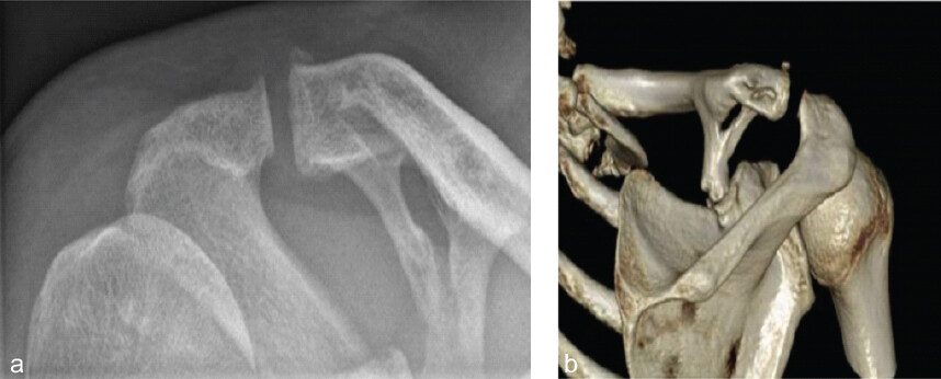
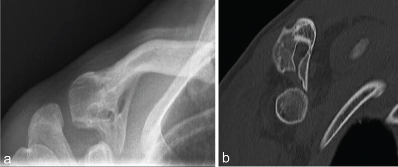
On plain film X-rays of the shoulder joint (i.e. the anteroposterior, supraspinatus outlet, axial and Zanca views), the suggestion of a bony bridge between the clavicle and the coracoid process was visible in all cases (Figs. 2a and 3a). A CT scan revealed the clear ossification of the CC ligament (Figs. 2b and 3b).
Surgical intervention
Each patient underwent an open surgical intervention through a deltopectoral approach, opening the deltopectoral interval. A small part of the deltoid origin on the anterolateral clavicle was detached to make it possible to approach the ossification. The ossification was then removed with an oscillating saw and osteotomes both anteriorly and posteriorly to the clavicle. After that, clavicular stability was assessed clinically by the manual palpation of the clavicle. When a dislocation of more than 5 mm in any direction was assessed, it was considered to be unstable. This was done in anticipation of AC joint complaints after such an operation. As with a clavicle stabilization for an AC luxation, an ACJ resection was therefore chosen.
In case 2, only slight vertical clavicular instability was assessed with an intact ACJ capsule providing sufficient stability to the clavicle. No additional stabilization was performed.
In cases 1, 3 and 4, the clavicle was assessed to be unstable after the removal of the ossification and lateral clavicle resection. Therefore, stabilization was performed using a hamstring autograft (semitendinosus or gracilis tendon). For the anatomical reconstruction of the CC ligaments, the autograft was looped around the coracoid process and passed through an anterolateral and a posteromedial drill hole (4.5 mm) in the clavicle, 4.5 cm from the distal end of the clavicle, and fixated on top with a non-dissolvable suture.
In cases 3 and 4, a suture tape (FiberTape®, Arthrex Inc. Erwin-Hielscher-Str. 9, 81249 Munich, Germany) was used along with the autograft. Because the clavicle was still assessed to be unstable in the horizontal plane, the ACJ was stabilized in these two cases by passing the gracilis tendon through an anteroposterior drill hole in the acromion and a superoinferior drill hole in the distal clavicle (4.5 mm). The remainder of the tendon was used to reinforce the ACJ capsule.
Outcome
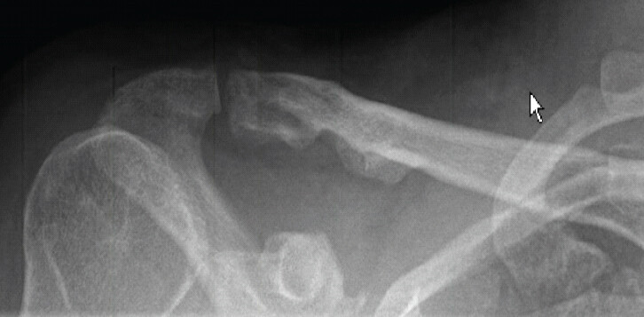
At the latest post-operative follow-up (range: 34–43 months), every patient completed an NRS pain and satisfaction questionnaire, the Oxford Shoulder Score, the QuickDASH score and the SACS after giving informed consent (Table 2). Post-operative X-rays show the near-complete removal of the ossified ligaments (Fig. 4).
PROMs at latest follow-up PROMs (scores are shown)
| Variables | Case 1 | Case 2 | Case 3 | Case 4 | Median (range) |
|---|---|---|---|---|---|
| Time after surgery (months) | 34 | 43 | 37 | 48 | 40 (34–48) |
| Mean NRS pain* | 2 | 2 | 3 | 0 | 2 (0–3) |
| Worst NRS pain* | 7 | 8 | 5 | 0 | 6 (0–8) |
| Overall satisfaction* | 9 | 7 | 8 | 10 | 8.5 (7–10) |
| Oxford shoulder score~ | 16 | 18 | 19 | 12 | 17 (12–19) |
| QuickDASH limitation# | 0 | 9 | 11 | 0 | 4.5 (0–11) |
| QuickDASH work# | 19 | 25 | 0 | N/A | 19 (0–25) |
| QuickDASH sports# | N/A | 6 | N/A | N/A | 6 (6–6) |
| SACS# | 16 | 26 | 13 | 2 | 14.5 (2–26) |
*
Scale 0–10;
~
Scale 12–60;
#
Scale 0–100. PROMs: Patient-reported outcome measures, NRS: Numeric pain rating scale, QuickDASH: Quick Disabilities of the Arm, Shoulder and Hand, SACS: Specific acromioclavicular score
Each patient had a relatively low score for average pain and a relatively high score for overall satisfaction. The ROM measurements at the latest follow-up are shown in Table 3. Case 1 showed better internal rotation but worse external rotation than the other cases. In addition, case 4 had worse flexion, abduction and external rotation than the other cases. None of the patients reported neurological symptoms postoperatively.
Pre- and post-operative range of motion at latest follow-up (range of degrees is shown)
| Variables | Case 1 | Case 2 | Case 3 | Case 4 | ||||
|---|---|---|---|---|---|---|---|---|
| Pre | Post | Pre | Post | Pre | Post | Pre | Post | |
| Flexion | 170 | 180 | 170 | 170 | 180 | 170 | 120 | 140 |
| Abduction | 170 | 180 | 170 | 170 | 180 | 170 | 120 | 140 |
| External rotation | 40 | 45 | 90 | 85 | 80 | 60 | 60 | 60 |
| Internal rotation* | Th 6 | Th8 | L2 | L2 | L2 | L2 | L3 | L3 |
*
The vertebrae which the patient could reach is shown
Discussion
This study describes four patients with ossification of the CC ligaments undergoing resection of the ossification. In three patients, the reconstruction of the CC ligaments was performed. Every patient experienced an improvement in functional outcome but still had some complaints regarding activities of daily living. In the inclusion period, three more patients were seen clinically but did not opt for surgery because their complaints were acceptable.
In healthy individuals, on average, the scapula internally rotates 8° during the elevation of the arm, and there are 11° of upward rotation and 19° of posterior tilting relative to the clavicle [2]. In case of the ossification of these ligaments, synostosis between the scapula and the clavicle occurs. This alters the dynamic forces acting on the lateral part of the clavicle and limits the scapular movements, changing the mechanics of the clavicle and, eventually, the entire shoulder girdle. This explains the restricted shoulder movement and/or pain in our patients. In addition, two patients exhibited neurological symptoms associated with brachial plexus neuropathy. Although neurological examination did not reveal neuropathy, reduced clavicular mobility could have led to the increased compression of the brachial plexus.
By the surgical removal of the ossification, the mobility of the scapula and ACJ can be restored [7,8]. However, the stabilizing function of the CC ligaments would be lost, and the reconstruction of these ligaments would be mandatory. In one patient (case 2), the ossifications consisted of an anterior part and a posterior part, where the anterior part resulted in near synostosis. After the removal of this anterior part, there was no impairment in function and stability, as the posterior part was not resected. As the CC ligaments were located more posteriorly, in this case, no resection of the conoid ligament was performed. Hence, no instability was found. In each of the other three cases, an extensive surgical approach was adopted in order to remove the ossification, including the CC ligament. In these cases, the reconstruction of the CC ligaments was mandatory. In our patients, the perioperative assessment of the pathology and instability was decisive for additive stabilization. Different techniques of CC reconstruction are described [9]. In our clinics, a reconstructive procedure with an autologous hamstring graft stabilized with suture tape was the procedure of choice.
Small differences were found in the ROM. Hence, case 4 showed worse flexion, abduction and external rotation than the other cases. This might be related to the older age of the patient and the age-related increase in stiffness [10].
Case 2 differed from cases 1, 3, and 4 in terms of lower overall satisfaction and higher pain levels. This observation may suggest the consideration of a reconstructive procedure at a lower threshold in similar cases. However, larger sample sizes are required to draw reliable conclusions regarding this potential association.
Two patients reported neurological complaints before surgery. Retrospectively, both cases may have suffered from neurogenic TOS caused by the ossification of the CC ligaments, which occupied space and impaired the movement of the clavicle and scapula. Postoperatively, these neurological complaints resolved.
Limitations
This study has several limitations that should be considered when interpreting the findings. First, pre-operative clinical data were not available; they included baseline outcome measures such as pain scores. As a result, it was not possible to directly compare the post-operative outcomes with the patients’ pre-operative clinical statuses. Second, the retrospective design of this study introduced the inherent risks of selection bias and information bias, as the data were collected from existing medical records rather than through a standardized prospective protocol. Third, the sample size was small, which limited the generalizability of the findings and prevented robust statistical analysis. This is inherent to the nature of a case report-based study. Finally, there was heterogeneity in the surgical techniques used, which may have influenced the outcomes and made it difficult to attribute the results to a single standardized surgical approach.
Conclusion
All the patients who underwent the resection of ossified CC ligaments experienced improvements in pain and functional outcome, with mild complaints regarding activities of daily living. We believe that the stability of the clavicle should be assessed during surgery and restored if needed. Hopefully, this article will provide insight to shoulder surgeons who encounter this condition and motivate them to publish the results of their treatment of CC ossification.
Clinical Message
An extensive search in the current literature shows that this is the first case report describing a surgical procedure to address CC ossification with patient-reported outcome measures. Despite the small cohort of patients and the fact that no questionnaires were sent before the surgery, we believe that this case report will be of interest to orthopedic surgeons, physiotherapists and other healthcare providers who treat shoulder problems.
Conflict of Interest:
Nil
Source of Support:
Nil
Consent:
The authors confirm that informed consent was obtained from the patient for publication of this article
References
- Blanco Salado R. Anatomy Learning - 3D Anatomy Atlas Explore Human Body in Real Time Available from: https://anatomylearning.com. [Last accessed on 2025 Jun 30]. Google Scholar | PubMed
- Ludewig PM, Phadke V, Braman JP, Hassett DR, Cieminski CJ, Laprade RF. Motion of the shoulder complex during multiplanar humeral elevation J Bone Joint Surg 2009;91:378-89. Google Scholar | PubMed
- Chen YM, Bohrer SP. Coracoclavicular and coracoacromial ligament calcification and ossification Skeletal Radiol 1990;19:263-6. Google Scholar | PubMed
- Schubert F. 'Duplication' of the clavicle or ossification in coracoclavicular ligament Aust Radiol 1997;41:70-2. Google Scholar | PubMed
- Qureshi AZ, AlSaleh AJ, AlHalemi AA. Ossification of coracoclavicular ligament in complete paraplegia: A case report Spinal Cord Ser Cases 2015;1:15008. Google Scholar | PubMed
- Ertogrul R, Sahin K, Celik H, Kapicioglu M, Ersen A, Bilsel K. Is coracoclavicular ossification a complication or a good prognostic factor after surgical treatment of acromioclavicular joint injury? JSES Int 2022;6:769-74. Google Scholar | PubMed
- Fleurette J, Solignac N, Werthel J D. Post-traumatic Coracoclavicular Ligament Ossification: A Case Report and Surgical Technique JBJS case connector 2024;14 3 10.2106/JBJS.CC.24.00136 Google Scholar | PubMed | CrossRef
- Moon K, Hwang YS, Kim KT, Kim JW, Chae JH. Ossification of the coracoacromial ligament in subacromial impingement syndrome: A case report Clin Shoulder Elb 2017;20:167-71. Google Scholar | PubMed
- Saraglis G, Prinja A, To K, Khan W, Singh J. Surgical treatments for acute unstable acromioclavicular joint dislocations SICOT J 2022;8:38. Google Scholar | PubMed
- Lee Y, Ashton-Miller JA. Effects of age, gender and level of co-contraction on elbow and shoulder rotational stiffness and damping in the impulsively end-loaded upper extremity Ann Biomed Eng 2015;43:1112-22. Google Scholar | PubMed
© 2026 Journal of Orthopaedic Case Reports - Published by Indian Orthopaedic Research Group
About the Authors
-

Dr. Borg Leijtens
-

Mr. Luc Dirk Pieter Tilman
-

Dr. Marieke Ostendorf
-

Dr. Frederik Okke Lambers Heerspink
-

Dr. Oscar Dorrestijn
Related Articles in Journal of Orthopaedic Case Reports
 March 1, 2025 A Retrospective Study of Functional Outcomes in Rockwood Type III and V Acromioclavicular Joint Dislocations Managed with Double Endobutton Technique
 October 10, 2022 Symptomatic Coracoclavicular Joint – A Case Report of an Important but Lesser known Differential Diagnosis of Shoulder Pain
 November 10, 2023 Uncemented Total Hip Arthroplasty in an Adult without Osteotomy for Neglected Developmental Dysplasia of Hip
 November 28, 2019 A Novel Unidirectional Porous ß-tricalcium Phosphate Grafting Impregnated with Bone Marrow Blood Collected from Unfractured Vertebral Bodies for Non-union of Osteoporotic Vertebral Fracture: A Preliminary Case Report