


Introduction
Anterior cruciate ligament (ACL) injury is a type of ligamentous injury in the knee that is very frequent among young, physically active people and results in functional instability, performance deterioration, and premature degenerative alterations in the case of untreated injury [1]. Primary ACL reconstruction (ACLR) is the standard treatment of the symptomatic rupture of the ACL, with the intended aim of restoring knee stability, biomechanics and functional capacity through the use of autografts or allografts. However, even with the improvement of surgical and rehabilitation protocols, the inconsistency of the overall outcome of patients is a significant clinical issue, and some patients recover fully, whereas others show chronic instability, graft failure, or slow recovery. As such, the biological recovery of the graft and its connection with functional recovery has become a hot discipline of study [2].
After ACLR, the graft is subjected to a complex bio-remodeling process termed as ligamentization that involves necrosis, revascularization, cellular proliferation, and collagen reorganization. In the process, the structural and biomechanical characteristics of the graft over time become similar to the native “ACL [3]. The most popular parameter that has been assessed is the graft signal intensity using magnetic resonance imaging (MRI) as a non-invasive method to analyze graft maturation. In general, the higher the signal intensity on MRI, the higher the water content, incomplete collagen structure, and the continued remodeling, the lower the signal intensity, the more mature and organized the graft structure [4].
Serial MRI evaluation enables the clinician to determine temporal variations in graft signal intensity that occur over time and give input into the healing process. The early post-operative grafts tend to show intermediate to high signal intensity owing to the vascular ingrowth and tissue remodeling that may continue up to 2 years [5]. Nonetheless, continued high signal intensity later than anticipated can be a sign of graft degeneration, impingement, or partial failure. In this way, MRI does not just aid in the evaluation of the integrity of the grafts; it also contributes to the detection of complications that can undermine the functional outcomes [6].
A number of studies have tried to relate MRI-based graft signal intensity with clinical and functional outcomes after ACLR. The functional recovery can be evaluated by use of parameters like knee stability (instrumented laxity testing), patient-reported outcome measures (e.g., international knee documentation committee [IKDC], Lysholm scores), and return-to-sport criteria [7]. There is some evidence that greater graft signal intensity, indicating a biological immaturity, correlates with a worse functional stability and knee laxity. MRI signal intensity and functional outcomes have been reported to have a negative relationship with each other, and patients who have more mature grafts (lower signal intensity) are more likely to have better clinical recovery [8].
Moreover, quantitative MRI parameters such as signal-to-noise quotient (SNQ) and graft volume have been identified to forecast the long-term outcome. Indicatively, it has been shown that simultaneous measurement of graft volume and signal intensity can predict clinical and patient outcomes several years post-surgery, indicating that MRI is prognostic in ACLR follow-up [9].
Nevertheless, the topic of correlating MRI results with functional recovery is not completely uniform throughout the literature. Although there are studies that provide substantial correlation between graft signal strength and clinical outcome, other studies have shown no dependable association, especially when it comes to return-to-sport outcome. Recent systematic review has concluded that despite the usefulness of MRI to provide information about graft maturation, presently, no single radiologic parameter can be reliably used to predict return to sport after ACLR [10].
Moreover, it is also possible that an overabundance of graft signal intensity at later phases of grafts, including 12 months after surgery, can be linked to such undesirable consequences as graft retear, which is a sign of poor biological healing. This emphasizes the significance of serial MRI assessment as compared to single-time-point assessment because longitudinal assessments give a better picture of graft maturation and its clinical consequences.
Although serial MRI has emerged as a useful non-invasive tool for evaluating graft maturation after ACLR, the relationship between MRI graft signal intensity and functional recovery remains inconsistent across the literature. Furthermore, most previous studies have focused on isolated imaging parameters or long-term outcomes, with limited prospective evidence evaluating serial MRI changes alongside validated functional outcome measures during the early post-operative period. Therefore, the present study was undertaken to prospectively evaluate serial changes in MRI graft signal intensity and determine their correlation with functional recovery, as assessed by the IKDC and Lysholm scores, following primary ACLR. We hypothesized that progressive reduction in graft signal intensity would be associated with superior functional outcomes, thereby supporting the clinical utility of serial MRI in monitoring graft maturation and guiding post-operative rehabilitation.
Materials and Methods
Study design
This prospective observational study was conducted in the Department of Orthopaedics of a tertiary care teaching hospital equipped with advanced arthroscopic surgical and MRI imaging facilities over a period of 18 months. The study aimed to evaluate serial MRI graft signal intensity and its relationship with functional recovery following primary ACLR. The study protocol was approved by the Institutional Ethics Committee, and written informed consent was obtained from all participants before enrollment in the study.
Participants
Inclusion criteria
-
Age group: 18–45 years.
Clinically and radiologically confirmed isolated ACL tear patients.
-
Patients who are having primary ACLR
-
Patients who are willing to take part and give informed consent
-
Patients who adhere to follow-up.
Exclusion criteria
-
Knee patients who have multi-ligament injury
-
Knee surgery patients who have had knee surgery on the affected side
-
Patients with associated fractures around the knee joint
-
Patients with systemic diseases affecting healing (e.g., uncontrolled diabetes, rheumatoid arthritis)
-
Lost to follow-up or non-compliant patients with rehabilitation.
Study sampling
This study used a consecutive sampling technique. All patients who were eligible and presented to the orthopedic department within the study period and met the inclusion criteria were recruited until the desired sample size was reached. This approach reduced selection bias and made the approach possible within the timeframe.
Study sample size (n = 50)
A total of 50 patients were included in this prospective observational study. The sample size was determined based on the anticipated number of eligible patients presenting during the 18-month study period and the feasibility of completing serial MRI evaluations at predefined follow-up intervals. As this was an exploratory prospective observational study, no formal a priori sample size calculation was performed. The study was intended to generate preliminary evidence regarding the association between serial MRI graft signal intensity and functional recovery following primary ACLR.
Study parameters
The main parameters that were evaluated in the study were the MRI graft signal intensity and the functional outcome scores. Parameters that were measured using MRI involved qualitative measurement of graft signal intensity and calculation of SNQ, where required. The standardized scoring systems that were used to assess functional recovery included the IKDC score and Lysholm knee score. Secondary parameters were the stability of the knee, which had been evaluated clinically through the Lachman test and anterior drawer test.
Study procedure
All patients underwent arthroscopic primary ACLR using a standardized surgical technique under spinal or general anesthesia. A uniform graft selection and fixation protocol was followed, and all patients completed a standardized supervised rehabilitation program. MRI examinations were performed at 3, 6, and 12 months postoperatively. Graft signal intensity was evaluated on standardized MRI sequences by an experienced musculoskeletal radiologist blinded to the clinical outcomes and was qualitatively classified as low, intermediate, or high based on the intraligamentous signal relative to the posterior cruciate ligament and surrounding soft tissues. Clinical and functional outcomes were assessed at each follow-up using the IKDC and Lysholm knee scores.
Study data collection
A pre-designed structured pro forma was used to collect data. At the time of admission, baseline demographic information, clinical history, and pre-operative findings were documented. The operations included intraoperative information (type of graft, graft fixation method). Follow-up data were comprised MRI data, functional scores, and clinical examination data. All MRI scans were graded by a qualified radiologist who did not have any clinical outcome information to reduce observer bias.
Data analysis
Data obtained were put in a Microsoft Excel and analyzed with the help of relevant statistical software. The demographic and clinical variables were summarized using descriptive statistics like mean, standard deviation, and percentages. An inferential statistics were used to determine the correlation between MRI graft signal intensities and functional outcome scores. Correlation coefficients were Pearson or Spearman based on the distribution of data. A P-value was taken to be statistically significant below 0.05.
Results
A total of 50 patients undergoing primary ACLR were included in the study. The majority of patients were aged 26–35 years (40%), followed by 36–45 years (32%) and 18–25 years (28%). Males constituted 76% of the study population. Sports-related injuries were the most common mechanism of injury (60%), followed by road traffic accidents (24%) and falls (16%) (Table 1).
Baseline characteristics of study participants (n=50)
| Variable | Frequency (%) |
|---|---|
| Age 18–25 years | 14 (28) |
| Age 26–35 years | 20 (40) |
| Age 36–45 years | 16 (32) |
| Male | 38 (76) |
| Female | 12 (24) |
| Sports-related injury | 30 (60) |
| Road traffic accident | 12 (24) |
| Fall | 8 (16) |
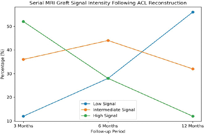
Serial MRI evaluation demonstrated progressive graft maturation over time. At 3 months, high graft signal intensity was observed in 52% of patients, decreasing to 28% at 6 months and 12% at 12 months. Conversely, the proportion of patients with low signal intensity increased from 12% at 3 months to 56% at 12 months, indicating progressive ligamentization and graft remodeling (Table 2 and Fig. 1).
Serial MRI graft signal intensity following ACL reconstruction
| Signal intensity | 3 months (%) | 6 months (%) | 12 months (%) |
|---|---|---|---|
| Low | 6 (12) | 14 (28) | 28 (56) |
| Intermediate | 18 (36) | 22 (44) | 16 (32) |
| High | 26 (52) | 14 (28) | 6 (12) |
ACL: Anterior cruciate ligament, MRI: Magnetic resonance imaging
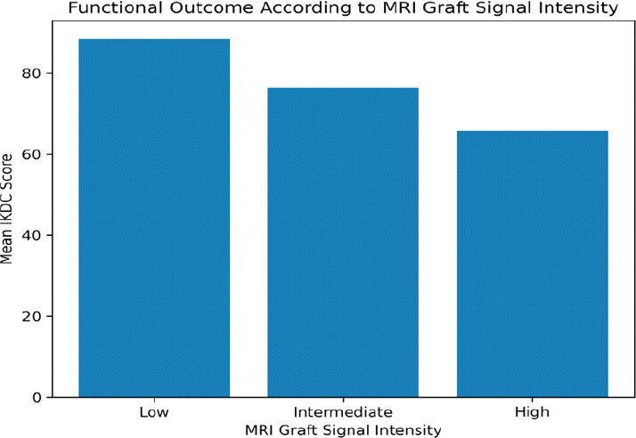
Functional assessment revealed favorable outcomes, with 40% of patients achieving excellent and 36% good IKDC scores. Similarly, Lysholm scores were excellent in 44% and good in 32% of patients, indicating satisfactory recovery in the majority of cases. At 12 months, patients with low graft signal intensity demonstrated significantly higher mean IKDC scores (88.5 ± 4.2) compared with those exhibiting intermediate (76.3 ± 5.1) and high signal intensity (65.8 ± 6.4). Correlation analysis demonstrated a significant negative association between MRI graft signal intensity and functional outcomes, with signal intensity showing inverse correlations with both IKDC scores (r = −0.62, P < 0.001) and Lysholm scores (r = −0.58, P < 0.001) (Table 3 and Fig. 2).
Correlation between MRI graft signal intensity and functional outcomes
| Parameter | Value |
|---|---|
| Mean IKDC score (low signal) | 88.5±4.2 |
| Mean IKDC score (intermediate signal) | 76.3±5.1 |
| Mean IKDC score (high signal) | 65.8±6.4 |
| Signal intensity versus IKDC score | r=−0.62, P<0.001 |
| Signal intensity versus lysholm score | r=−0.58, P<0.001 |
MRI: Magnetic resonance imaging, IKDC: International knee documentation committee
Discussion
The current prospective observational trial compared serial graft signal intensity at MRI and its relationship with primary ACLR functional recovery in 50 cases, and the results showed that there is a definite pattern of graft signal maturation with time and a statistically significant correlation between MRI signal characteristics and clinical outcomes. The demographic data of the study revealed that most patients were aged between 26 and 35 years (40%), 36–45 years (32%), and 18–25 years (28), which is indicative of the fact that ACL injuries are high among young and active people. This was also facilitated by the fact that the majority of them were males (76%) as opposed to females (24%), probably due to higher exposure to sports and high-impact activities. The mode of injury analysis showed that the most common (60%) were sports-related injuries compared to road traffic injuries (24%), and falls (16), which supports the already known fact that ACL injuries are mostly related to sporting activities.
The current study demonstrated serial MRI results of progressive reduction in graft signal intensity, which signified biological graft maturation with time. Most of the patients (52%), at 3 months post-operative, exhibited high signal intensity, and 36% had intermediate and 12% low signal intensity, which were indicators of early graft remodeling with high vascularity and water content. The transition to maturation was seen by the 6 months, with 44% of patients having intermediate signal intensities, as well as 50% (28% each) in low and high signal groups. By 12 months, this change to graft maturity was pronounced, with 56% of the patients having low signal intensity, 32% intermediate, and only 12% high signal intensity. This is in line with the biological mechanism of ligamentization, wherein the graft slowly becomes more like the native ACL. The same temporal variation in graft signal intensity was reported in by with the mean signal intensity ratio (SIR) recorded at 1 year of 2.7, and in most patients, the graft signal did not vary significantly after 1 year, indicating the stabilization of graft healing. Nonetheless, the patients with high initial signal intensity (SIR >4) showed a significant signal reduction over time and an increase in sporting level, which showed a further graft maturation later than the initial year. This result confirms the observation made in the current study that patients with a strong signal at earlier stages can still progress through with maturation, as determined by the decreasing number of high signal cases at 52% at 3 months and 12% at 12 months.
The present study demonstrated that a functional outcome assessment indicates that 40% of patients experienced excellent IKDC scores, 36% good scores, 16% fair scores, and 8% poor scores, whereas the Lysholm scoring indicates that 44% had excellent results, 32% good results, and 16% fair results, though a few had poor results. The results show that about 76–80% of patients recovered well, with some having excellent functional recovery, which implies the effectiveness of ACLR. Notably, MRI graft signal intensity was significantly negatively correlated with functional outcomes, with the group of patients with low signal intensity at 12 months showing the highest mean of the IKDC score (88.5 ± 4.2), intermediate (76.3 ± 5.1), and high signal intensity groups (65.8 ± 6.4). Correlation analysis also indicated that signal intensity and IKDC score (r = −0.62, P < 0.001) and Lysholm score (r = −0.58, P < 0.001) are statistically significantly negatively correlated and thus higher signal intensity is associated with worse functional outcomes. These results are consistent with the study by Biercevicz et al and Miyawaki M., [3,11] also showed that MRI-based graft parameters (signal intensity and graft volume) predicted functional outcomes (hop test performance and patient-reported outcome measures at 3 and 5 years) and were significantly predictive (R = 0.62, P =0.003 of hop test at 5 years) of the outcome. It is helpful in arguing that MRI signal strength is an indicator of the biomechanical integrity of the graft, and it can be used to predict the functional recovery.
The results of the current study, however, are not consistent with Saupe et al. [12] have stated that there is no significant relation between graft signal intensity and IKDC scores (P = 0.667), arthrometric laxity, or time since surgery at long-term follow-up of 412 years. In their research, a large proportion of patients (70%) had increased intrasubstance signal intensity that was not associated with functional impairment or instability. This can possibly be explained by the differences in the follow-up period since the current study was based on early to mid-term outcomes (up to 12 months), whereas Saupe et al. considered long-term outcomes where the graft remodeling can have reached a plateau, and the compensatory mechanisms can affect the functional performance.
Equally, Miyawaki et al. [11,13] have found that signal intensity in graft bundles differed (posterolateral bundle exhibited higher signal intensity than anteromedial bundle, 2.5 ± 1.7 versus 1.7 ± 1.5 on proton density-weighted imaging, P < 0.05), and that signal intensity was only weak to moderately correlated with time since surgery (r = 0.38 – This implies that although the MRI signal intensity is an indicator of graft remodeling, its dependence on time and functional outcomes might not be linear or even consistent across all graft components. Conversely, the current research showed a more reliable temporal attenuation of signal intensity and a closer relationship with functional outcomes, perhaps due to homogeneous graft type and homogeneous rehabilitation guidelines.
Moreover, Flannery et al. [14] found that the MRI-based measures of cross-sectional area, volume, and estimated failure load were predictive of functional improvement (r = 0.44–0.48, P = 0.01); however, signal intensity alone was not significantly correlated with IKDC” scores or knee laxity when covariate with demographic variables. The result of this finding is that, although structural parameters are more likely to be predictive, the intensity of signals alone would not be a complete predictor of graft functionality. The current study, however, showed a significant association between signal intensity and functional scores, and this could be attributed to variations in study design, sample size, imaging methods, and outcome measures.
On the whole, the current study demonstrates that serial MRI graft signal intensity is an effective non-invasive biomarker to evaluate graft maturation and predict functional recovery during the initial post-operative period. The gradual reduction of the level of high signal intensity at 52% at 3 months–12% at 12 months, and the corresponding increases observed in the IKDC and Lysholm scores confirm the importance of MRI in the process of graft healing monitoring [14]. The correlation is very negative (r = −0.62 and −0.58), which underlines that reduced signal strength correlates with improved clinical results. Although there are some studies that show that there is limited correlation between MRI signal and long-term functionality, the current results show that during the early stages of recovery, the signal intensity is a key indicator of graft biology and functional performance. Thus, serial MRI assessment could be integrated with clinical and functional assessment to assist in shaping the rehabilitation plans and enhancing patient outcomes after ACLR.
The strengths of the present study include its prospective design, standardized serial MRI evaluation at multiple post-operative intervals, and the use of validated functional outcome measures such as the IKDC and Lysholm scores. However, certain limitations should be acknowledged. The study was conducted at a single center with a relatively small sample size, which may limit the generalizability of the findings. The follow-up period was limited to 12 months and therefore may not fully reflect long-term graft maturation and clinical outcomes. In addition, MRI graft signal intensity was assessed primarily using qualitative grading rather than advanced quantitative imaging parameters. Future multicenter studies with larger sample sizes, longer follow-up durations, and quantitative MRI assessment are warranted to further validate these findings.
Conclusion
Serial MRI graft signal intensity is a useful indicator of graft maturation following primary ACLR and demonstrates a significant correlation with functional recovery. Progressive reduction in graft signal intensity over 12 months reflected ongoing ligamentization, while lower signal intensity was associated with superior IKDC and Lysholm scores. These findings suggest that serial MRI assessment may serve as a valuable non-invasive tool for monitoring graft healing and guiding post-operative rehabilitation.
Clinical Message
Non-invasive graft signal intensity MRI can be utilized to assess graft healing in the post-operative period and patients at risk of slower recovery. It can assist clinicians to customize rehabilitation plans and make effective decision-making on return to activity or sports.
Conflict of Interest:
Nil
Source of Support:
Nil
Consent:
The authors confirm that informed consent was obtained from the patient for publication of this article
References
- Ahmad SS, Meyer JC, Krismer AM, Ahmad SS, Evangelopoulos DS, Hoppe S, et al. Outcome measures in clinical ACL studies: An analysis of highly cited level I trials Knee Surg Sports Traumatol Arthrosc 2017;25:1517-27. [Google Scholar] | [PubMed]
- Barnett SC, Murray MM, Badger GJ, Yen YM, Kramer DE, Sanborn R, et al. Earlier resolution of symptoms and return of function after bridge-enhanced anterior cruciate ligament repair as compared with anterior cruciate ligament reconstruction Orthop J Sports Med 2021;9:23259671211052530. [Google Scholar] | [PubMed]
- Biercevicz AM, Akelman MR, Fadale PD, Hulstyn MJ, Shalvoy RM, Badger GJ, et al. MRI volume and signal intensity of ACL graft predict clinical, functional, and patient-oriented outcome measures after ACL reconstruction Am J Sports Med 2015;43:693-9. [Google Scholar] | [PubMed]
- Biercevicz AM, Miranda DL, Machan JT, Murray MM, Fleming BC. In situ, noninvasive, T2*-weighted MRI-derived parameters predict ex vivo structural properties of an anterior cruciate ligament reconstruction or bioenhanced primary repair in a porcine model Am J Sports Med 2013;41:560-6. [Google Scholar] | [PubMed]
- Biercevicz AM, Murray MM, Walsh EG, Miranda DL, Machan JT, Fleming BC. T2* MR relaxometry and ligament volume are associated with the structural properties of the healing ACL J Orthop Res 2014;32:492-9. [Google Scholar] | [PubMed]
- Biercevicz AM, Proffen BL, Murray MM, Walsh EG, Fleming BC. T2* relaxometry and volume predict semi-quantitative histological scoring of an ACL bridge-enhanced primary repair in a porcine model J Orthop Res 2015;33:1180-7. [Google Scholar] | [PubMed]
- Chu CR, Williams AA, Erhart-Hledik JC, Titchenal MR, Qian Y, Andriacchi TP. Visualizing pre-osteoarthritis: Integrating MRI UTE-T2* with mechanics and biology to combat osteoarthritis- -the 2019 Elizabeth Winston Lanier kappa delta award J Orthop Res 2021;39:1585-95. [Google Scholar] | [PubMed]
- Chu CR, Williams AA, West RV, Qian Y, Fu FH, Do BH, et al. Quantitative magnetic resonance imaging UTE-T2* mapping of cartilage and meniscus healing after anatomic anterior cruciate ligament reconstruction Am J Sports Med 2014;42:1847-56. [Google Scholar] | [PubMed]
- Davies WT, Myer GD, Read PJ. Is it time we better understood the tests we are using for return to sport decision making following ACL reconstruction? A critical review of the hop tests Sports Med 2020;50:485-95. [Google Scholar] | [PubMed]
- Irrgang JJ, Ho H, Harner CD, Fu FH. Use of the International knee documentation committee guidelines to assess outcome following anterior cruciate ligament reconstruction Knee Surg Sports Traumatol Arthrosc 1998;6:107-14. [Google Scholar] | [PubMed]
- Miyawaki M, Hensler D, Illingworth KD, Irrgang JJ, Fu FH. Signal intensity on magnetic resonance imaging after allograft double-bundle anterior cruciate ligament reconstruction Knee Surg Sports Traumatol Arthrosc 2014;22:1002-8. [Google Scholar] | [PubMed]
- Saupe N, White LM, Chiavaras MM, Essue J, Weller I, Kunz M, et al. Anterior cruciate ligament reconstruction grafts: MR imaging features at long-term follow-up--correlation with functional and clinical evaluation Radiology 2008;249:581-90. [Google Scholar] | [PubMed]
- Putnis SE, Klasan A, Oshima T, Grasso S, Neri T, Coolican MR, et al. Magnetic resonance imaging assessment of hamstring graft healing and integration 1 and minimum 2 years after ACL reconstruction Am J Sports Med 2022;50:2102-10. [Google Scholar] | [PubMed]
- Flannery SW, Murray MM, Badger GJ, Ecklund K, BEAR Trial Team, Kramer DE, et al. Early MRI-based quantitative outcomes are associated with a positive functional performance trajectory from 6 to 24 months post-ACL surgery Knee Surgery, Sports Traumatology, Arthroscopy 2023;31:1690-8. [Google Scholar] | [PubMed]
© 2026 Journal of Orthopaedic Case Reports - Published by Indian Orthopaedic Research Group
About the Authors
-

Dr. Siddharth Malik
-

Dr. Piyush Suresh Vasaikar
-

Dr. Prajwal Ganesh Senthilnathan
-

Dr. Ayush Gupta
-

Dr. Manvir Singh
-

Dr. Shubham Gupta
Related Articles in Journal of Orthopaedic Case Reports
 September 1, 2026 Comparative Analysis of Functional Outcomes of Accelerated versus Conventional Rehabilitation after Anterior Cruciate Ligament Reconstruction Surgery
 August 1, 2026 Comparative Outcomes of Hamstring Tendon versus Peroneus Longus Tendon Autograft in Arthroscopic Single-Bundle Anterior Cruciate Ligament Reconstruction: A Prospective Comparative Study
 July 1, 2026 Role of Meniscal Ramp Lesion Repair in Preventing Residual Instability after Anterior Cruciate Ligament Reconstruction
 May 1, 2026 Anterior Cruciate Ligament Reconstruction Using Tibial Attachment – Preserving Hamstring Grafts: Early Clinical and Magnetic Resonance Imaging Assessment of Graft Ligamentization