


Introduction
Acute bacterial arthritis of the pediatric hip is an orthopedic emergency because delay in source control can damage articular cartilage, disturb proximal femoral growth, and lead to avascular necrosis, chondrolysis, or permanent loss of function [1]. Clinical presentation, however, ranges from a toxic, febrile child who refuses weight-bearing to an ambulant child with only a persistent limp. Current pediatric infectious-disease guidance therefore emphasizes integration of history, serial examination, imaging rather than dependence on a single blood test [1].
Kocher et al., derived four predictors to distinguish septic arthritis from transient synovitis: A history of fever above 38.5°C, inability to bear weight, erythrocyte sedimentation rate (ESR) of at least 40 mm/h, and peripheral white blood cell count above 12,000/mm³ [2]. The four-predictor rule was subsequently validated in a separate cohort [3]. Caird et al. added CRP of at least 20 mg/L as a fifth predictor [4]. External evaluations have reported materially different probabilities for identical scores, highlighting spectrum, referral and pathogen effects [5, 6, 7]. More recent appraisal has also questioned whether a rule developed largely in the pre-Kingella era can be applied unchanged across ages and microbiological settings [8].
Children referred to tertiary hospitals in India have frequently received undocumented antipyretics, non-steroidal anti-inflammatory drugs, or antibiotics. These exposures may attenuate fever, alter inflammatory-marker trajectories, and reduce culture yield without ensuring source control. North Indian studies have demonstrated a predominance of Staphylococcus aureus among culture-positive pediatric septic arthritis [9], while a local late-presentation series described prior antibiotic exposure, culture negativity, and substantial hip morbidity [10].
Evidence from Northern India is limited on the child who presents after treatment elsewhere with a persistent limp, a sonographic hip collection and normal total leukocyte count (TLC), ESR, and C-reactive protein (CRP). If a low score is treated as an absolute exclusion test, drainage may be delayed despite ongoing intra-articular infection. Defining this clinically discordant phenotype is important because it identifies when careful history, repeated examination, and imaging should outweigh reassuring blood markers, while avoiding the unsupported claim that every hip effusion requires surgery.
This study aimed to describe the presentation and outcomes of partially treated children with operatively confirmed septic arthritis of the hip despite normal admission inflammatory markers, and to assess their Kocher–Caird score distribution. We hypothesized that most would have a score of ≤1.
Materials and Methods
Study design, setting, and ethics
This was a single-center retrospective descriptive case series of consecutive operatively confirmed cases treated at a tertiary pediatric orthopedic referral center in Northern India from January 1, 2023, to December 31, 2025. The manuscript was structured according to STROBE principles. The study was approved by the Institutional Ethics Committee before data extraction (129th ECM IIA/P15/98/Ethics/2024).
Source population and screening
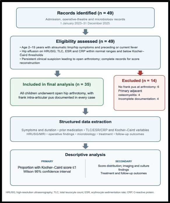
Records were reviewed for presenting symptoms and their duration, imaging findings, inflammatory marker values, operative details, and culture results. Forty-nine potentially eligible patients were identified; 35 fulfilled the inclusion criteria and were included in the final analysis, as shown in Fig. 1.
Sample-size considerations
Because this retrospective study used a census of all consecutive eligible cases treated during the prespecified 3-year period, no a priori sample-size calculation was used to restrict enrolment. As no reliable prior estimate existed for this selected low-marker, operatively confirmed phenotype, all eligible cases (n = 35) were included, and the precision of the primary proportion was reported using a 95% Wilson confidence interval (CI).
Eligibility criteria and case definition
Children aged 2–15 years were included if they had an atraumatic limp, preceding or current fever, hip effusion on high-resolution ultrasonography (HRUSG), normal pre-operative inflammatory markers below Kocher–Caird thresholds, and frank intra-articular pus at open arthrotomy. Complete records permitting score reconstruction were required. Patients with secondary hip infection, immunosuppression, inflammatory arthritis, alternative musculoskeletal diagnoses, adjacent infection explaining the presentation, established septic sequelae, absent operative pus, or incomplete records were excluded from the study.
Culture-positive bacterial arthritis was defined by growth of a pathogenic organism from joint fluid, synovium, or blood in a clinically compatible case. Culture-negative cases were classified as presumed bacterial arthritis only when operative purulence, compatible clinical, or synovial inflammation where available, exclusion of an alternative diagnosis, and resolution after drainage plus antibacterial therapy were all documented. Purulence was therefore an operative inclusion criterion, not a claim that every case was microbiologically confirmed.
Data sources and extraction
A structured case-record form captured age, sex, side, comorbidity, symptom duration, maximum documented temperature, weight-bearing status, prior healthcare visits, and pre-referral medication. Previous antibiotic exposure was defined as at least one systemic antimicrobial dose before blood or joint sampling; drug name, route, duration, and last dose were recorded when available. Antipyretic and non-steroidal anti-inflammatory drug exposure was recorded separately. Eligibility, score variables, and treatment details were abstracted from the source records.
Clinical assessment and score reconstruction
The original Kocher score assigned one point each for a documented temperature above 38.5°C before or at referral, inability to bear weight, ESR of at least 40 mm/h, and TLC above 12,000/mm³ [2]. The modified Kocher–Caird score added one point for CRP of at least 20 mg/L [4]. A parental report of fever without a recorded value was retained as a clinical feature but was not scored as temperature above 38.5°C. Weight-bearing was classified from the examination at referral as normal, antalgic but possible, or impossible; only complete inability or refusal to bear weight generated a score point. Scores were reconstructed using information documented before definitive drainage, thereby avoiding post-operative values.
Laboratory and imaging assessment
The first available pre-operative TLC, neutrophil percentage, ESR and quantitative CRP at the referral center were recorded as continuous values and then dichotomized at both the institutional upper reference limit and the published Kocher–Caird thresholds. Blood cultures were obtained before new empirical antibiotics whenever clinical stability permitted. No later or lowest value was substituted for the admission result.
All children underwent anteroposterior pelvis and lateral hip radiography. HRUSG was performed by a radiologist using an anterior approach and a high-frequency transducer appropriate to body habitus. The maximal anterior capsule-to-femoral-neck fluid depth was measured perpendicular to the femoral neck and compared with the contralateral side when available. For descriptive analysis, 5–10 mm was labeled a moderate effusion. Internal echoes or debris, synovial thickening, and Doppler hyperemia were recorded separately. Magnetic resonance imaging (MRI) was obtained for symptoms longer than 7 days, abnormal radiographs, disproportionate pain, poor early response or concern for adjacent osteomyelitis, and pyomyositis or abscess. Ultrasound was used to identify an effusion, not to establish its bacterial etiology.
Operative, microbiological, and antimicrobial management
After resuscitation and blood culture where feasible, children underwent open drainage, lavage, and debridement by hip arthrotomy through the standard anterior approach. Arthrotomy was undertaken when persistent atraumatic hip symptoms and painful restriction of motion continued despite prior treatment, and HRUSG confirmed a hip effusion; internal echoes or debris, synovial thickening or hyperemia, and supportive MRI findings when available strengthened the indication. No fixed Kocher–Caird score threshold was used, and a low score did not preclude drainage when the overall clinical and imaging findings remained concerning. Every included child underwent arthrotomy, and frank intra-articular pus was documented in all 35 operative notes. The volume and gross appearance of fluid, synovial condition, and associated cartilage or bone changes were documented. Joint fluid and synovial tissue were sent for Gram staining, aerobic culture, mycobacterial cultures, and antimicrobial susceptibility testing. Anaerobic, fungal, and molecular tests were performed when clinically indicated. Empirical anti-staphylococcal treatment was selected according to the local antibiogram and modified after culture results and infectious-disease review. Decisions regarding repeat washout were based on persistent or recurrent fever, pain, restricted motion, wound drainage, rising inflammatory markers, or persistent collection rather than on the prediction score.
Outcomes and follow-up
The primary outcome was the proportion of children with a Kocher–Caird score of 1 or less at referral. Secondary outcomes were the complete score distribution, HRUSG and MRI findings, culture positivity, organisms and susceptibility, number of washouts, intravenous and total antibiotic duration, time to comfortable full weight-bearing, hospital stay, and readmission. Follow-up assessments at approximately 2 and 6 weeks, 3 and 6 months, 12 months, and the last available visit were reviewed for pain, limp, hip range of motion, recurrence, and radiographic avascular necrosis, chondrolysis, growth disturbance, or joint-space loss. Recurrence was defined as renewed clinical infection requiring antibiotics or drainage after completion of the index treatment.
Statistical analysis
Continuous variables were assessed for distribution and reported as median with interquartile range (IQR); categorical variables were reported as number and percentage. The primary proportion was accompanied by a two-sided Wilson 95% CI. Culture positivity after documented pre-sampling antibiotics versus no documented exposure was compared using Fisher’s exact test. A two-sided P value below 0.05 was considered statistically significant.
Results
Cohort characteristics
Forty-nine records were screened; 14 were excluded (six lacked operative purulence, four had primary adjacent osteomyelitis, and four had insufficient documentation). Thirty-five children formed the study cohort (Fig. 1). Their demographic and presentation characteristics are detailed in Table 1. Median age was 8 years (IQR 5–11), 23 (65.7%) were boys and 19 (54.3%) hips were right-sided. Median symptom duration before referral was 9 days (IQR 8–12). Twenty-nine children (82.9%) had documented previous antibiotics, and all had received an antipyretic or non-steroidal anti-inflammatory drug. Twelve children (34.3%) were unable to bear weight; the remainder walked with an antalgic limp. Only 6 (17.1%) had a documented temperature above 38.5 °C during the preceding illness, and 2 (5.7%) were febrile at admission.
Demographic and presentation characteristics
| Characteristic | Value | Characteristic | Value |
|---|---|---|---|
| Included children | 35 | Age, years | 8 (5–11) |
| Boys | 23 (65.7%) | Right hip | 19 (54.3%) |
| Symptom duration, days | 9 (8–12) | Prior antibiotics | 29 (82.9%) |
| Prior antipyretic/NSAID | 35 (100%) | Unable to bear weight | 12 (34.3%) |
| Documented fever >38.5°C | 6 (17.1%) | Febrile at admission | 2 (5.7%) |
| Admission TLC, /mm3 | 8,600 (7,400–9,700) | Admission ESR, mm/h | 17 (12-23) |
| Admission CRP, mg/L | 5.9 (3.7–8.1) | Follow-up, months | 16 (12–24) |
NSAID: Non-steroidal anti-inflammatory drug, TLC: Total leukocyte count, ESR: Erythrocyte sedimentation rate, CRP: C-reactive protein
Laboratory values and scores
Admission TLC was 8,600/mm³ (IQR 7,400–9,700), ESR was 17 mm/h (IQR 12–23), and CRP was 5.9 mg/L (IQR 3.7–8.1). No child crossed the published TLC, ESR, or CRP thresholds. Twenty children (57.1%) had a Kocher score of 0, 12 (34.3%) scored 1, and 3 (8.6%) scored 2; the Kocher–Caird distribution was identical because CRP contributed no additional point. The component predictors and complete score distribution are shown in Table 2. Thus, 32 of 35 children (91.4%; 95% CI 77.6–97.0%) scored 1 or less. The three children who scored 2 had both documented fever above 38.5 °C and inability to bear weight.
Kocher–Caird predictors and score distribution
| Variable | n | % | Interpretation |
|---|---|---|---|
| Fever >38.5°C | 6 | 17.1 | Quantified pre-referral or admission temperature |
| Unable to bear weight | 12 | 34.3 | At referral examination |
| TLC >12,000/mm3 | 0 | 0 | No laboratory point |
| ESR ≥40 mm/h | 0 | 0 | No laboratory point |
| CRP ≥20 mg/L | 0 | 0 | No additional Caird point |
| Score 0 | 20 | 57.1 | Neither quantified fever nor inability to bear weight |
| Score 1 | 12 | 34.3 | One clinical predictor |
| Score 2 | 3 | 8.6 | Both clinical predictors |
| Score ≤1 | 32 | 91.4 | 95% CI 77.6–97.0% |
TLC: Total leukocyte count, ESR: Erythrocyte sedimentation rate, CRP: C-reactive protein, CI: Confidence interval
Imaging, microbiology, and treatment
HRUSG showed a median effusion depth of 7.2 mm (IQR 5.8–8.6); internal echoes were present in 28 (80.0%), synovial thickening in 24 (68.6%), and Doppler hyperemia in 17 (48.6%). Twelve children underwent MRI, which confirmed effusion or synovitis without adjacent osteomyelitis or abscess. As summarized in Table 3, all 35 children underwent arthrotomy, and frank intra-articular pus was recorded in every case. Joint culture was positive in 14 (40.0%): Methicillin-sensitive S. aureus in 9, methicillin-resistant S. aureus in 3, Streptococcus pyogenes in 1, and Streptococcus pneumoniae in 1. Blood cultures were positive in two children and matched the joint isolate. Culture positivity was 9 of 29 (31.0%) after documented prior antibiotics and 5 of 6 (83.3%) without documented exposure (P = 0.028).
Imaging and microbiological findings
| Finding | n (%) or median (IQR) | Details |
|---|---|---|
| HRUSG effusion depth | 7.2 mm (5.8–8.6) | Moderate collection defined descriptively as 5-10 mm |
| Internal echoes | 28 (80.0) | Complex or particulate fluid |
| Synovial thickening | 24 (68.6) | Reported on HRUSG |
| Doppler hyperemia | 17 (48.6) | Perisynovial vascularity |
| MRI performed | 12 (34.3) | No adjacent osteomyelitis or abscess |
| Frank pus at arthrotomy | 35 (100) | Operative inclusion criterion |
| Joint culture positive | 14 (40.0) | 9 MSSA, 3 MRSA, 1 S. pyogenes, 1 S. pneumoniae |
| Blood culture positive | 2 (5.7) | Both matched the joint isolate |
HRUSG: High-resolution ultrasonography, IQR: Interquartile range, MRI: Magnetic resonance imaging, MSSA: Methicillin-sensitive Staphylococcus aureus, MRSA: Methicillin-resistant Staphylococcus aureus
Clinical outcomes
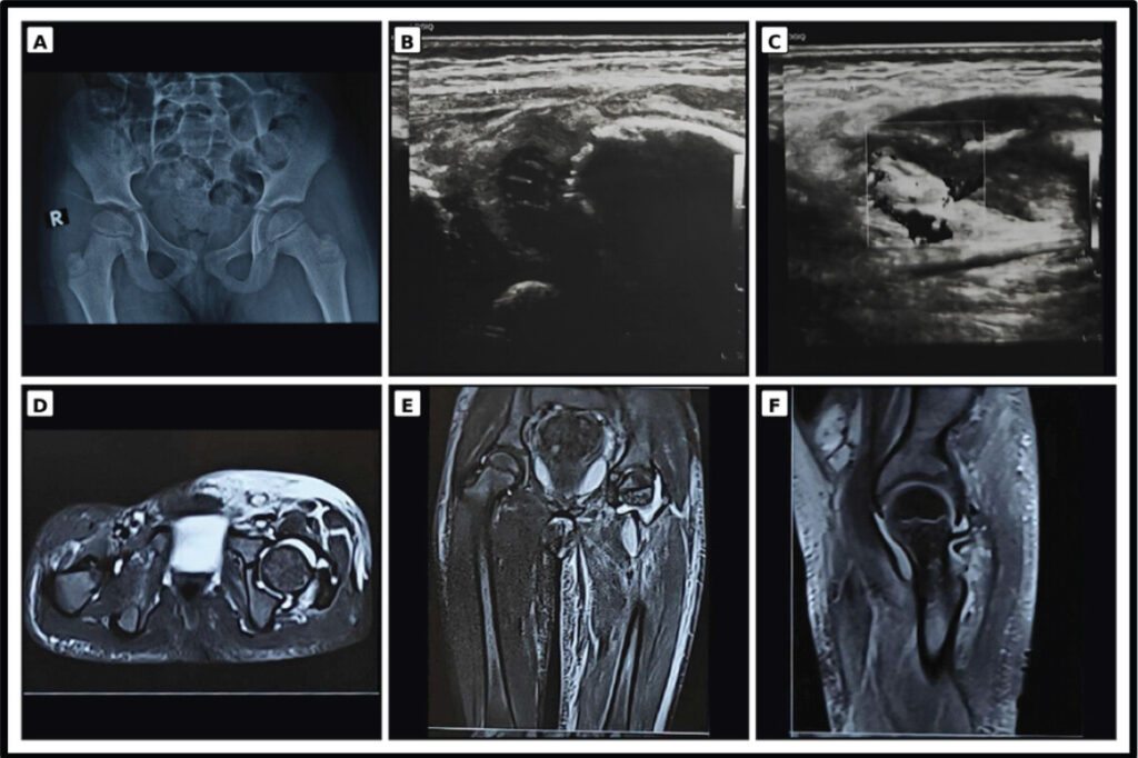
Thirty-three children required one arthrotomy, and two with methicillin-resistant S. aureus required repeat washout. Median intravenous antibiotic duration was 15 days (IQR 14–16), total treatment duration was 42 days (IQR 40–45), and hospital stay was 14 days (IQR 13–16). Comfortable full weight-bearing returned after a median of 8 days (IQR 7–10). At a median follow-up of 16 months (IQR 12–24), infection had resolved in all children. Three had a residual limp at 6 weeks that resolved by the final visit. No recurrence, avascular necrosis, chondrolysis, growth disturbance, or radiographic joint-space loss was observed during the available follow-up; treatment and outcome measures are summarized in Table 4. The representative case example is seen in Fig. 2.
Treatment and outcomes
| Outcome | Value | Outcome | Value |
|---|---|---|---|
| Single arthrotomy | 33 (94.3%) | Repeat washout | 2 (5.7%) |
| IV antibiotics, days | 15 (14–16) | Total antibiotics, days | 42 (40–45) |
| Hospital stay, days | 14 (13–16) | Full weight-bearing, days | 8 (7–10) |
| Culture positive after antibiotics | 9/29 (31.0%) | Culture positive without antibiotics | 5/6 (83.3%) |
| Residual limp at 6 weeks | 3 (8.6%) | Residual limp at final follow-up | 0 |
| Recurrent infection | 0 | Radiographic sequelae | 0 |
Discussion
The central observation was that 91.4% of this highly selected cohort had a Kocher–Caird score of 1 or less despite frank pus at arthrotomy. Laboratory variables contributed no points; most children were afebrile and could still walk with a limp. This does not refute Kocher or Caird: their rules estimated probability among acutely irritable hips, principally against transient synovitis [2, 3,4]. Our cohort was selected after prior treatment, clinical escalation, and operative confirmation and, therefore, represented a different clinical spectrum.
The principal diagnostic studies are compared in Table 5. Kocher’s derivation cohort linked four predictors to probabilities ranging from less than 0.2% with no predictor to 99.6% with all four [2]. Discrimination diminished during same-center validation [3]. External performance was less stable: Luhmann et al. reported 59% probability with all four predictors [5], and Sultan and Hughes reported 59.9% with all five in a low-prevalence referral population [6]. Thus, score-linked probabilities depend on case definition, referral pathway, and disease prevalence. A systematic review of 11 studies involving 391 children also highlighted substantial heterogeneity in diagnosis, management, and reported outcomes [11].
Comparison with major studies evaluating Kocher–Caird variables in pediatric irritable hip
| Study | Design and cohort | Principal finding | Relationship to the present study |
|---|---|---|---|
| Kocher et al., 1999 [2] | Retrospective derivation; 168 children (82 septic arthritis, 86 transient synovitis). | Four predictors. Probability rose from <0.2% with 0 predictors to 99.6% with all 4. | Established the rule in an acutely irritable-hip population with a non-septic comparator. |
| Kocher et al., 2004 [3] | Prospective same-center validation; 154 children (51 septic arthritis, 103 transient synovitis). | Same four predictors retained; AUC decreased from 0.96 in derivation to 0.86. | Demonstrated diminished performance even during formal validation. |
| Luhmann et al., 2004 [5] | Retrospective external cohort; 163 children/165 hips (47 septic, 118 transient synovitis). | All 4 Kocher predictors gave only 59% probability; fever, WBC and prior healthcare visit formed an alternative triad. | Shows transportability and referral-spectrum effects. |
| Caird et al., 2006 [4] | Prospective aspiration-selected cohort; 53 enrolled and 48 analyzed. | Added CRP ≥20 mg/L. Estimated probability was 83% with 3 factors, 93% with 4 and 98% with all 5. | CRP was important in a cohort already considered suspicious enough for aspiration. |
| Sultan and Hughes, 2010 [6] | Retrospective primary-referral cohort; 96 included (5 septic arthritis, and 91 transient synovitis). | All 5 factors yielded 59.9% probability; fever was the strongest predictor. | Illustrates prevalence-dependent positive predictive value. |
| Singhal et al., 2011 [7] | Retrospective ultrasound-effusion cohort; 311 children (29 confirmed septic arthritis, 282 transient synovitis). | CRP and inability to bear weight were independent; neither gave <1% probability and both gave 74%. | Supports a compact rule, but the present cohort contains surgically purulent low-CRP cases after prior treatment. |
| Olandres et al., 2023 [12] | Five-year retrospective cohort; 101 children with acute atraumatic hip pain. | CRP ≥20 mg/L plus US effusion ≥7 mm: sensitivity 71%, specificity 97%; Kocher/Caird sensitivity was 0% in that dataset. | Relevant imaging threshold, but treatment timing and case spectrum differ from the current referral cohort. |
| QingSong et al., 2024 [13] | Systematic review/meta-analysis; 11 studies, 1,810 cases. | Fever, non-weight-bearing, ESR and WBC were significant; CRP was not significant in pooled analysis, with high heterogeneity. | Confirms variable performance and identifies timing and prior treatment as potential modifiers. |
| Present synthetic cohort | Retrospective series; 35 partially treated purulent hips with normal admission markers; no comparator. | 91.4% had score ≤1; median HRUSG effusion 7.2 mm; culture positivity 40%. | Defines a post-treatment low-marker failure phenotype; cannot estimate diagnostic accuracy or invalidate the criteria. |
AUC: Area under the receiver operating characteristic curve, CRP: C-reactive protein, ESR: Erythrocyte sedimentation rate, HRUSG/US: High-resolution ultrasonography/ultrasound, WBC: White blood cell count
Caird et al. identified CRP above 20 mg/L as a strong independent predictor in children already selected for aspiration [4]. Singhal et al. found CRP and inability to bear weight independently informative [7]. Olandres et al. reported 71% sensitivity and 97% specificity when CRP at least 20 mg/L was combined with an effusion at least 7 mm [12]. Our median effusion depth was similar, but CRP remained below the Caird threshold. This is not a head-to-head accuracy comparison; it identifies a possible post-treatment phenotype that a high-CRP rule could miss.
A 2024 systematic review and meta-analysis of 11 studies and 1,810 cases found pooled associations for fever, non-weight-bearing, ESR, and white blood cell count, with substantial heterogeneity; CRP was not significant in pooled analysis [13]. Presentation timing and prior treatment were potential sources of variability. Prediction variables are therefore useful risk markers, not absolute exclusion tests.
Within the studies summarized in Table 5, none was designed specifically to characterize children from Northern India who had received treatment before referral, retained normal admission inflammatory markers and nevertheless had frank pus at arthrotomy. This series does not propose a new threshold; it defines an important failure phenotype in which limp and a drainable effusion persist after fever and blood markers settle. It links the score to pre-referral medication, examination, HRUSG, operative findings, microbiology, and outcome.
This regional perspective matters because indiscriminate use of a low score to label such children as transient synovitis may delay drainage, whereas surgery for effusion alone may also cause harm. A staged approach is required: Reconstruct the pre-treatment history, repeat examination, characterize the effusion, exclude adjacent infection, and obtain joint fluid when symptoms remain discordant with blood tests. These findings justify a prospective multi-center Northern Indian cohort with a non-septic comparator.
Prior treatment is a plausible contributor to the low-marker phenotype, but causation cannot be inferred from this design. More than four-fifths had documented antibiotics and all received fever- or pain-suppressing medication. Culture positivity was lower after antibiotics, consistent with culture-negative pediatric septic arthritis [14]. Interpretation is limited by incomplete drug and timing data and only six unexposed children. Molecular testing was not routinely available [15].
Among positive cultures, the predominance of S. aureus was consistent with previous North Indian experience [9]. The culture-negative fraction resembles referral cohorts sampled after antimicrobial treatment [10, 14]. Presumed cases required operative pus, compatible inflammation, exclusion of chronic or non-infective mimics, and response to drainage plus antibiotics; nevertheless, absent routine broad-range molecular testing leaves possible misclassification.
HRUSG provided the decisive objective finding when peripheral markers were unrevealing. Ultrasound is sensitive for effusion but cannot determine bacterial etiology; echoes, synovial thickening, and hyperemia are concerning but not pathognomonic [16]. Guidance supports ultrasound to identify effusion, joint-fluid testing when infection remains suspected and MRI for possible adjacent infection [1]. In a persistently symptomatic, partially treated child, effusion should trigger reassessment rather than an automatic diagnosis of transient synovitis.
Early source control was followed by resolution in all children, with two repeat washouts and no radiographic sequelae during available follow-up. These outcomes differ from late-presenting cohorts with established structural change [10] and support timely drainage and antibiotics [17]. However, 16 months of median follow-up may miss growth-related complications; longer surveillance remains appropriate.
Study strengths include consecutive multi-register screening, explicit score reconstruction from pre-operative documentation, retention of continuous laboratory values, quantified HRUSG features, and a strict operative case definition. The analysis also avoids claiming accuracy measures that the available denominator cannot support. Limitations are substantial: The study is retrospective, single-center, and small; medication histories and pre-treatment temperatures were incompletely documented; operative purulence is partly subjective; routine molecular testing was unavailable; and follow-up was limited. Conditioning inclusion on effusion, surgeon concern, and arthrotomy creates verification and selection bias. Most importantly, there was no transient-synovitis comparator, so this study cannot estimate sensitivity, specificity, predictive value, calibration, or overall validity of either score. These findings define a warning phenotype and should not be generalized as proof that the Kocher–Caird criteria are invalid in Northern India.
Conclusion
In this selected cohort of partially treated children with persistent limp and HRUSG-proven hip effusion, normal peripheral inflammatory markers and a low Kocher–Caird score did not exclude operative septic arthritis. Prediction rules should support rather than replace careful pre-treatment history, repeated examination, appropriate imaging, and joint-fluid evaluation. The study’s importance lies in identifying a clinically relevant post-treatment, low-marker phenotype for prospective regional validation, not in proposing that every low-score irritable hip requires surgery.
Clinical Message
A low Kocher–Caird score is not a stand-alone rule-out test in a partially treated child with persistent limp. When concern continues, reconstruct the pre-treatment history, repeat the examination, define the hip effusion with ultrasonography, consider adjacent infection, and obtain joint fluid rather than allowing normal blood markers alone to delay source control.
Conflict of Interest:
Nil
Source of Support:
Nil
Consent:
The authors confirm that informed consent was obtained from the patient for publication of this article
References
- Woods CR, Bradley JS, Chatterjee A, Kronman MP, Arnold SR, Robinson J, et al. Clinical practice guideline by the pediatric infectious diseases society (PIDS) and the Infectious Diseases Society of America (IDSA): 2023 guideline on diagnosis and management of acute bacterial arthritis in pediatrics J Pediatric Infect Dis Soc 2024;13:1-59. Google Scholar | PubMed
- Kocher MS, Zurakowski D, Kasser JR. Differentiating between septic arthritis and transient synovitis of the hip in children: An evidence-based clinical prediction algorithm J Bone Joint Surg Am 1999;81:1662-70. Google Scholar | PubMed
- Kocher MS, Mandiga R, Zurakowski D, Barnewolt C, Kasser JR. Validation of a clinical prediction rule for the differentiation between septic arthritis and transient synovitis of the hip in children J Bone Joint Surg Am 2004;86:1629-35. Google Scholar | PubMed
- Caird MS, Flynn JM, Leung YL, Millman JE, D'Italia JG, Dormans JP. Factors distinguishing septic arthritis from transient synovitis of the hip in children. A prospective study J Bone Joint Surg Am 2006;88:1251-7. Google Scholar | PubMed
- Luhmann SJ, Jones A, Schootman M, Gordon JE, Schoenecker PL, Luhmann JD. Differentiation between septic arthritis and transient synovitis of the hip in children with clinical prediction algorithms J Bone Joint Surg Am 2004;86:956-62. Google Scholar | PubMed
- Sultan J, Hughes PJ. Septic arthritis or transient synovitis of the hip in children: The value of clinical prediction algorithms J Bone Joint Surg Br 2010;92:1289-93. Google Scholar | PubMed
- Singhal R, Perry DC, Khan FN, Cohen D, Stevenson HL, James LA, et al. The use of CRP within a clinical prediction algorithm for the differentiation of septic arthritis and transient synovitis in children J Bone Joint Surg Br 2011;93:1556-61. Google Scholar | PubMed
- Valisena S, De Marco G, Vazquez O, Cochard B, Steiger C, Dayer R, et al. The Kocher-Caird criteria for pediatric septic arthritis of the hip: Time for a change in the Kingella era? Microorganisms 2024;12:550. Google Scholar | PubMed
- Yadav S, Dhillon MS, Aggrawal S, Tripathy SK. Microorganisms and their sensitivity pattern in septic arthritis of north Indian children: A prospective study from tertiary care level hospital ISRN Orthop 2013;2013:583013. Google Scholar | PubMed
- 10 Chand S, Srivastava S, Afaque SF, Yadav A, Verma V, Qidwai S, et al. Late-presenting septic arthritis of the hip in children: Variations in presentation and a review of 25 hips after surgical debridement Cureus 2023;15:e47717. Google Scholar | PubMed
- Nannini A, Giorgino R, Bianco Prevot L, Bobba A, Curci D, Cecchinato R, et al. Septic arthritis in the pediatric hip joint: A systematic review of diagnosis, management, and outcomes Front Pediatr 2023;11:1311862. Google Scholar | PubMed
- Olandres RA, Seng DW, Seneviratna A, Hamouda ES, Foong BC, Wong KP, et al. C-reactive protein of ≥20 mg/L and ultrasound finding of an effusion ≥7 mm has a high specificity and sensitivity in diagnosing paediatric hip septic arthritis Arch Orthop Trauma Surg 2023;143:7027-33. Google Scholar | PubMed
- QingSong T, XinLing M, Xiang R, Kang Z, Jie H. Clinical indicators for distinguishing septic arthritis from paediatric transient synovitis of the hip: A systematic review and meta-analysis BMC Infect Dis 2024;24:1432. Google Scholar | PubMed
- Pääkkönen M. Septic arthritis in children: Diagnosis and treatment Pediatric Health Med Ther 2017;8:65-8. Google Scholar | PubMed
- Carter K, Doern C, Jo CH, Copley LA. The clinical usefulness of polymerase chain reaction as a supplemental diagnostic tool in the evaluation and the treatment of children with septic arthritis J Pediatr Orthop 2016;36:167-72. Google Scholar | PubMed
- Zamzam MM. The role of ultrasound in differentiating septic arthritis from transient synovitis of the hip in children J Pediatr Orthop B 2006;15:418-22. Google Scholar | PubMed
- Pääkkönen M, Kallio MJ, Peltola H, Kallio PE. Pediatric septic hip with or without arthrotomy: Retrospective analysis of 62 consecutive nonneonatal culture-positive cases J Pediatr Orthop B 2010;19:264-9. Google Scholar | PubMed
© 2026 Journal of Orthopaedic Case Reports - Published by Indian Orthopaedic Research Group
About the Authors
-

Dr. Syed Faisal Afaque
-

Dr. Udit Agrawal
-

Dr. Vikas Verma
Related Articles in Journal of Orthopaedic Case Reports
 September 1, 2026 Clinical Presentation, Orthopedic Outcomes, and Microbiological Spectrum in Pediatric Septic Arthritis: A Prospective Study
 January 1, 2026 From Hip Effusion to Hemophilia: Unveiling the Cause of Pain in 6-Month-Old Infant. A Case Report
 January 1, 2026 Meniscal Repair Technique for Bicompartmental Displaced Bucket Handle Meniscal Tear in a Previously Reconstructed Anterior Cruciate Ligament: Trampoline-related Knee Injury – A Case Report
 April 1, 2026 Unusual Presentation of Spinal Osteoid Osteoma: A Case Report