


Introduction
Anterior cruciate ligament (ACL) rupture is one of the most common and debilitating ligamentous injuries of the knee, particularly among young, physically active individuals and athletes participating in pivoting sports. The ACL plays a fundamental role in maintaining knee stability by preventing anterior translation of the tibia relative to the femur while also contributing to rotational control during dynamic activities. Injury to the ACL frequently results from non-contact mechanisms involving sudden deceleration, cutting, or pivoting movements, although direct trauma may also be responsible [1]. Globally, the incidence of ACL injuries continues to increase due to greater participation in recreational and competitive sports, with substantial socioeconomic implications resulting from healthcare costs, prolonged rehabilitation, and loss of productivity. If left untreated, ACL deficiency may lead to recurrent instability, secondary meniscal tears, cartilage degeneration, and the early development of post-traumatic osteoarthritis, thereby significantly affecting long-term knee function and quality of life [2].
ACR reconstruction (ACLR) has long been regarded as the gold standard surgical treatment for symptomatic ACL tears, particularly in young and active patients seeking to return to sports. Conventional ACLR typically employs autografts harvested from the bone–patellar tendon–bone (BPTB), hamstring tendons, or quadriceps tendon [3]. These grafts have demonstrated excellent long-term stability and functional outcomes, with reported return-to-sport rates ranging between 65% and 90%. Nevertheless, autograft reconstruction is associated with several disadvantages, including donor-site morbidity, post-operative pain, muscle weakness, sensory disturbances, longer operative time, and the inability to restore the native ACL anatomy and proprioceptive function completely. Furthermore, reconstructed grafts undergo a prolonged process of ligamentization rather than true biological healing, and patients remain at risk of graft rupture, contralateral ACL injury, persistent instability, and delayed return to high-level athletic activities [4].
Recent advances in regenerative orthopedics have shifted attention toward biologic preservation and repair of the native ACL rather than complete replacement. Historically, primary ACL repair yielded disappointing results because of inadequate healing capacity, poor patient selection, and limitations of earlier surgical techniques. However, improved understanding of ligament biology, enhanced arthroscopic instrumentation, and the development of biologically augmented repair strategies have renewed interest in ACL preservation [5]. The bridge-enhanced ACL (BEAR) repair procedure represents one of the most promising innovations in this field. Rather than replacing the torn ligament, BEAR utilizes a bioengineered, absorbable extracellular matrix scaffold saturated with the patient’s autologous blood to bridge the gap between the torn ligament ends, thereby promoting intrinsic tissue healing. The scaffold gradually resorbs while supporting cellular migration, vascularization, collagen deposition, and restoration of the native ligament architecture, offering the potential to preserve proprioception and normal knee biomechanics [6].
Biomechanical investigations have demonstrated encouraging findings regarding the healing characteristics of BEAR repair. Preclinical animal studies have shown that repaired ligaments treated with the BEAR scaffold achieve biomechanical properties approaching those of reconstructed ligaments while exhibiting superior preservation of native tissue organization and proprioceptive nerve endings. Experimental studies have also demonstrated improved collagen alignment, enhanced vascularization, and reduced inflammatory response compared with traditional reconstruction techniques. These biological advantages may translate into better restoration of neuromuscular control, reduced muscle atrophy, and improved functional recovery during rehabilitation [7].
Clinical evidence supporting BEAR repair has expanded rapidly over the past decade. Early feasibility studies and prospective clinical trials have demonstrated comparable short-term patient-reported outcomes between BEAR repair and conventional autograft ACLR. Patients undergoing BEAR repair have shown significant improvements in International Knee Documentation Committee (IKDC) scores, Knee Injury and Osteoarthritis Outcome Score (KOOS), Tegner activity levels, and overall knee function [8]. Importantly, BEAR has been associated with less post-operative pain, preservation of hamstring muscle strength due to avoidance of tendon harvest, and earlier restoration of quadriceps function. Several randomized controlled trials have further reported that knee stability, measured using instrumented laxity testing, remains comparable between BEAR and autograft reconstruction at 2-year follow-up, although long-term durability continues to be evaluated [9].
Despite these encouraging findings, several important questions remain unanswered regarding the relative biomechanical performance and long-term clinical effectiveness of BEAR repair. The indications for BEAR are currently limited to acute proximal ACL tears with adequate tissue quality, and concerns persist regarding repair failure, re-tear rates, patient selection criteria, cost-effectiveness, and outcomes beyond 5 years. In addition, while autograft reconstruction has decades of evidence supporting its durability, BEAR remains a relatively novel technology with limited long-term comparative data. Variability in rehabilitation protocols, surgical expertise, and outcome reporting across published studies further complicates direct comparisons between the two approaches [10].
Given the growing interest in biologic ligament preservation and personalized treatment strategies, a comprehensive comparison of BEAR repair and standard autograft ACLR is clinically relevant. Evaluating both biomechanical outcomes, such as graft or ligament strength, knee stability, and structural healing, alongside clinical outcomes including patient-reported function, return to sports, complication rates, revision surgery, and quality of life, will provide valuable evidence to guide surgical decision-making. Such comparisons may help determine whether BEAR repair can offer equivalent or superior functional outcomes while minimizing donor-site morbidity and preserving native ligament biology. Ultimately, understanding the relative advantages and limitations of these two treatment modalities is essential for optimizing patient selection and improving long-term outcomes following ACL injury.
Methods
Study design
This comparative observational study was conducted in the Department of Orthopedics over a period of 6 months, from January 2026 to June 2026. Patients with complete ACL tears who fulfilled the predefined eligibility criteria and underwent either BEAR repair or standard autograft ACLR were included in the study. The study protocol was approved by the Institutional Ethics Committee, and written informed consent was obtained from all patients before inclusion in the study.
Participants
Inclusion criteria
-
Published randomized controlled trials
-
Prospective or retrospective comparative studies
-
Cohort studies evaluating BEAR repair or autograft ACLR
-
Adult and adolescent patients with complete ACL rupture
-
Studies reporting biomechanical outcomes
-
Studies reporting clinical outcomes with at least 12 months of follow-up
-
Articles published in the English language
-
Full-text articles available for review.
Exclusion criteria
-
Case reports and case series with fewer than ten patients
-
Review articles, editorials, conference abstracts, and expert opinions
-
Cadaveric studies without clinical outcome assessment
-
Animal studies
-
Studies involving allograft reconstruction only
-
Duplicate publications
-
Studies lacking sufficient outcome data
-
Non-English publications.
Study sampling
Patients presenting to the Department of Orthopedics with a confirmed diagnosis of ACL rupture and fulfilling the predefined eligibility criteria were consecutively enrolled during the study period. Eligible patients underwent either BEAR repair or standard autograft ACLR based on the treating surgeon’s clinical assessment and patient suitability. Baseline demographic and clinical characteristics were recorded before surgery. All patients received standardized post-operative rehabilitation and were followed at regular intervals for clinical and functional evaluation. Outcome measures, including IKDC score, KOOS score, Lysholm score, Tegner activity score, knee stability assessment, muscle strength recovery, return-to-sport status, and post-operative complications, were recorded and compared between the two treatment groups.
Study sample size
A total of 50 patients fulfilling the predefined inclusion and exclusion criteria were enrolled in the study. Patients were allocated into two groups: the BEAR repair group (n = 25) and the standard autograft ACLR group (n = 25). The sample size was selected based on the expected patient availability during the study period and was considered adequate to compare clinical and biomechanical outcomes between the two treatment groups. Outcome measures evaluated included functional recovery, patient-reported outcome scores, knee stability, muscle strength recovery, return-to-sport rate, post-operative complications, graft or repair failure, and revision surgery.
Study groups
The included studies were categorized into two comparison groups:
-
Group A: Patients who underwent BEAR repair
-
Group B: Patients who underwent standard autograft ACLR using hamstring tendon, BPTB, or quadriceps tendon autografts.
Comparative analyses were performed between these groups for biomechanical, radiological, and clinical outcome measures.
Study parameters
The study parameters included demographic characteristics (age, sex, and body mass index [BMI]), mechanism of injury, time from injury to surgery, and type of surgical intervention (BEAR repair or standard autograft ACLR). Clinical and functional outcomes were assessed using the IKDC score, KOOS, Lysholm Knee Score, and Tegner activity scale. Biomechanical outcomes included knee laxity assessment, instrumented anterior tibial translation, quadriceps and hamstring muscle strength recovery, and magnetic resonance imaging (MRI) evidence of ligament healing. Additional outcome measures included return-to-sport rate, post-operative complications, repair or graft failure, revision surgery, and duration of follow-up.
Study procedure
After obtaining approval from the Institutional Ethics Committee and written informed consent from all participants, eligible patients fulfilling the inclusion criteria were enrolled in the study. Baseline demographic and clinical characteristics, including age, sex, BMI, mechanism of injury, and duration of symptoms, were recorded. Patients underwent either BEAR repair or standard autograft ACLR according to the planned surgical protocol. All surgical procedures were performed under standard aseptic conditions by experienced orthopedic surgeons. Postoperatively, all patients followed a standardized rehabilitation protocol, including progressive range-of-motion exercises, muscle-strengthening exercises, and functional rehabilitation. Patients were evaluated at scheduled follow-up visits for clinical and functional outcomes. Outcome measures included the IKDC score, KOOS, Lysholm Knee Score, Tegner activity score, knee stability assessment, muscle strength recovery, return-to-sport status, and post-operative complications. The collected data were recorded in a structured case record form and analyzed to compare clinical and biomechanical outcomes between the BEAR repair and autograft ACLR groups.
Study data collection
Data were collected prospectively using a standardized case record form for all enrolled patients. Baseline demographic and clinical information, including age, sex, BMI, mechanism of injury, duration of symptoms, and affected side, was recorded at the time of enrollment. Intraoperative details, including the type of surgical procedure performed (BEAR repair or standard autograft ACLR), were documented. Patients were followed at predefined post-operative intervals, and clinical assessments were performed during each follow-up visit. Outcome measures included the IKDC score, KOOS, Lysholm Knee Score, Tegner Activity Score, knee stability assessment, muscle strength recovery, return-to-sport status, and post-operative complications. All data were entered into a standardized data collection sheet, verified for completeness and accuracy, and subsequently transferred to Microsoft Excel before statistical analysis.
Data analysis
Data were analyzed using IBM Statistical Package for the Social Sciences Statistics version 27.0. Continuous variables were expressed as mean ± standard deviation, while categorical variables were presented as frequencies and percentages. Continuous outcomes were compared between the BEAR and autograft groups using the independent-samples t-test or Mann–Whitney U test, as appropriate. Categorical variables were compared using the Chi-square test or Fisher’s exact test. Effect estimates with 95% confidence intervals were reported where appropriate. A two-sided P < 0.05 was considered statistically significant.
Results
The study included patients who underwent either BEAR repair or standard autograft ACLR. Baseline demographic characteristics were comparable between the two groups. The mean age was 24.8 ± 5.6 years in the BEAR group and 25.3 ± 5.9 years in the autograft group. The majority of patients in both groups were male, and no statistically significant differences were observed with respect to age, sex distribution, BMI, or duration of follow-up (Table 1).
Baseline characteristics of patients
| Variable | BEAR repair | Autograft ACL reconstruction | P-value |
|---|---|---|---|
| Mean age (years) | 24.8±5.6 | 25.3±5.9 | 0.54 |
| Male (%) | 58.7 | 60.2 | 0.71 |
| Female (%) | 41.3 | 39.8 | 0.71 |
| Mean BMI (kg/m2) | 24.5 ± 2.8 | 24.8 ± 3.1 | 0.63 |
| Mean follow-up (months) | 24.6 ± 8.2 | 25.3 ± 8.8 | 0.59 |
BEAR: Bridge-enhanced anterior cruciate ligament, ACL: Anterior cruciate ligament, BMI: Body mass index
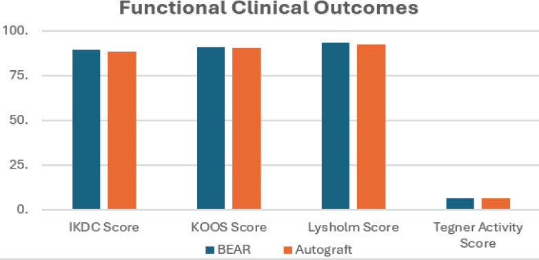
Functional outcome assessment demonstrated excellent post-operative recovery in both groups. The mean IKDC, KOOS, Lysholm Knee Score, and Tegner Activity Score were comparable between the BEAR repair and autograft reconstruction groups, with no statistically significant differences (P > 0.05). These findings indicate that both surgical techniques provided similar improvements in knee function and patient-reported outcomes (Table 2, Fig. 1).
Functional clinical outcomes
| Variable | BEAR | Autograft | P-value |
|---|---|---|---|
| IKDC score | 89.6±6.8 | 88.9±7.2 | 0.42 |
| KOOS score | 91.2±5.7 | 90.6±6.0 | 0.48 |
| Lysholm score | 93.5±4.9 | 92.7±5.2 | 0.37 |
| Tegner activity score | 6.8±1.3 | 6.7±1.4 | 0.65 |
BEAR: Bridge-enhanced anterior cruciate ligament, IKDC: International Knee Documentation Committee, KOOS: Knee injury and Osteoarthritis Outcome Score
Evaluation of biomechanical outcomes revealed comparable anterior tibial translation, pivot-shift grading, Lachman test findings, and MRI evidence of ligament healing between the two treatment groups. No statistically significant differences were observed in objective knee stability or structural healing; suggesting that BEAR repair achieved biomechanical outcomes comparable to those of conventional autograft ACLR (Table 3,Fig. 2).
Biomechanical outcomes
| Variable | BEAR | Autograft | P-value |
|---|---|---|---|
| Anterior tibial translation (mm) | 2.1±0.9 | 2.0±0.8 | 0.58 |
| Pivot shift grade 0 (%) | 90.8 | 91.4 | 0.76 |
| Lachman grade 0 (%) | 92.6 | 93.2 | 0.81 |
| MRI complete healing (%) | 94.2 | 91.5 | 0.29 |
BEAR: Bridge-enhanced anterior cruciate ligament, MRI: Magnetic resonance imaging
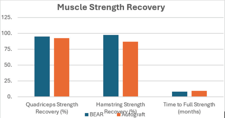
Muscle strength recovery was significantly better in patients who underwent BEAR repair. Quadriceps strength recovery was significantly higher in the BEAR group than in the autograft group (95.2 ± 5.3% vs. 92.6 ± 6.1%; P = 0.04). Similarly, hamstring strength recovery was significantly greater following BEAR repair (97.4 ± 4.2% vs. 86.8 ± 7.5%; P < 0.001). Patients in the BEAR group also achieved full muscle strength significantly earlier than those who underwent autograft reconstruction (Table 4, Fig. 3).
Muscle strength recovery
| Parameter | BEAR | Autograft | P-value |
|---|---|---|---|
| Quadriceps strength recovery (%) | 95.2±5.3 | 92.6±6.1 | 0.04* |
| Hamstring strength recovery (%) | 97.4±4.2 | 86.8±7.5 | <0.001* |
| Time to full strength (months) | 8.3±1.4 | 9.6±1.8 | 0.01* |
BEAR: Bridge-enhanced anterior cruciate ligament
*
P < 0.05, statistically significant.
Return-to-sport outcomes were comparable between the two groups. Approximately 82.6% of patients in the BEAR group and 84.3% in the autograft group returned to their pre-injury level of sporting activity. The mean time to return to sports and the proportion of patients participating in high-level competitive sports were also similar, with no statistically significant differences observed between the groups (Table 5).
Return-to-sport outcomes
| Variable | BEAR | Autograft | P-value |
|---|---|---|---|
| Returned to pre-injury sports (%) | 82.6 | 84.3 | 0.59 |
| Mean return time (months) | 9.2±1.8 | 9.5±2.1 | 0.44 |
| High-level competitive sports (%) | 69.8 | 71.1 | 0.67 |
BEAR: Bridge-enhanced anterior cruciate ligament
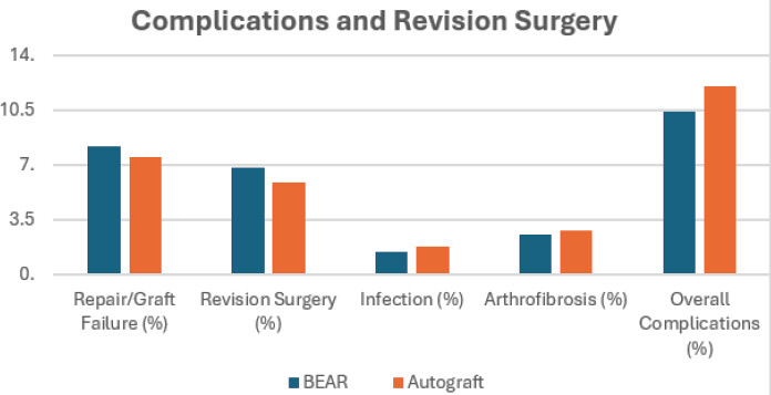
Post-operative complications were infrequent in both treatment groups. The rates of repair or graft failure, revision surgery, infection, arthrofibrosis, and overall complications were comparable between patients undergoing BEAR repair and those undergoing standard autograft ACLR, with no statistically significant differences (P > 0.05), indicating similar short-term safety profiles for both surgical procedures (Table 6).
Complications and revision surgery
| Outcome | BEAR | Autograft | P-value |
|---|---|---|---|
| Repair/graft failure (%) | 8.2 | 7.5 | 0.81 |
| Revision surgery (%) | 6.8 | 5.9 | 0.73 |
| Infection (%) | 1.4 | 1.8 | 0.69 |
| Arthrofibrosis (%) | 2.5 | 2.8 | 0.82 |
| Overall complications (%) | 10.4 | 12.1 | 0.62 |
BEAR: Bridge-enhanced anterior cruciate ligament
Discussion
The present study compared the biomechanical and clinical outcomes of BEAR repair with standard autograft ACLR. The findings demonstrated that BEAR repair achieved clinical and biomechanical outcomes comparable to conventional autograft reconstruction while providing superior preservation of hamstring and quadriceps muscle strength. Functional outcome scores, objective knee stability, return-to-sport rates, revision surgery, and complication rates were similar between the two treatment groups, suggesting that BEAR repair is an effective biological alternative to conventional ACLR in appropriately selected patients.
One of the principal findings of the present study was that patient-reported functional outcomes were comparable between BEAR repair and autograft ACLR. The present study demonstrated similar post-operative IKDC, KOOS, Lysholm, and Tegner activity scores in both groups without statistically significant differences. These findings are consistent with the randomized controlled trial conducted by Murray et al. [11], who evaluated 100 patients with complete midsubstance ACL injuries and reported that BEAR successfully met the predefined non-inferiority criteria for IKDC subjective scores. At 2 years of follow-up, the mean IKDC score was 88.9 points in the BEAR group compared with 84.8 points in the ACLR group, confirming equivalent patient-reported outcomes. Likewise, Murray et al. [12] observed significant improvement in IKDC subjective scores in both treatment groups, with no clinically meaningful difference between the interventions. These findings support the results of the present study, indicating that preservation of the native ACL through BEAR does not compromise post-operative knee function.
Objective biomechanical stability was another important outcome evaluated in the present study. Comparable anterior tibial translation, Lachman test findings, pivot-shift grading, and MRI evidence of ligament healing were observed between the BEAR and autograft reconstruction groups. Similar findings were reported by Murray et al. [11], who demonstrated equivalent restoration of knee stability between the two procedures. Murray et al. [12] also reported satisfactory mechanical stability following both BEAR repair and ACLR at 24 months. These findings suggest that BEAR repair can restore knee stability comparable to conventional autograft reconstruction.
A major advantage observed in the present study was the significantly superior preservation of hamstring and quadriceps muscle strength following BEAR repair. Patients treated with BEAR demonstrated significantly greater quadriceps and hamstring strength recovery than those undergoing autograft reconstruction. These findings agree with Murray et al. [11], who reported significantly greater hamstring strength following BEAR repair. Similarly, Murray et al. [12] and Barnett et al. [13] demonstrated superior post-operative hamstring strength among patients treated with BEAR. The preservation of the native hamstring tendons and avoidance of graft harvesting likely explain this advantage.
The present study also demonstrated comparable return-to-sport outcomes between the two treatment groups. Most patients returned successfully to their pre-injury level of activity irrespective of the surgical procedure. Barnett et al. [13] similarly reported comparable return-to-sport rates between BEAR repair and ACLR. Murray et al. [12] also observed similar functional hop test performance between the two groups, indicating comparable restoration of dynamic knee function.
Another important finding of the present study was the comparable incidence of post-operative complications, repair or graft failure, and revision surgery between the two groups. Murray et al. [11] also reported no significant difference in subsequent ACL surgery between BEAR and ACLR. Likewise, Fleming et al. [14] demonstrated similar long-term revision rates and functional outcomes up to 6 years after surgery, supporting the durability of BEAR repair.
The findings of the present study are further supported by the meta-analysis conducted by Mansour et al. [15], which concluded that BEAR repair provides superior hamstring strength while maintaining post-operative IKDC, KOOS, quadriceps strength, knee laxity, and overall functional outcomes comparable to ACLR. These observations strengthen the evidence supporting BEAR as a biological alternative to conventional ACLR.
The clinical implications of the present study are important. Preservation of the native ACL tissue and hamstring tendons may facilitate improved muscle recovery while avoiding donor-site morbidity associated with tendon harvesting. At the same time, comparable knee stability, functional recovery, return-to-sport rates, and complication profiles indicate that BEAR repair can be considered an effective treatment option in carefully selected patients with acute ACL tears.
This study has several limitations. The small sample size (n = 50) may have limited statistical power, particularly for infrequent outcomes. The approximately 2-year follow-up was insufficient to assess long-term ligament durability, recurrent instability, osteoarthritis, and sustained return to sport. The single-center, non-randomized design may have introduced selection bias and limits generalizability. BEAR findings are restricted to carefully selected patients with acute ACL tears and adequate tissue quality. Heterogeneity of autograft types may have influenced outcomes, while lack of blinding may have introduced observer and reporting bias. Cost-effectiveness, quality-of-life measures, and advanced biological, biomechanical, and proprioceptive assessments were not evaluated. Larger multicenter randomized studies with longer follow-up are warranted.
Overall, the findings of the present study demonstrate that BEAR repair provides clinical outcomes and biomechanical stability comparable to standard autograft ACLR while offering significantly better post-operative hamstring and quadriceps muscle strength recovery. These findings support the use of BEAR repair as a promising biological treatment option for selected patients with acute ACL injuries.
Conclusion
The present study demonstrated that BEAR repair is a safe and effective biological alternative to conventional autograft ACLR for the management of acute ACL injuries. BEAR repair achieved functional outcomes, patient-reported knee function, objective knee stability, MRI healing, return-to-sport rates, revision surgery, and post-operative complication rates comparable to those of standard autograft reconstruction. A significant advantage of BEAR repair was the superior preservation of hamstring and quadriceps muscle strength, which may contribute to improved post-operative rehabilitation and functional recovery. These findings suggest that BEAR repair is an effective treatment option for appropriately selected patients with acute ACL tears. However, further large-scale prospective studies with longer follow-up are required to evaluate its long-term durability and clinical effectiveness.
Clinical Message
BEAR repair is an effective alternative to standard autograft ACLR, offering equivalent knee stability, functional recovery, and return-to-sport outcomes with the added benefit of better post-operative muscle strength preservation. Appropriate patient selection and adherence to standardized rehabilitation protocols are essential to optimize long-term clinical outcomes.
Conflict of Interest:
Nil
Source of Support:
Nil
Consent:
The authors confirm that informed consent was obtained from the patient for publication of this article
References
- Abourezk MN, Ithurburn MP, McNally MP, Thoma LM, Briggs MS, Hewett TE, et al. Hamstring strength asymmetry at 3 years after anterior cruciate ligament reconstruction alters knee mechanics during gait and jogging Am J Sports Med 2017;45:97-105. Google Scholar | PubMed
- Andernord D, Desai N, Bjornsson H, Ylander M, Karlsson J, Samuelsson K. Patient predictors of early revision surgery after anterior cruciate ligament reconstruction: A cohort study of 16,930 patients with 2-year follow-up Am J Sports Med 2015;43:121-7. Google Scholar | PubMed
- Anderson AF, Irrgang JJ, Kocher MS, Mann BJ, Harrast JJInternational Knee Documentation Committee The international knee documentation committee subjective knee evaluation form: Normative data Am J Sports Med 2006;34:128-35. Google Scholar | PubMed
- Arneja S, Leith J. Review article: Validity of the KT-1000 knee ligament arthrometer J Orthop Surg (Hong Kong) 2009;17:77-9. Google Scholar | PubMed
- Astur DC, Cachoeira CM, Da Silva Vieira T, Debieux P, Kaleka CC, Cohen M. Increased incidence of anterior cruciate ligament revision surgery in paediatric verses adult population Knee Surg Sports Traumatol Arthrosc 2018;26:1362-6. Google Scholar | PubMed
- Beck NA, Lawrence JT, Nordin JD, DeFor TA, Tompkins M. ACL tears in school-aged children and adolescents over 20 years Pediatrics 2017;139:e20161877. Google Scholar | PubMed
- Cordasco FA, Black SR, Price M, Wixted C, Heller M, Asaro LA, et al. Return to sport and reoperation rates in patients under the age of 20 after primary anterior cruciate ligament reconstruction: Risk profile comparing 3 patient groups predicated upon skeletal age Am J Sports Med 2019;47:628-39. Google Scholar | PubMed
- Cox CL, Huston LJ, Dunn WR, Reinke EK, Nwosu SK, Parker RD, et al. Are articular cartilage lesions and meniscus tears predictive of IKDC, KOOS, and Marx activity level outcomes after anterior cruciate ligament reconstruction? A 6-year multicenter cohort study Am J Sports Med 2014;42:1058-67. Google Scholar | PubMed
- Feagin JA Jr, Curl WW. Isolated tear of the anterior cruciate ligament: 5-year follow-up study Am J Sports Med 1976;4:95-100. Google Scholar | PubMed
- Feller JA, Webster KE. A randomized comparison of patellar tendon and hamstring tendon anterior cruciate ligament reconstruction Am J Sports Med 2003;31:564-73. Google Scholar | PubMed
- Murray MM, Fleming BC, Badger GJ, Freiberger C, Henderson R, Barnett S, et al. Bridge-enhanced anterior cruciate ligament repair is not inferior to autograft anterior cruciate ligament reconstruction at 2 years: Results of a prospective randomized clinical trial Am J Sports Med 2020;48:1305-15. Google Scholar | PubMed
- Murray MM, Kalish LA, Fleming BC, Flutie B, Freiberger C, Henderson RN, et al. Bridge-enhanced anterior cruciate ligament repair: Two-year results of a first-in-human study Orthop J Sports Med 2019;7:2325967118824356. Google Scholar | PubMed
- Barnett SC, Murray MM, Badger GJ, Sanborn R, Kiapour A, Proffen B, et al. Earlier resolution of symptoms and return of function after bridge-enhanced anterior cruciate ligament repair as compared with anterior cruciate ligament reconstruction Orthop J Sports Med 2021;9:23259671211052530. Google Scholar | PubMed
- Fleming BC, Baranker B, Badger GJ, Kiapour AM, Ecklund K, Micheli LJ, et al. Bridge-enhanced anterior cruciate ligament restoration: 6-year results from the first-in-human cohort study Orthop J Sports Med 2024;12:23259671241260632. Google Scholar | PubMed
- Mansour J, Ghanimeh J, Ghoul A, Estephan M, Khoury A, Daher M. Bridge enhanced ACL repair vs. ACL reconstruction for ACL tears: A systematic review and meta-analysis of comparative studies SICOT J 2023;9:8. Google Scholar | PubMed
© 2026 Journal of Orthopaedic Case Reports - Published by Indian Orthopaedic Research Group
About the Authors
-

Dr. Elvis Benjamin
-

Dr. Deepak Khandelwal
-

Dr. Anuragdeep Syal
-

Dr. Abhishek Singhal
-

Dr. Nisanth Velu Selvaraj
-

Dr. Shubham
Related Articles in Journal of Orthopaedic Case Reports
 September 1, 2026 Bridge-Enhanced Anterior Cruciate Ligament Repair versus Autograft Reconstruction in Anterior Cruciate Ligament Surgery
 September 1, 2026 Arthroscopic ACL Tibial Spine Avulsion Fixation Using Suture Pull-Through Double-Tunnel Technique
 August 1, 2026 Comparative Outcomes of Hamstring Tendon versus Peroneus Longus Tendon Autograft in Arthroscopic Single-Bundle Anterior Cruciate Ligament Reconstruction: A Prospective Comparative Study
 May 1, 2026 Comparative Evaluation of Peroneus Longus Tendon and Hamstring Tendon Autografts in Primary Anterior Cruciate Ligament Reconstruction: A Randomized Controlled Trial