


Introduction
Anterior cruciate ligament (ACL) injury is a high-impact musculoskeletal problem, in that it occurs at a time when children are still maturing (education, work, sport), and because failure to treat can lead to instability, meniscal injury, recurrent surgery, and reduced quality of life. Current practice guidelines from the American Academy of Orthopaedic Surgeons support both skeletally mature and immature patients. They are based on the best available evidence, but there are ongoing gaps [1]. Consensus building internationally also views treatment as a compromise between structured rehabilitation and surgery, based on the degree of instability, activity goals, associated injuries, and patient priorities, not on imaging alone [2].
Autograft ACL reconstruction (ACLR) is the most popular surgical procedure due to its ability to provide gross sagittal and rotational stability in various acute, chronic, midsubstance, proximal, distal, partial, and revision scenarios. Treatment recommendations are based on evidence and involve diagnosis using history, physical examination, and magnetic resonance imaging (MRI); individualized rehabilitation is recommended; early reconstruction is recommended when instability or high-demand pivoting activity is anticipated; rehabilitation with delayed reconstruction is recommended for selected copers. Reconstruction does not repair the native ligament, however, and necessitates a harvest of tendons or bone-tendon, which disrupts the native proprioceptive tissue and has not been shown to prevent post-traumatic osteoarthritis (PTOA) [3] consistently.
Bridge-enhanced ACL repair (BEAR) has broken the mould of the belief that the intra-articular ACL is not suitable for repair. The process involves inserting a resorbable extracellular-matrix scaffold between the torn ACL ends, soaking the scaffold with the patient’s own blood and adding the biologic bridge to the patient’s own tissue with sutures to protect a provisional clot [4]. The arthroscopic visualisation of the primary ACL has also contributed to this renewed interest in its repair, along with the advent of modern fixation techniques and the knowledge of internal augmentation, which was largely due to the idea of a more conservative definition of what constitutes a favorable tissue biology for repair, in an acute tear [5]. The main question now is not whether BEAR can be used in specific patients, but whether it offers long-term, widespread, and clinically significant benefits over autograft reconstruction that would alter the standard approach to ACL surgery.
This controversy is compounded by differing results that are desired by the patient, the surgeon, the payer, and the team. It is also time-sensitive, as swelling, stiffness, tissue retraction, and scheduling delays may diminish the time window for repair before a definitive operative plan. A competitive athlete might seek rotational stability and predictable clearance. At the same time, a recreational patient may value less pain at the donor site, faster recovery of early strength, and retention of native anatomy. A pediatric surgeon will also need to take into account the growth plates, remnant quality, and long-term effects of meniscal loss. There is a need for health systems to consider implant cost, revision burden, and whether early functional gains result in fewer lifelong morbidities [1,2]. These competing priorities make a balanced narrative synthesis more useful than a binary declaration of superiority, particularly for surgeons counseling families who are required to make a choice between biologic novelty and reconstructive familiarity when making their decision under time pressure and incomplete long-term comparative evidence for individual patient subgroups. This narrative review is intended to bring together existing data to compare BEAR with autograft ACLR as well as to suggest an algorithmic framework for modern clinical practice.
Materials and Methods
Structured review of studies on autograft ACLR, BEAR, and biologically augmented ACL repair was conducted. The PubMed, Embase, Cochrane Library, and Scopus databases were searched from January 2019 to June 2026. Published studies as part of the selection of landmark translational studies and early feasibility studies before 2019 were included when they provided a clear biological rationale or clinical pathway for BEAR. The combined controlled vocabulary and free text search terms were: “bridge-enhanced ACL repair,” “bridge-enhanced anterior cruciate ligament repair,” “BEAR implant,” “anterior cruciate ligament repair,” “primary ACL repair,” “autograft anterior cruciate ligament reconstruction,” “bone-patellar tendon-bone,” “hamstring autograft,” “quadriceps tendon autograft,” “KT-1000,” “IKDC,” “KOOS,” “MRI,” “return to sport,” “revision,” “registry,” “pediatric,” “skeletally immature,” “InternalBrace,” and “dynamic intraligamentary stabilization.”
Randomized controlled trials, prospective comparative cohorts, post-market registries, national ACL registry analyses, systematic reviews and meta-analyses, consensus statements, rehabilitation guidelines, and mechanistic large-animal studies relevant to the biological repair of the ACL were also prioritized for inclusion. Articles were omitted if they lacked peer review, were not PubMed traceable, were not about the use of the acronym “BEAR” in connection to any related issues, or were about synthetic grafts that had nothing to do with autograft decision making. The data extracted encompassed the tear indication, time to surgery, tissue quality, surgical technique, rehabilitation, patient-reported outcomes, KT-1000 or arthrometer laxity, retear/revision, MRI maturation, return to sport, donor-site morbidity, and PTOA. The review was done on SANRA domains, including importance and aims, search strategies, evidence used, reference to claims, and clinically relevant endpoints.
Results
Biological rationale
There is a biological reason why isolated ACL suture repair has so far proved unsuccessful. The ACL is located in the synovial fluid, where the provisional fibrin-platelet clot is not stable, either mechanically or enzymatically, which is not the case for extra-articular ligaments. BEAR was designed to preserve that bridge; a scaffold made from the bovine extracellular matrix is placed in the interstump space and infused with the patient’s own blood to provide a “biologic space” that can be used to help the ligament heal [6]. Bench-to-bedside testing was described as iterative preclinical testing, scaffold refinement, and regulatory translation preceding first-in-human use [7]. In the feasibility cohort, 10 BEAR patients were not randomized to have had a joint infection, had no clinically significant inflammatory reaction, had continuous ACL tissue on MRI, and had better early hamstring strength than 10 patients undergoing hamstring-autograft ACLR at 3 months. The rationale is very different between ACLR and BEAR; ACLR will replace the ligament with a tendon graft, while BEAR will allow the original ligament to heal in a protected environment. Successful native insertion geometry, remnant mechanoreceptors, and graft-harvest avoidance may be maintained, but these theoretical benefits will need to be validated over time [8].
Indications for BEAR (tear pattern, time-to-surgery, length of tibial stump), BEAR step by step, and differences in rehabilitation
BEAR is not an all-purpose repair. The first in human study and subsequent clinical trials investigated only complete tears with adequate tibial remnant to receive sutures and form a bridge; in the small number of patients at 2 years of follow-up, no repair failures or graft failures were observed, there was no difference in the International Knee Documentation Committee (IKDC) scores, there was a similar laxity found by arthrometer, and the strength of the hamstrings was found to be greater than the hamstring autograft ACLR [9]. The pivotal randomized trial compared BEAR with ACLR, which involved sutures through the blood-soaked scaffold and across the tibial stump, femoral fixation, and the blood-soaked scaffold, and autograft, respectively, mostly quadrupled semitendinosus-gracilis. In that trial, BEAR was noninferior in 2-year IKDC score and anteroposterior AP laxity, greater hamstring strength index, and higher incidence of second ipsilateral ACL surgery (14% vs. 6% after ACLR) [10]. The BEAR cohort was analyzed separately by sex to determine if there was a sex difference in IKDC, Knee Injury and Osteoarthritis Outcome Score (KOOS), KT-1000 laxity, or ipsilateral reinjury at 2 years. This operation is only done once the arthroscopic surgeon has confirmed that the remnant can be reduced and tensioned, and is available if the quality of tissue is poorer than expected. This real-time choice is a distinction between BEAR and a purely preoperatively chosen implant [11].
Comparative outcomes – BEAR-II and BEAR-MOON trials: IKDC, KOOS, KT-1000 laxity, re-tear rate, graft hypertrophy, return to sport
There were no differences between the groups at 6-year follow-up after the first human studies, aside from preserved isometric hamstring strength after BEAR; however, the original 10-versus-10 design and loss to follow-up limit interpretation of these results [12]. BEAR-MOON is the key exportability trial, as it was a 6-center, 12-surgeon, 200-patient randomized noninferiority study comparing BEAR to bone-patellar tendon-bone (BPTB) ACLR with IKDC and 13.61-kg arthrometer side-to-side laxity margins [13]. In a review in 2025, the short-term patient-reported and functional outcomes were found to be comparable between BEAR and ACLR in clinical trials, with an overall retear rate of 15% from the three BEAR trials [14]. Registry follow-up is still early and uncontrolled – but the first 100 post-commercial registry patients had a mean follow-up of 15.3 months, an 8.3% 1-year reoperation rate, 0% ACL retear at 1 year, and 2.1% ACL retear at final follow-up [15]. Comparative signal is thus best for short-term function, laxity, and strength, and less for return to preinjury sport, traumatic retear during high-risk exposure, contralateral injury, and radiographic osteoarthritis. BEAR-MOON outcomes will be significant due to BPTB ACLR avoiding hamstring weakness and potentially decreasing the strength advantage that favored BEAR over hamstring autograft. Key procedural comparisons are listed in Table 1, with key details of the concepts and limitations.
BEAR versus autograft reconstruction
| Domain | BEAR | BPTB autograft | Quadrupled hamstring autograft |
|---|---|---|---|
| Best indication | Acute repairable ACL tear, good stump | Young pivoting athlete, high stability demand | Broad ACLR use, lower anterior-knee-pain concern |
| Biology | Native ligament healing scaffold | Bone-tendon-bone graft incorporation | Tendon graft ligamentization |
| Technique | Scaffold, autologous blood, remnant sutures | Bone plug tunnels and fixation | Soft-tissue graft fixation |
| 2-year outcomes | Noninferior IKDC/AP laxity in selected RCT | Strong stability benchmark | Reliable outcomes but donor hamstring deficit |
| 6-year data | Small cohort, similar outcomes | Extensive mature ACLR literature | Extensive mature ACLR literature |
| Retear/revision | Trial aggregate about 15% | Often lower revision in registries | Higher risk in young/high-risk cohorts |
| Revision pathway | Usually standard ACLR possible | Revision may need tunnel strategy | Revision may need graft/tunnel strategy |
| Cost drivers | Implant, MRI, protected rehab | Donor morbidity, operative time | Donor weakness, graft-size risk |
AP: Anteroposterior, ACL: Anterior cruciate ligament, ACLR: Anterior cruciate ligament reconstruction, BEAR: Bridge-enhanced ACL repair, BPTB: Bone-patellar tendon-bone, IKDC: International knee documentation committee, MRI: Magnetic resonance imaging, RCT: Randomized controlled trial
Follow-up imaging – MRI signal evolution, ligamentization comparison
Imaging post-BEAR should NOT be thought of as imaging post ACLR. The healing ACL volume and cross-sectional area decreased over 24 months with serial MRI in 64 BEAR patients. They remained larger than the contralateral ACL, while the normalized signal intensity decreased over time, and sagittal orientation was similar to the native contralateral ligament. These findings are consistent with a maturation sequence that involves repair of native tissue instead of remodeling of tendon grafts. Conventional ACLR interpretation focuses on the position of the tunnel, graft continuity, graft signal, cyclops lesion and graft incorporation; BEAR interpretation needs to consider remnant morphology, bridge continuity and bulk reduction that might be expected due to the scaffold. They also warn against making a binary judgment of “no” when there is a high early signal and the bulkiness of the tissue is acceptable. On the other hand, if the knee is discontinuous, lax, recurrent pivot shift, or the meniscus is injured, the knee should be evaluated as a “failed knee” and not be treated with “reassurance” due to the presence of scar-like tissue [16].
Patient selection
The current literature on the BEAR technique focuses on complete ACL rupture, an adequate tibial stump, good tissue quality, and the ability to repair the acute pattern [17]. Technique modification is not necessarily an indication for more widespread use; internal bracing or alternative suture-anchor techniques may be considered a technique evolution [18]. The rehabilitation for repair differs in that a repaired ACL-scaffold construct is reliant on biologic healing instead of immediate tendon-graft substitution. Rehabilitation for repair reviews emphasise bracing, range-of-motion protection, delayed loading progression in some protocols, and caution with accelerated return to sport [19,20]. Heterogeneity and relatively short follow-up in the available randomized repair versus reconstruction studies suggest similar early scores in selected acute repairs, but further study is needed to make inferences [21]. Strict indication control and expert technique are advocated by proximal ACL repair, as this is the area of the strongest consensus by global experts and is where repair is most defensible. At the same time, poor-quality midsubstance tears are reconstructed in the territory [22].
Failure modes, revision strategy following failed BEAR, and economic factors
Post-BEAR failure can happen because of traumatic retear, biological nonhealing, progressive elongation, inadequate remnant capture, coincident pathology, or early return to pivoting sports. However, internal brace augmentation has demonstrated reasonable early failure rates in proximal ACL repairs but has been heterogeneous, and it should not be assumed to be equivalent to BEAR [23]. A systematic review of dynamic intraligamentary stabilisation found failure rates of 4–13.6% (best in proximal ruptures) [24]. In a subsequent review, dynamic intraligamentary stabilisation was clinically comparable to hamstring autograft ACLR at short- to mid-term follow-up, but the findings were technique-dependent and were not found to be superior to BEAR trials. If the BEAR failed, then the revision is typically conventional ACLR. This could be easier than the revision of a failed graft, but there is a lack of robust comparative cost-effectiveness data. The costs encompass the implant itself, operative time, MRI surveillance, rehabilitation time, reoperation, and the cost of avoiding autograft morbidity. Productivity loss, supervised therapy visits, bracing, secondary meniscal procedures, revision complexity, and potentially saving graft-harvest symptoms should also be considered in a cost model. However, given the lack of long-term data on retear, osteoarthritis, and revision at this time, no economic) A conclusion can be drawn [25].
Knowledge gaps related to long-term OA development, ACL repair augmentation, or BEAR
The main comparator is autograft, as there are different autograft trade-offs for BPTB, hamstring tendon, and quadriceps tendon. Some studies suggest the quadriceps tendon is a suitable autograft, with similar outcomes to BPTB and hamstring grafts, with the results varying depending on study design and follow-up [26, 27]. A 2025 systematic review of athletes reported similar patient-reported outcomes and compared return to sport and graft failure rate between BPTB and hamstring autografts, emphasising the importance of failure tolerance and activity demands in making central decisions, not just on the score line [28]. In the Scandinavian data and Kaiser Permanente data, both younger age and hamstring graft use were common revision risk factors by registry [29]. A review of the Norwegian Knee Ligament Register found an overall revision rate of 7.1% over 15 years, with the highest revision rates coming from new trauma as the surgeon-reported indication and a hamstring graft as the surgical procedure used [30]. In addition, Swedish and Norwegian registry data suggest that the hamstring graft size below 8 mm has an increased risk for revision [31]. Similar revision rates were found for the quadriceps tendon (3.6%), the hamstring tendon (2.5%), and the BPTB (1.2%) in 2-year revision rates in the Norwegian data [32].
Recommendations for current practice, and an algorithmic decision framework
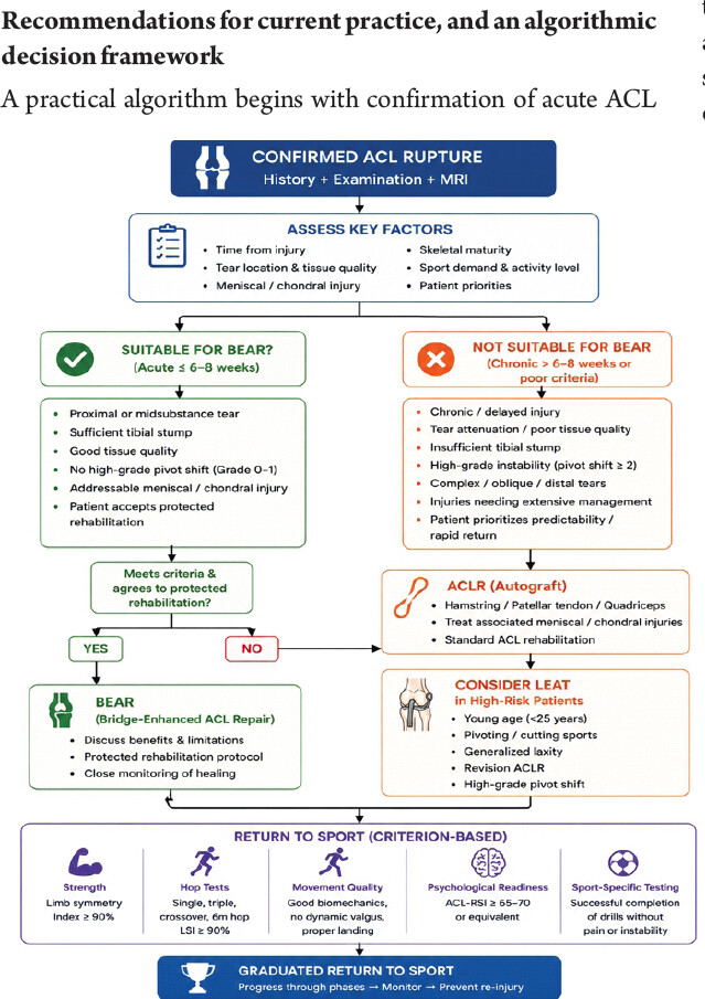
A practical algorithm begins with confirmation of acute ACL rupture, associated meniscal and chondral injury, skeletal maturity, sport demands, and patient acceptance of the uncertainty associated with repair. Early surgery is possible if MRI and arthroscopy demonstrate an acute midsubstance or proximal tear with good tissue quality and sufficient tibial remnant: BEAR can be discussed as a biologic option for repair. When the tear is chronic, tissue is thin, remnant is small, there is high-grade pivot shift, or when predictability of the revision is the goal, autograft ACLR is still preferred. In young high-risk pivoting athletes, validated graft-risk modeling allows lateral extra-articular tenodesis or BPTB compared to isolated hamstring in selected patients [33]. In patients under 25 years of age who had high-risk features, the STABILITY randomized trial demonstrated that lateral extra-articular tenodesis in combination with hamstring autograft ACLR decreased the failure rate [34]. A wider meta-analysis does support that other surgical factors besides high activity, younger age, and graft type have an impact on rerupture/revision [35]. Return to sport counseling should be conservative, as 20% of athletes younger than 20 experience a reinjury after return to high-risk sport [36] and a return prior to 9 months increases the likelihood of a second injury by 7-fold [37]. Rehabilitation should be based on criterion-based strength, hop, movement-quality, psychological-readiness and sport-specific progression (not time) [38]. The risk of PTOA should also be considered when making long-term decisions; there are still unresolved issues with both BEAR and ACLR [39]. The algorithm for BEAR versus ACLR is depicted in Fig. 1.
Discussion
The present evidence, however, is in favor of BEAR as the best-developed biologic ACL repair technique, but not as a substitute for autograft reconstruction in all patients, as shown in Table 2. The most compelling argument for it is the selective noninferiority to autograft ACLR for short-term IKDC and instrumented AP laxity for young, acute, repairable tears, and the added benefit of the preservation of hamstring strength when the comparator is hamstring autograft. This has a significant biological and clinical relevance due to the fact that morbidity associated with tendon harvesting is not innocuous, and preservation of the native tissue may preserve the anatomic, osteoarthritis, and proprioceptive benefits. The same RCT revealed a numerically greater number of second ipsilateral ACL surgeries after BEAR, though, and the evidence base for the procedure is currently limited to 6 years. Therefore, the BEAR-MOON trial is crucial as BPTB autograft is a more stable comparator than hamstring autograft in many high-risk athletes [10, 12, 13].
Practical selection checkpoints
| Checkpoint | BEAR-favorable | ACLR-favorable |
|---|---|---|
| Tear age | Acute, early surgery feasible | Chronic or delayed presentation |
| Tear pattern | Proximal/midsubstance repairable tear | Poor tissue, distal avulsion, attenuation |
| Tibial stump | Adequate length and quality | Inadequate remnant capture |
| Athlete profile | Accepts protected repair rehabilitation | High-risk pivoting/collision priority |
| Skeletal status | Physeal-safe tunnel plan possible | Reconstruction strategy better defined |
| Revision priority | Preserve autograft options | Maximize proven stability now |
ACLR: Anterior cruciate ligament reconstruction, BEAR: Bridge-enhanced ACL repair
Furthermore, interpreting the results of noninferiority should be approached with caution. A noninferior mean IKDC score does not guarantee that all the subgroups are equally safe, and comparable arthrometer laxity is not a complete indicator of pivot shift, neuromuscular readiness, fear of reinjury, and sport-specific exposure. The opposite would be true if a higher revision rate occurred in an early repair group, but if revision is easy and long-term joint preservation is later verified. This choice is therefore preference sensitive: some patients may be willing to risk the complication of repair, but not have a graft harvested to avoid the risk, while others may reasonably opt for the most proven reconstruction route [2, 3, 10], as illustrated in Table 3.
Pivotal BEAR and ACLR evidence
| Author, year | Design | n | Intervention | Comparator | Primary outcome | Follow-up | Key finding | References |
|---|---|---|---|---|---|---|---|---|
| Murray et al., 2016 | Feasibility cohort | 20 | BEAR | Hamstring ACLR | Safety/MRI continuity | 3 months | No infection or inflammatory reaction; continuous ACL on MRI | [8] |
| Murray et al, 2019 | First-in-human cohort | 20 | BEAR | Hamstring ACLR | IKDC, laxity, function | 24 months | Similar outcomes; higher hamstring strength after BEAR | [9] |
| Murray et al, 2020 | Randomized trial | 100 | BEAR | Autograft ACLR | IKDC, AP laxity | 24 months | BEAR noninferior; higher hamstring strength | [10] |
| Fleming et al., 2024 | Cohort follow-up | 20 | BEAR | Hamstring ACLR | IKDC, KOOS, strength | 6 years | Similar outcomes except preserved hamstring strength | [12] |
| Bear-Moon et al., 2022 | Multicenter RCT design | 200 planned | BEAR | BPTB ACLR | IKDC, arthrometer laxity | 2 years planned | Tests exportability against stronger autograft comparator | [13] |
| Wittstein et al., 2026 | Postcommercial cohort | 100 | BEAR | None | Adverse events, IKDC, KOOS | 15.3 months | 2.1% ACL retear at final follow-up | [15] |
ACLR: Anterior cruciate ligament reconstruction, AP: Anteroposterior, BEAR: Bridge-enhanced ACL repair, BPTB: Bone-patellar tendon-bone, IKDC: International knee documentation committee, KOOS: Knee injury and osteoarthritis outcome score, RCT: Randomized controlled trial
This synthesis makes BEAR stand out from other repair technologies when compared to previous reviews about ACL repair. Selected acute ACL tears can be repaired and have been supported by several articles in the literature regarding internal brace and dynamic intraligamentary stabilisation. Still, the constructs are mechanically and biologically different from a bovine extracellular-matrix scaffold with autologous blood. They may lead to overconfidence, a lack of appreciation of failure modes, and misguided indications for surgery. On the other hand, if BEAR is not used, the biological scaffold, trial design, and indication criteria are overlooked, which are all components that make modern BEAR different from historical primary repair [5, 6, 23].
Patient selection is the principal issue. Adolescents and young adults would like BEAR for its lack of graft harvest and for the fact that it leaves a future graft option; however, it would also be these same age groups where exposure to reinjury is greatest. Skeletally immature patients may be at risk for misplacement of tunnels, physeal injuries, concomitant meniscal repair, and family awareness of limitations to rehabilitation. There is evidence that supports BEAR in acute complete tears with a long enough tibial stump and good tissue quality, but partial tears and proximal avulsions may be salvageable. Explicit counseling is required for contact athletes, collision athletes and patients returning to high-risk pivoting sport, where early improvements of symptoms do not equal biologic maturation [14, 22, 36].
One of the most significant translational benefits is the revision strategy. When the BEAR fails, the surgeon is typically faced with only standard primary ACLR options, while when the ACLR fails, there are several further options that could be considered: tunnel widening, hardware, staged bone grafting, graft-choice depletion, meniscal and chondral progression. This potential benefit must not be exaggerated without a cost-effectiveness analysis that incorporates implant cost, rehabilitation visits, imaging, time off sport/employment, reoperation, and lifetime osteoarthritis burden. The economic value will vary among different health systems and is dependent on whether BEAR decreases donor-site morbidity, decreases the time to functional recovery, or decreases late degenerative disease [4, 14, 29].
Clinical translation should be staged, therefore. Surgeons early in their learning curve should have a narrow indication, informed consent with a clear distinction between the options of repair and reconstruction, and prospective outcome collection. Centers implementing BEAR should take the same operative images of the remnant, record the length of the stump and quality of the tissue, and define failure before return to sport exposure. Variables that may influence the success or failure of BEAR are tear location, time from injury, sex, skeletal maturity, sport, concomitant meniscal repair, and rehabilitation protocol, and should be stratified by these variables. Absolute scores and clinically important differences should be reported in comparative studies, because a similar IKDC score can inadvertently overlook clinically meaningful differences in knee pain, hamstring performance, sport confidence, or burden of revision [13, 14, 22].
BEAR outcome studies are still relatively scarce and narrative, and the evidence of emerging post-commercial studies has been limited. Comparative autograft data are derived from registries and meta-analyses, which suffer from selection bias, surgical learning curve, graft-size variation, and even exposure to return-to-sport. However, the overall direction of the treatment is evident that BEAR is to be used within a well-defined indication and autograft ACLR is still the reference surgery for conditions where tear biology, chronicity, high-risk sport, or revision reliability would seem to benefit from reconstruction [1, 15, 29].
Limitations
This review has several limitations. It is a narrative review rather than a systematic review or meta-analysis, which increases its susceptibility to selection and interpretation bias, and no formal assessment of study quality or risk of bias, such as Cochrane RoB 2, ROBINS-I, or AMSTAR, was performed for the included studies. The included studies were also heterogeneous with respect to patient selection, tear characteristics, rehabilitation protocols, outcome measures, and follow-up duration, which limited direct comparison across investigations, and no quantitative meta-analysis was performed to calculate pooled estimates for clinical outcomes, failure rates, or return-to-sport outcomes.
Long-term clinical evidence for BEAR remains limited, as most published studies report only short- to mid-term outcomes of 2–6 years, and the evidence base relies heavily on a small number of prospective trials, many originating from the same research groups, which may limit external validity. Registry data included in this review have relatively short follow-up, preventing robust conclusions regarding long-term durability and revision risk, and long-term outcomes such as PTOA, graft maturation, and survivorship remain inadequately studied, preventing definitive conclusions regarding joint preservation. Cost-effectiveness could not be critically evaluated because comprehensive economic analyses comparing BEAR and autograft reconstruction are lacking, and publication bias cannot be excluded, as early positive studies and industry-supported investigations may be overrepresented in the available literature.
Most available evidence focuses on carefully selected acute, repairable ACL tears, which limits applicability to chronic tears, poor tissue quality, revision cases, and multiligament injuries. Differences in rehabilitation protocols across studies may have influenced reported functional outcomes and return-to-sport rates, and variability in graft choice for ACLR, including hamstring, BPTB, and quadriceps tendon autografts, complicates direct comparison with BEAR. Patient-reported outcomes and return-to-sport criteria were not standardized across the included studies, further limiting comparisons between investigations. Given these limitations, current evidence does not establish the superiority of BEAR over autograft ACLR; therefore, larger multicenter randomized trials with long-term follow-up, standardized rehabilitation protocols, and cost-effectiveness analyses are required before recommending widespread adoption.
Conclusion
BEAR is a valid next-generation biologic approach to ACL surgery based on translation research, a randomized trial, early durability evidence and increasing registry experience. It has the most compelling benefits in acute tears that can be repaired, where there is a good stump on the tibia, when tissue quality is good, and when patients understand protected rehabilitation and potential long-term durability. Autograft ACLR is the standard of care for widespread use, high-risk athletes, chronic tears, insufficient remnants, and predictability. Autograft selection should be based on age, sport, laxity, donor-site risk, graft size, and the need for extra-articular augmentation laterally. The current practice should not be an either-or scenario: BEAR should not be viewed as an historical repair, but it should not be recommended for conditions for which there is a lack of evidence or indications. Future trials should identify the long-term risk of osteoarthritis, return to sport safety, cost-effectiveness, pediatric performance, revision results, and determine if long-term sport and health benefits resulting from native ligament preservation are measurable and relevant to the patient, surgeon, and health system over decades.
Clinical Message
BEAR may be offered to carefully selected patients with acute repairable ACL tears, adequate tibial stump, and good tissue quality
Autograft reconstruction remains preferred for chronic tears, poor remnants, high-risk instability, and revision certainty
Shared decision-making should compare biology, retear risk, graft morbidity, rehabilitation restrictions, return-to-sport timing, cost, and revision pathways.
Conflict of Interest:
Nil
Source of Support:
Nil
Consent:
The authors confirm that informed consent was obtained from the patient for publication of this article
References
- Brophy RH, Lowry KJ. American academy of orthopaedic surgeons clinical practice guideline summary: Management of anterior cruciate ligament injuries J Am Acad Orthop Surg 2023;31:531-7. Google Scholar | PubMed
- Diermeier T, Rothrauff BB, Engebretsen L, Lynch AD, Ayeni OR, Paterno MV, et al. Treatment after anterior cruciate ligament injury: Panther symposium ACL treatment consensus group Knee Surg Sports Traumatol Arthrosc 2020;28:2390-402. Google Scholar | PubMed
- Filbay SR, Grindem H. Evidence-based recommendations for the management of anterior cruciate ligament (ACL) rupture Best Pract Res Clin Rheumatol 2019;33:33-47. Google Scholar | PubMed
- Murray MM. Optimizing outcomes of ACL surgery-Is autograft reconstruction the only reasonable option? J Orthop Res 2021;39:1843-50. Google Scholar | PubMed
- Robinson JD Jr, Williamson T, Carson T, Whelan RJ, Abelow SP, Gilmer BB. Primary anterior cruciate ligament repair: Current concepts J ISAKOS 2023;8:456-66. Google Scholar | PubMed
- Proffen BL, Perrone GS, Roberts G, Murray MM. Bridge-enhanced ACL repair: A review of the science and the pathway through FDA investigational device approval Ann Biomed Eng 2015;43:805-18. Google Scholar | PubMed
- Perrone GS, Proffen BL, Kiapour AM, Sieker JT, Fleming BC, Murray MM. Bench-to-bedside: Bridge-enhanced anterior cruciate ligament repair J Orthop Res 2017;35:2606-12. Google Scholar | PubMed
- Murray MM, Flutie BM, Kalish LA, Ecklund K, Fleming BC, Proffen BL, et al. The bridge-enhanced anterior cruciate ligament repair (BEAR) procedure: An early feasibility cohort study Orthop J Sports Med 2016;4:2325967116672176. Google Scholar | PubMed
- Murray MM, Kalish LA, Fleming BC, BEAR Trial Team, Flutie B, Freiberger C, et al. Bridge-enhanced anterior cruciate ligament repair: Two-year results of a first-in-human study Orthop J Sports Med 2019;7:2325967118824356. Google Scholar | PubMed
- Murray MM, Fleming BC, Badger GJ, BEAR Trial Team, Freiberger C, Henderson R, et al. Bridge-enhanced anterior cruciate ligament repair is not inferior to autograft anterior cruciate ligament reconstruction at 2 years: Results of a prospective randomized clinical trial Am J Sports Med 2020;48:1305-15. Google Scholar | PubMed
- Barnett S, Badger GJ, Kiapour A, Yen YM, Henderson R, Freiberger C, et al. Females have earlier muscle strength and functional recovery after bridge-enhanced anterior cruciate ligament repair Tissue Eng Part A 2020;26:702-11. Google Scholar | PubMed
- Fleming BC, Baranker B, Badger GJ, Kiapour AM, Ecklund K, Micheli LJ, et al. Bridge-enhanced anterior cruciate ligament restoration: 6-year results from the first-in-human cohort study Orthop J Sports Med 2024;12:23259671241260632. Google Scholar | PubMed
- Bear-Moon Design Group, Spindler KP, Imrey PB, Yalcin S, Beck GJ, Calbrese G, et al. Design features and rationale of the BEAR-MOON (bridge-enhanced ACL restoration multicenter orthopaedic outcomes network) randomized clinical trial Orthop J Sports Med 2022;10:23259671211065447. Google Scholar | PubMed
- Shah AK, Neijna AG, Retzky JS, Gomoll AH, Strickland SM. Indications, techniques, and outcomes of bridge-enhanced ACL restoration (BEAR) Curr Rev Musculoskelet Med 2025;18:140-8. Google Scholar | PubMed
- Wittstein J, Strickland S, Gomoll A, Sherman S, Lau B, Taylor D, et al. Postcommercialisation outcomes of bridge-enhanced anterior cruciate ligament restoration: The first 100 Bridge registry patients Knee Surg Sports Traumatol Arthrosc 2026;34:2013-24. Google Scholar | PubMed
- Pruneski JA, Kaushal SG, Ecklund K, Kramer DE, Yen YM, Micheli LJ, et al. Magnetic resonance imaging assessment of bridge-enhanced ACL restoration: An imaging atlas of morphological features and healing trajectories Orthop J Sports Med 2026;14:23259671261445974. Google Scholar | PubMed
- Bond E, Wu KA, Mills FB, O'Donnell R, Cochran G, Lau BC. Bridge-enhanced anterior cruciate ligament restoration: Surgical technique Video J Sports Med 2024;4:26350254231218749. Google Scholar | PubMed
- Kantrowitz DE, Darden CN, Haunschild ED, Gladstone JN, Anthony SG. Modified bridge-enhanced anterior cruciate ligament repair Arthrosc Tech 2024;13:103034. Google Scholar | PubMed
- Hourston GJ, Kankam HK, McDonnell SM. A systematic review of anterior cruciate ligament primary repair rehabilitation J Clin Orthop Trauma 2022;25:101774. Google Scholar | PubMed
- Wu J, Kator JL, Zarro M, Leong NL. Rehabilitation principles to consider for anterior cruciate ligament repair Sports Health 2022;14:424-32. Google Scholar | PubMed
- Shen Z, Chen H, Ye M, Gao Z, Li H, Lu H, et al. Early outcomes of primary repair versus reconstruction for acute anterior cruciate ligament injury: A systematic review and meta-analysis Medicine (Baltimore) 2022;101:e32411. Google Scholar | PubMed
- Zhang S, Xia T, Dai X, DiFelice GS, Engebretsen L, Gobbi A, et al. Primary repair of proximal anterior cruciate ligament injury: A global expert consensus statement Burns Trauma 2025;13 tkae079. Google Scholar | PubMed
- Wilson WT, Hopper GP, Banger MS, Blyth MJ, Riches PE, MacKay GM. Anterior cruciate ligament repair with internal brace augmentation: A systematic review Knee 2022;35:192-200. Google Scholar | PubMed
- Ahmad SS, Schreiner AJ, Hirschmann MT, Schröter S, Döbele S, Ahrend MD, et al. Dynamic intraligamentary stabilization for ACL repair: A systematic review Knee Surg Sports Traumatol Arthrosc 2019;27:13-20. Google Scholar | PubMed
- Ambrosio L, Franco D, Vadalà G, Russo F, Papalia R. Anterior cruciate ligament repair augmented with dynamic intraligamentary stabilization is equivalent to hamstring autograft reconstruction at short- and mid-term follow-up: A systematic review Arthrosc J Arthrosc Relat Surg 2024;40:2121-31.e1. Google Scholar | PubMed
- Dai W, Leng X, Wang J, Cheng J, Hu X, Ao Y. Quadriceps tendon autograft versus bone-patellar tendon-bone and hamstring tendon autografts for anterior cruciate ligament reconstruction: A systematic review and meta-analysis Am J Sports Med 2022;50:3425-39. Google Scholar | PubMed
- Mouarbes D, Menetrey J, Marot V, Courtot L, Berard E, Cavaignac E. Anterior cruciate ligament reconstruction: A systematic review and meta-analysis of outcomes for quadriceps tendon autograft versus bone-patellar tendon-bone and hamstring-tendon autografts Am J Sports Med 2019;47:3531-40. Google Scholar | PubMed
- Connors JP, Cusano A, Saleet J, Hao KA, Efremov K, Parisien RL, et al. Return to sport and graft failure rates after primary anterior cruciate ligament reconstruction with a bone-patellar tendon-bone versus hamstring tendon autograft: A systematic review and meta-analysis Am J Sports Med 2025;53:1996-2006. Google Scholar | PubMed
- Rahardja R, Zhu M, Love H, Clatworthy MG, Monk AP, Young SW. Factors associated with revision following anterior cruciate ligament reconstruction: A systematic review of registry data Knee 2020;27:287-99. Google Scholar | PubMed
- Vindfeld S, Persson A, Lindanger L, Fenstad AM, Visnes H, Inderhaug E. Revision anterior cruciate ligament reconstruction: Surgeon-reported causes of failure from the Norwegian knee ligament register Am J Sports Med 2025;53:801-8. Google Scholar | PubMed
- Snaebjörnsson T, Hamrin-Senorski E, Svantesson E, Karlsson L, Engebretsen L, Karlsson J, et al. Graft diameter and graft type as predictors of anterior cruciate ligament revision: A cohort study including 18,425 patients from the Swedish and Norwegian national knee ligament registries J Bone Joint Surg Am 2019;101:1812-20. Google Scholar | PubMed
- Zegzdryn M, Moatshe G, Engebretsen L, Drogset JO, Lygre SHL, Visnes H, et al. Increased risk for early revision with quadriceps graft compared with patellar tendon graft in primary ACL reconstructions Knee Surg Sports Traumatol Arthrosc 2024;32:656-65. Google Scholar | PubMed
- Marmura H, Getgood AM, Spindler KP, Kattan MW, Briskin I, Bryant DM. Validation of a risk calculator to personalize graft choice and reduce rupture rates for anterior cruciate ligament reconstruction Am J Sports Med 2021;49:1777-85. Google Scholar | PubMed
- Getgood AM, Bryant D, Litchfield RB, et al[1.1. Lateral extra-articular tenodesis reduces failure of hamstring tendon autograft ACL reconstruction -two year outcomes from the STABILITY study randomized clinical trial Orthop J Sports Med 2019;7:2325967119S00280. Google Scholar | PubMed
- Zhao D, Pan JK, Lin FZ, Luo MH, Liang GH, Zeng LF, et al. Risk factors for revision or rerupture after anterior cruciate ligament reconstruction: A systematic review and meta-analysis Am J Sports Med 2023;51:3053-75. Google Scholar | PubMed
- Barber-Westin S, Noyes FR. One in 5 Athletes sustain reinjury upon return to high-risk sports after ACL reconstruction: A systematic review in 1239 Athletes younger than 20 years Sports Health 2020;12:587-97. Google Scholar | PubMed
- Beischer S, Gustavsson L, Senorski EH, Karlsson J, Thomeé C, Samuelsson K, et al. Young Athletes who return to sport before 9 months after anterior cruciate ligament reconstruction have a rate of new injury 7 times that of those who delay return J Orthop Sports Phys Ther 2020;50:83-90. Google Scholar | PubMed
- Kotsifaki R, Korakakis V, King E, Barbosa O, Maree D, Pantouveris M, et al. Aspetar clinical practice guideline on rehabilitation after anterior cruciate ligament reconstruction Br J Sports Med 2023;57:500-14. Google Scholar | PubMed
- Whittaker JL, Losciale JM, Juhl CB, Thorlund JB, Lundberg M, Truong LK, et al. Risk factors for knee osteoarthritis after traumatic knee injury: A systematic review and meta-analysis of randomised controlled trials and cohort studies for the OPTIKNEE Consensus Br J Sports Med 2022;56:1406-21. Google Scholar | PubMed
© 2026 Journal of Orthopaedic Case Reports - Published by Indian Orthopaedic Research Group
About the Authors
-
Dr. Sanjeevi Bharadwaj
-

Dr. Naveen Jeyaraman
-

Dr. Subasri Balasubramanian
-

Dr. Arunagiri Gunasekar
-

Dr. Arulkumar Nallakumarasamy
-

Dr. Madhan Jeyaraman
Related Articles in Journal of Orthopaedic Case Reports
 September 1, 2026 Comparing Bridge-Enhanced Anterior Cruciate Ligament Repair to Standard Autograft for Anterior Cruciate Ligament Reconstruction: Biomechanical and Clinical Outcomes
 September 1, 2026 Arthroscopic ACL Tibial Spine Avulsion Fixation Using Suture Pull-Through Double-Tunnel Technique
 July 1, 2026 Clinical and Donor-site Outcomes of Full-thickness Peroneus Longus Autograft in Arthroscopic Anterior Cruciate Ligament Reconstruction: A Prospective Indian Cohort Study
 June 1, 2026 Artelon Graft Augmentation in Revision Anterior Cruciate Ligament Reconstruction: Surgical Technique Guide