


Introduction
In 1814, Giovanni Battista Monteggia defined a Monteggia fracture as a fracture of the proximal ulna with a dislocation of the radial head [1, 2]. Further classification of the fracture came in 1967 by Jose Luis Bado, who described four types [3]. Type I is a fracture of the proximal ulna with anterior radial head dislocation [1]. Type I fractures are the most common in children and account for approximately 70% of cases. The mechanism of injury includes forced forearm pronation during hyperextension of the elbow [3]. Type II is a fracture of the ulna with a posterior radial head dislocation [1]. Type II fractures are the most common type occurring in adults, accounting for approximately 80% of cases [3]. The injury is usually caused by axial loading on a partially flexed elbow [3]. Type III fractures consist of an ulnar fracture with a lateral radial head dislocation, which is also commonly seen in children [1]. Mechanism of injury results from a fall on the elbow with hyperextension and pronation combined with forced abduction or a varus stress [3]. Finally, a type IV fracture is a fracture of both the ulnar and radial shafts with an anterior radial head dislocation [1]. Bado Type I and Type IV fractures have similar mechanisms of injury, but a Type IV is usually the result of higher energy or greater impact [3].
Further classification of Bado Type II fractures came in 1991 by Jesse Jupiter. Type IIA fractures involve the proximal ulna and coronoid process [4]. Type IIB fractures occur at the meta-diaphyseal junction, distal to the coronoid [5]. Type IIC fractures occur at the diaphyseal level, distal to the coronoid [4]. Type IID fractures are often comminuted and extend from the olecranon to the ulnar diaphysis [4].
Overall, Monteggia fractures account for <2% of forearm fractures. Malunion and nonunion occur in approximately 2–10% of cases, which is higher than the average forearm fracture nonunion rate of 2% [3]. Causes of nonunion are multifactorial and include lack of mechanical stability and impaired bone healing due to factors such as infection or metabolic disorders. Other factors that play a role in nonunion include the fracture pattern such as comminuted fractures, open fractures, soft-tissue stripping, and location of fracture [6]. Fractures of the distal and middle third junction have the least blood supply, contributing to poor healing. Management of nonunion includes excision of the nonunion site, anatomic reduction, and stabilization. In addition, bone grafting can stimulate bone formation [7].
Case Report
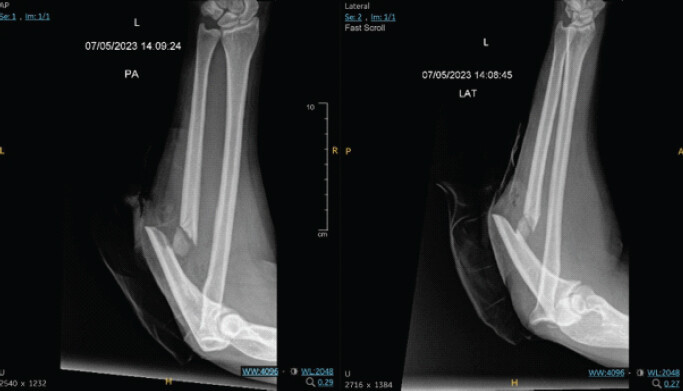
Informed consent was obtained from the patient before starting this manuscript. A 42-year-old female with no significant past medical history presented to the emergency department with an open fracture of the left forearm following a motor vehicle accident. On examination, the patient was neurovascularly intact. X-rays of the left forearm revealed a proximal third comminuted highly displaced ulnar shaft fracture with a dislocation of the radial head at the elbow, signifying an open Monteggia Type IIC injury (Fig. 1). The patient was brought to the operating room (OR) for emergent incision and drainage of her open fracture along with open reduction internal fixation of the ulna and closed reduction of the radial head. The wound was cleaned with saline and vancomycin. During the procedure, the proximal and distal ends were reduced to their comminuted fragments and then to each other using towel reduction clamps. A 9-hole Synthes plate was applied with cortical and locking screws proximal and distal to the fracture. Following confirmation of the plate using AP and lateral images, a series of locking screws was placed both proximal and distal. An additional non-locking screw was placed distally. After placement of screws, one fragment appeared mobile, so an additional 3.5 cortical screw was placed through the plate and into this volar fragment. Radiograph confirmed reduction and placement of hardware at the ulnar fracture site along with reduction of the radial head into the radiocapitellar joint. Patient did well postoperatively and was discharged.
At 2 weeks post-operative, the patient reported improvement in her pain and was advised to begin physical therapy. The patient continued to present for follow-up visits; however, 3 months postoperatively, the fracture site still showed nonunion per radiographs (Fig. 2). Patient underwent bloodwork for potential infection; however, results showed no significant findings.
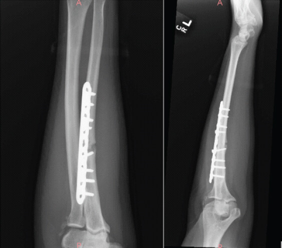
4 months postoperatively, a gap of 2.5 mm at the fracture site was found on imaging. Patient was interested in trying an external bone stimulator; however, was unable to see any progression in the fracture after several weeks. A computed tomography scan was performed to evaluate for any bony bridging across the fracture site; however, result showed an ulnar nonunion. Further surgical treatment was planned due to lack of healing.
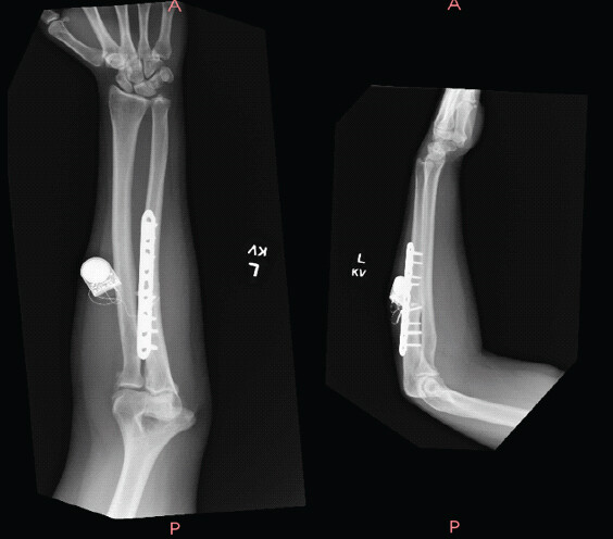
Surgical course included cultures to evaluate for any sign of infection, allograft bone grafting, platelet-rich plasma (PRP) augmentation, and an implantable bone stimulator placement.
Approximately 6 weeks postoperatively, anteroposterior and lateral images showed some interval healing at her ulnar shaft fracture (Fig. 3). Six months postoperatively, X-rays showed full healing of her fracture. Her bone stimulator was removed 2 weeks later.
Discussion
The current standard of care in the evaluation of a Monteggia fractures include careful visual inspection, gentle palpation, and a detailed neurovascular exam [3]. Careful attention should be given to identifying any open wounds overlying fracture sites, which require urgent surgical intervention [8]. The preferred treatment is operative repair with open reduction internal fixation (ORIF) [3]. Bado Type II fractures can be associated with more complex management, as was observed in this case [5].
This patient sustained a Type IIC Monteggia fracture that failed to heal within the standard time frame. According to the Food and Drug Administration, a fracture is classified as a nonunion if it has failed to heal after 9 months with no signs of radiographic healing for at least 3 months [7]. Two principal causes of nonunion include compromised blood supply and inadequate mechanical stability at the fracture site [6]. High energy fractures with soft tissue compromise and open fractures can limit blood supply, which was observed in this case [6]. Soft tissue damage can disrupt local vascularity, impairing the delivery of osteogenic cells and growth factors essential for bone regeneration. In addition, disruption of the osteoconductive scaffold increases the distance across the fracture gap, limiting new bone formation [6]. In this case, proper surgical technique was utilized, and sufficient time was allowed for healing before further imaging and laboratory evaluation were sought to rule out infection [7].
Given the absence of infection and appropriate mechanical stabilization, impaired vascular supply was likely the main contributing factor to nonunion. To promote healing, a combination of bone allograft, PRP, and an implantable bone stimulator were utilized. The bone allograft, derived from bovine Achilles tendon and rich in Type I collagen, served as an osteoconductive scaffold [9, 10]. This graft is designed to resorb and be replaced by bone over time, restoring the structural integrity and biological function [9]. After this treatment, the patient was able to achieve full healing. This patient had multiple follow-up visits due to the nonunion. The importance of this case shows that adjuncts, such as a bone graft, might be beneficial when a patient presents with a Type IIC Monteggia fracture to avoid future frequent visits and even potential return to the OR.
Conclusion
Monteggia fractures account for <2% of all forearm fractures with malunion or nonunion occurring in 2–10% of those cases [3]. Given this patient’s poor healing despite proper surgical technique and time allotted for healing, a multimodal approach was utilized to provide the patient with the best outcome. Since Bado Type II fractures are associated with poorer outcomes and fracture patterns can affect vascularity, additional modalities might be beneficial in addition to ORIF to prevent nonunion [5]. Use of additional modalities, such as the bone grafting, can help decrease future healthcare costs associated with nonunion [9]. This patient required multiple subsequent visits and even a second surgery that increased her healthcare costs. Special consideration might be needed going forward with Monteggia Type IIC fracture repairs in the future to avoid these unnecessary extra healthcare costs.
Clinical Message
Chronic Monteggia fracture-dislocations can be challenging to manage, particularly when malunion or nonunion has developed. Successful reconstruction requires careful assessment of the proximal radioulnar relationship, restoration of ulnar alignment and length, and consideration of biologic augmentation when healing is compromised. This case highlights the importance of a comprehensive, multimodal approach to achieve union and restore functional alignment in a complex Monteggia fracture nonunion.
Conflict of Interest:
Nil
Source of Support:
Nil
Consent:
The authors confirm that informed consent was obtained from the patient for publication of this article
References
- Delpont M, Louahem D, Cottalorda J. Monteggia injuries. Orthop Traumatol Surg Res 2018;104:S113-20. Google Scholar | PubMed
- Robles EL, Rolfs H, Hargett D. Monteggia variant with posterior elbow dislocation and radial shaft fracture: A case report. Int J Surg Case Rep 2022;99:107705. Google Scholar | PubMed
- Johnson NP, Silberman M. Monteggia Fractures. StatPearls. Available from: https://www.ncbi.nlm.nih.gov/books/nbk470575 [Last accessed on 2023 Jul 31]. Google Scholar | PubMed
- Soderlund T, Zipperstein J, Athwal GS, Hoekzema N. Monteggia fracture dislocation. J Orthop Trauma 2024;38:S26-30. Google Scholar | PubMed
- Siebenlist S, Buchholz A, Braun KF. Fractures of the proximal ulna: Current concepts in surgical management. EFORT Open Rev 2019;4:1-9. Google Scholar | PubMed
- Thomas JD, Kehoe JL. Bone Nonunion. StatPearls. Available from: https://www.ncbi.nlm.nih.gov/books/nbk554385 [Last accessed on 2023 Mar 06]. Google Scholar | PubMed
- Dimartino S, Pavone V, Carnazza M, Cuffaro ER, Sergi F, Testa G. Forearm fracture nonunion with and without bone loss: An overview of adult and child populations. J Clin Med 2022;11:4106. Google Scholar | PubMed
- Small R, Taqi M, Yaish AM. Radius and Ulnar Shaft Fractures. StatPearls. Available from: https://www.ncbi.nlm.nih.gov/books/nbk557681 [Last accessed on 2022 Dec 28]. Google Scholar | PubMed
- Prosidyan, Inc. FIBERGRAFT BG Matrix Instructions for Use [Brochure]. United States: Prosidyan, Inc.; 2023. Google Scholar | PubMed
- Rafi BM, Tiwari V. Forearm Fractures. StatPearls. Available from: https://www.ncbi.nlm.nih.gov/books/nbk574580 [Last accessed on 2023 Aug 08]. Google Scholar | PubMed
© 2026 Journal of Orthopaedic Case Reports - Published by Indian Orthopaedic Research Group
About the Authors
-

Miss. Kiana Zanganeh
-

Dr. Jennifer Madden
Related Articles in Journal of Orthopaedic Case Reports
 April 1, 2025 A Rare Case of Xanthogranulomatous Osteomyelitis of Radius Presenting as Pathological Fracture – A Case Report
 December 1, 2024 Alternative Management Approach for Infected Gap Non-Union of Both Bone Forearms – A Case Report
 May 10, 2023 Old Monteggia Fracture-Dislocation Treated with Plating and Forearm Fascial Slip Annular Ligament Reconstruction: A Rare Method of Treatment and Review of Literature
 August 1, 2026 Plastic Bowing of the Pediatric Tibia is Extraordinarily Rare and Not Represented in the Literature, the Unicorn of Pediatric Fractures: A Case Report