


Introduction
Osteoblastomas are rare, benign, osteoid-producing primary bone tumors, occurring most commonly in adolescents and young adults in the second and third decades of life. First described as “giant osteoid osteoma” by Dahlin and Johnson in 1954 [1] and later renamed by Lichtenstein and Jaffe in 1956 [2,3]. They constitute only 1% of all bone tumors and are exceptionally rare in the hand and wrist [4,5]. Predominantly affecting adolescents and young adults, approximately 30–40% arise from the posterior elements of the spine, including the sacrum [6, 7]. Due to their rarity in the hand and wrist, osteoblastomas are seldom considered in the differential diagnosis of bone tumors in these locations. Literature reports only a few cases in carpal bones, mostly in the scaphoid.
We present a rare case of hamate osteoblastoma in late adulthood. He underwent extended curettage with cancellous bone grafting, resulting in a favorable functional outcome with complete healing and restoration of the full range of movements of the wrist joint.
Case Report
A 51-year-old right-handed male with no significant medical history presented with a 2.5-year history of pain over the dorsal aspect of the ulnar side of the left wrist. The pain, unrelieved by painkillers and worsened after minor trauma, led to progressive swelling. Clinical examination revealed a firm, tender, and poorly defined swelling on the dorsal aspect of the ulnar side of the wrist, accompanied by wrist joint stiffness.
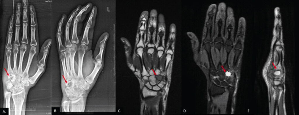
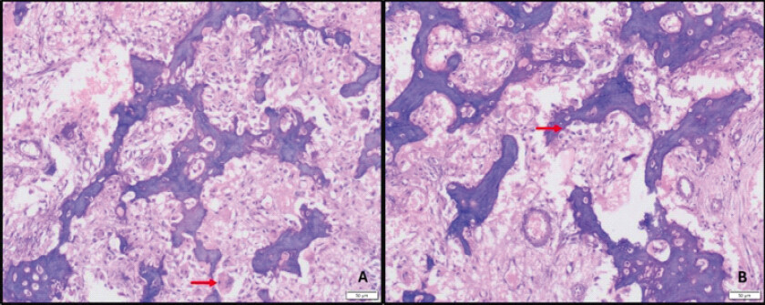
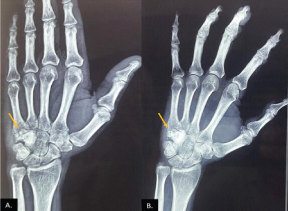
Radiographs showed a well-defined lytic lesion with a sclerotic rim in the hamate, which was mildly expansile and occupied half of the bone, with a cortical breach (Fig. 1a and b). Magnetic resonance imaging (MRI) demonstrated a well-defined, T1-hypointense, T2/PD-hyperintense expansile lesion with a sclerotic rim and blood-fluid levels, with no soft tissue extension (Fig. 1c, d, e). Histopathology confirmed osteoblastoma, characterized by interconnecting, delicate woven bone trabeculae rimmed by plump osteoblasts. The stroma was loose, richly vascularized, and contained stellate and plasmacytoid cells, along with interspersed osteoclastic giant cells. No anaplasia or significant mitotic activity was observed (Fig. 2).
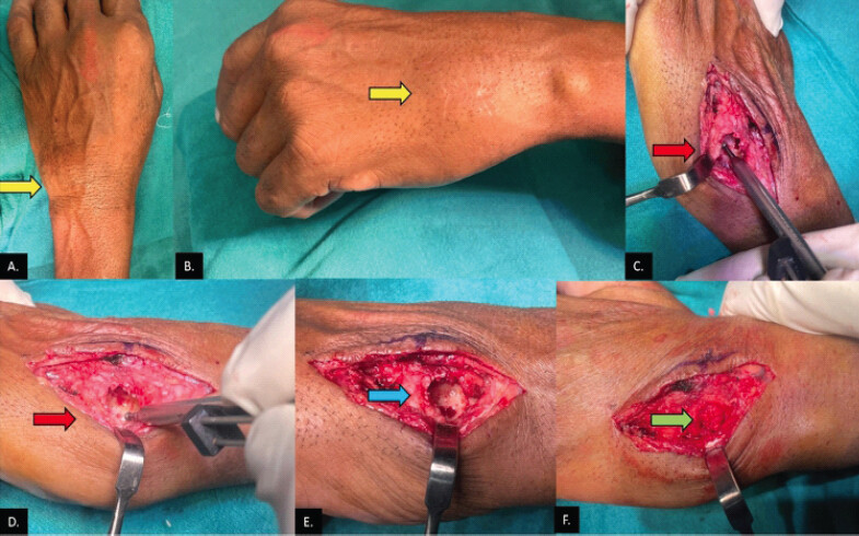
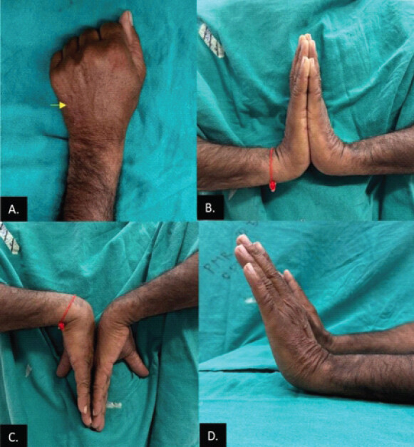
Extended curettage using a high-speed burr was performed by dorsal incision, carefully preserving the ulnar neurovascular bundle (Fig. 3), followed by filling the defect with autologous cancellous bone graft harvested from the ipsilateral iliac crest. Following surgery, the patient experienced rapid symptomatic relief, with a significant reduction in pain, swelling, and stiffness. The incision exhibited good healing (Fig. 4a). The wrist was immobilized in a splint for 6 weeks. The patient resumed routine daily activities within 2 months post-surgery with full range of motion at the wrist with unrestricted flexion-extension and supination-pronation (Fig. 4b, c, d). Plain radiographs were conducted during follow-ups, revealing no signs of recurrence (Fig. 5). Patient wrist function score as per the disabilities of the arm, shoulder and hand questionnaire was 1.7 at the latest 18-month follow-up.
Discussion
Osteoblastoma constitutes only 1% of all bone tumors and predominantly affects the spine, pelvis, and long bones. Carpal bone involvement is exceedingly rare. A review of the literature identified only nine reported cases of osteoblastoma involving the hamate (Table 1). Osteoblastoma typically arises in individuals aged 10–25 years, with a notable male predominance (2:1) [6]. Occurrence of this neoplasm in the 5th decade of life is extremely rare.
Literature review of cases with osteoblastoma of the hamate bone
| Authors | Age at presentation (in years)/Gender | Onset of symptoms to radiological diagnosis | Initial treatment | Recurrence | Revision surgery | Follow-up |
|---|---|---|---|---|---|---|
| Menon et al .(1988) [11] | Not known | Immediate | Curettage; grafting | 11 months | Excision of the hamatun | 48 months |
| Carpo-metacarpal arthrodesis | ||||||
| Apergis et al. (1993) [12] | 52/M | 15 months | Curettage; grafting | – | No | 12 months |
| Maréchal (1999) [13] | 54/M | Not known | Curettage; grafting | – | No | Not known |
| Van Dijk et al. (1999) [8] | 13/F | Not known | Curettage | 6 months | Wide resection | 3 months |
| Wrist arthrodesis | ||||||
| Gdoura et al. (2010) [6] | 28/N/K Manual labourer | 15 months | Curettage | – | No | 24 months |
| Dunda et al. (2013) [14] | 54/M | 48 months | Wide resection of the hamate, metacarpal bones, distal radius, and ulna | – | No | 6 months |
| Ayan and Serinsöz (2014) [10] | 39/F | 12 months | Curettage; grafting | – | No | 12 months |
| Repáraz Padrós et al. (2016) [15] | 28/M | 12 months | Curettage; grafting | – | No | 48 months |
| Zyluk et al. (2017) [7] | 26/F | 2 months | Resection of the hamate | – | No | 12 months |
| Our Case (2025) | 51/M | 30 months | Curettage; grafting | – | No | 18 months |
Unlike osteoid osteoma, it is not associated with nocturnal pain relieved by salicylates, a finding consistent with our case. The exact mechanism of osteoblastoma-associated pain remains unclear but is thought to be linked to the tumor’s rich vascularity and local inflammatory response.
Histopathologically, osteoblastoma is a locally aggressive benign neoplasm. It can be challenging to distinguish from osteosarcoma, particularly in its very aggressive variants. Osteoblastoma carries a recurrence rate of 10–19%. In rare cases, malignant transformation has been reported [8].
In our case, the patient presented with chronic wrist pain and swelling, initially misattributed to trauma. Delayed diagnosis is common, as early radiographic findings can be subtle. Radiographs revealed a well-defined lytic lesion with a sclerotic rim within the hamate, while MRI demonstrated a T1-hypointense, T2-hyperintense lesion with a blood-fluid level—features consistent with osteoblastoma.
Treatment strategies vary based on tumor aggressiveness and recurrence risk, ranging from curettage with bone grafting to en bloc resection [6,7,9]. While curettage remains the preferred approach for most cases, radical excision is advocated for aggressive or recurrent lesions [7, 10].
The long-term functional outcome following carpal osteoblastoma resection depends on the extent of bone loss and the reconstruction approach. Some authors recommend en bloc resection with proximal row carpectomy or four-corner fusion for scaphoid involvement [6,7,9]; however, these procedures can significantly impair wrist mobility. In our patient, meticulous curettage combined with bone grafting led to rapid symptom resolution, restoration of grip strength, and a favorable functional outcome, with no signs of recurrence at follow-up. This method not only provided symptomatic relief but also ensured structural stability.
Conclusion
Despite its rarity, osteoblastoma should be considered in the differential diagnosis of persistent, non-traumatic wrist pain, particularly in young adults. Early recognition and appropriate surgical management can provide excellent long-term outcomes, preserving wrist function while minimizing recurrence risk. Future studies and case reports will help refine treatment strategies and expand our understanding of this uncommon tumor.
Clinical Message
Although exceedingly rare in carpal bones and older adults, osteoblastoma must be considered in the differential diagnosis of chronic, non-traumatic wrist pain. Early recognition and meticulous extended curettage with autologous bone grafting can provide excellent long-term pain relief and structural stability while avoiding the risk of radical resections that compromise wrist mobility.
Conflict of Interest:
Nil
Source of Support:
Nil
Consent:
The authors confirm that informed consent was obtained from the patient for publication of this article
References
- Dahlin DC, Johnson EW. Giant osteoid osteoma. J Bone Joint Surg Am 1954;36:559-72. [Google Scholar] | [PubMed]
- Lichtenstein L. Benign osteoblastoma. A category of osteoid and bone-forming tumors other than classical osteoid osteoma, which may be mistaken for giant cell tumor or osteogenic sarcoma. Cancer 1956;9:1044-50. [Google Scholar] | [PubMed]
- Jaffe HL. Benign osteoblastoma. Bull Hosp Joint Dis 1956;17:141-51. [Google Scholar] | [PubMed]
- Healey HJ, Ghelman B. Osteoid osteoma and osteoblastoma. Clin Orthop 1986;204:76-85. [Google Scholar] | [PubMed]
- Manguini U. Tumors of the skeleton of the hand. Bull Hosp Joint Dis 1967;28:61-103. [Google Scholar] | [PubMed]
- Gdoura F, Trigui M, Ellouze Z, Hamed YB, Ayadi K, Keskes H. Hamatum osteoblastoma. Orthop Traumatol Surg Res 2010;96:712-6. [Google Scholar] | [PubMed]
- Zyluk A, Puchalski P, Szlosser Z. Osteoblastoma of the hamate bone: A case report. J Hand Surg Am 2017;49:350-1. [Google Scholar] | [PubMed]
- Van Dijk M, Winters HA, Wuisman PI. Recurrent osteoblastoma of the hamate bone. A two-stage reconstruction with a free vascularized iliac crest flap. J Hand Surg Br 1999;24:501-5. [Google Scholar] | [PubMed]
- Chughtai M, Orriols AM, Saengchote SA, Kim J, Wright TW, Dell PC. Distribution and characteristics of carpal bone tumors: A systematic review. J Wrist Surg 2023;13:534-49. [Google Scholar] | [PubMed]
- Ayan I, Serinsöz E. Osteoblastoma in the os hamatum: A rare case report. Eklem Hastalik Cerrahisi 2014;25:56-9. [Google Scholar] | [PubMed]
- Menon J, Rankin D, Jacobson C. Recurrent osteoblastoma of the carpal hamate. Orthopedics 1988;11:609-11. [Google Scholar] | [PubMed]
- Apergis E, Tsamouri M, Theodoratos G, Maris I, Antoniou N. Osteoblastoma of the hamate bone: A case report. J Hand Surg Am 1993;18:137-40. [Google Scholar] | [PubMed]
- Maréchal E. Recurrent osteoblastoma of the hamate bone. J Hand Surg Br 1999;24:757-8. [Google Scholar] | [PubMed]
- Dunda SE, Kauczok J, Demir E, Braunschweig T, Pallua N. A rare case of massive carpal osteoblastoma requiring complex reconstructive surgery. J Plast Reconstr Aesthet Surg 2013;66:e193-6. [Google Scholar] | [PubMed]
- Repáraz Padrós FJ, Eraso Lara I, Garbayo Marturet AJ, Tejero Ibáñez A, Castaño Serrano A, Panizo Santos A. Osteoblastoma del hueso ganchoso [Hamatum osteoblastom. An Sist Sanit Navar 2016;39:433-7. [Google Scholar] | [PubMed]
© 2026 Journal of Orthopaedic Case Reports - Published by Indian Orthopaedic Research Group
About the Authors
-

Dr. Prabodh Kantiwal
-

Dr. Syed Mohammed Adnan
-

Prof. Siddhi Chawla
-

Dr. Amir Suhail
-

Dr. Ritvik Sharma
-

Dr. Divya Aggarwal
Related Articles in Journal of Orthopaedic Case Reports
 October 1, 2026 Osteoblastoma of the Radial Shaft Mimicking Parosteal Osteosarcoma: A Case Report
 October 1, 2026 Clinicopathological Spectrum and Management of Aneurysmal Bone Cysts in Atypical Anatomical Location: A Comprehensive Review of Demographics, Presentation, Diagnosis, and Treatment Outcomes
 September 1, 2026 Unnoticed Giant Cell Tumor of the Left Distal Tibia in a 52-Year-Old Male: A Rare Case Report
 September 1, 2026 Acetabular Giant Cell Tumor Presenting with Protrusio Acetabuli: A Rare Case Report