


Introduction
Bosworth fractures represent an uncommon but clinically significant subset of ankle fracture-dislocations in which the proximal fibular fragment becomes locked behind the posterior tibial tubercle. First described by Bosworth in 1947, these injuries account for <1% of all ankle fractures and are frequently misdiagnosed on initial presentation due to subtle radiographic findings and their overall rarity [1,2]. The mechanism typically involves a supination-external rotation force that drives the fibula posteriorly, where it becomes entrapped behind the tibia, preventing successful closed reduction [3]. Once incarcerated, the fibula acts as a fixed posterior block, rendering the injury irreducible by closed means and distinguishing it from more common ankle fracture-dislocations. The resulting fixed displacement disrupts normal ankle biomechanics and is often accompanied by associated injuries, including posterior malleolar fractures, syndesmotic disruption, and ligamentous tearing [4].
Despite advances in imaging and clinical awareness, Bosworth fractures continue to pose diagnostic challenges. Standard radiographs may fail to clearly demonstrate posterior fibular displacement, and persistent malalignment after attempted reduction should raise suspicion for this injury pattern. In such cases, computed tomography (CT) has become essential for identifying the characteristic posterior entrapment and evaluating the extent of associated osseous and ligamentous injury.
Case Report
A 34-year-old man presented to the emergency department via ambulance after falling down three stairs while carrying furniture. He reported rolling his left ankle during the fall and denied other injuries. Vital signs were within normal limits. Physical examination revealed an obvious left ankle deformity with swelling and tenderness, intact dorsalis pedis and posterior tibial pulses, preserved sensation, and limited range of motion due to pain.
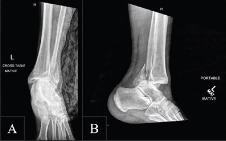
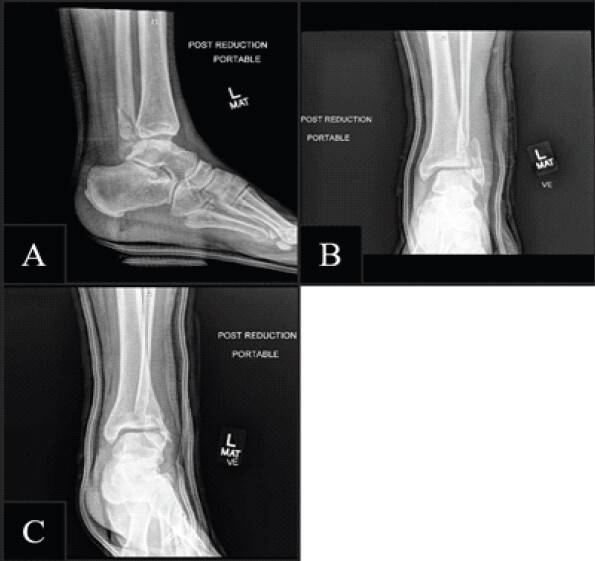
Radiographs demonstrated a posterior malleolus fracture, an angulated distal fibular fracture, and tibiotalar dislocation (Fig. 1). CT imaging further demonstrated posterior fibular entrapment (Fig. 2). An ankle nerve block was performed using 8 mL of 2% lidocaine targeting the tibial, sural, saphenous, deep, and superficial fibular nerves. The patient received 50 mcg of intravenous (IV) fentanyl, followed by closed reduction and splinting. Repeat radiographs showed improved alignment but persistent lateral displacement of the distal fibula (Fig. 3).
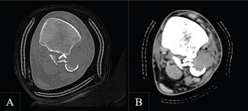
CT imaging revealed mortise asymmetry concerning for significant ligamentous injury and possible posterior fibular entrapment. Orthopedic surgery recommended operative management. A standard lateral approach exposed a defect where the fibula had dislocated posteriorly. There was significant comminution and complete transection of the anterior syndesmosis. A separate articular fragment was removed for later reconstruction. The posteriorly displaced fibular shaft was reduced using traction, and a mini-fragment plate was applied for provisional fixation (Fig. 4). The articular fragment was anatomically reduced and secured with a K-wire, followed by plate fixation of the fibular shaft with five screws. A lag screw was placed to stabilize the anterior articular fragment. The syndesmosis was reduced and stabilized with two lag screws. A sterile dressing was applied.
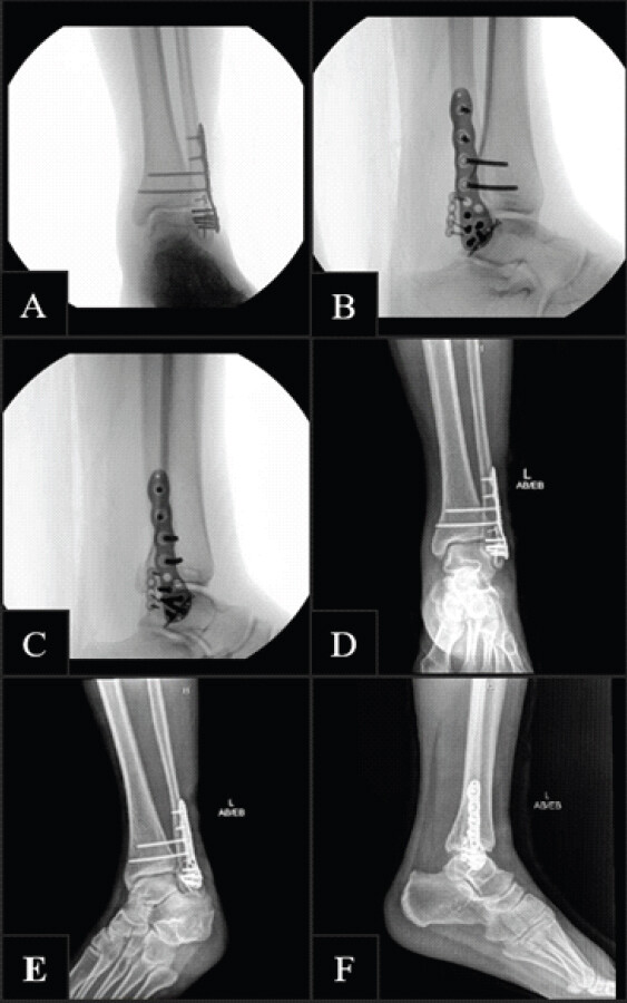
The patient received IV cefazolin for 24 h and began physical and occupational therapy. He was discharged the next morning in a controlled ankle motion boot with instructions for non-weight-bearing and aspirin 81 mg daily for 14 days for deep vein thrombosis prophylaxis. His post-operative course was followed for 16 weeks and was unremarkable. He declined elective removal of syndesmotic screws and later progressed to weight-bearing as tolerated.
Discussion
Bosworth fractures are rare, complex injuries characterized by posterior entrapment of the fibula behind the tibia, making them inherently irreducible by closed means. The literature consistently reports closed reduction failure rates exceeding 90% [5], underscoring the need for early operative intervention. These injuries are frequently misinterpreted on initial radiographs, and persistent malalignment after reduction attempts should prompt CT imaging, which is considered the diagnostic standard due to its ability to delineate the fracture pathoanatomy and associated ligamentous injuries [6].
Complications associated with Bosworth fractures include compartment syndrome, neurovascular injury, syndesmotic diastasis, and post-traumatic arthritis [7]. Early surgical management is critical, as delays and repeated closed reduction attempts are associated with poorer outcomes [8]. In this case, early recognition of persistent mortise asymmetry and prompt CT imaging facilitated timely operative intervention, leading to an uncomplicated recovery. This case aligns with the broader literature demonstrating that open reduction and internal fixation with syndesmotic stabilization is the standard of care and generally yields favorable outcomes when performed early [9].
Bosworth fractures are frequently missed on initial evaluation due to subtle or non-specific radiographic findings [10]. Standard ankle radiographs may demonstrate talar shift, loss of tibiofibular overlap, or a posterior malleolar fleck, but these signs are often overlooked in the acute setting. A key diagnostic clue is persistent mortise asymmetry or failure to achieve anatomic alignment after an apparently successful closed reduction attempt. In such cases, clinicians should maintain a high index of suspicion for posterior fibular entrapment. CT imaging plays a critical role in confirming the diagnosis, as it clearly delineates the posterior displacement of the fibula and associated osseous or ligamentous injuries, allowing for accurate preoperative planning [11].
Surgical management of Bosworth fractures presents unique technical challenges due to the mechanical incarceration of the fibula behind the tibia. Open reduction is mandatory, as repeated closed reduction attempts risk further soft-tissue injury and delay definitive care. A lateral or posterolateral approach typically provides adequate exposure to disengage the fibula and address associated comminution. Restoration of fibular length and rotation is essential for re-establishing ankle stability, particularly in the presence of syndesmotic disruption. Fixation strategies often include plate fixation of the fibula, lag screw stabilization of articular fragments, and syndesmotic screws to maintain reduction [12]. Attention to anatomic alignment and careful handling of soft tissues are critical to minimizing post-operative complications and optimizing functional outcomes.
Conclusion
Bosworth fracture-dislocations are rare and frequently irreducible injuries that require a high index of suspicion, particularly when closed reduction fails or mortise asymmetry persists. CT imaging is essential for identifying posterior fibular entrapment and associated syndesmotic disruption. Early operative intervention remains the definitive treatment to restore anatomic alignment and prevent long-term complications. This case reinforces the importance of timely recognition and surgical management in achieving favorable outcomes.
Clinical Message
Persistent malalignment after ankle reduction should prompt evaluation for a Bosworth fracture, as early CT imaging and timely operative management are critical for successful treatment.
Conflict of Interest:
Nil
Source of Support:
Nil
Consent:
The authors confirm that informed consent was obtained from the patient for publication of this article
How to Cite this Article
Wahhab J, Scofield SM, Turinske T. Posterior Fibular Entrapment in a Bosworth Fracture-Dislocation: A Case Report and Review of Diagnostic and Operative Considerations. Journal of Orthopaedic Case Reports 2026 October;16(10):247-251.
References
- Bosworth DM. Fracture-dislocation of the ankle with fixed displacement of the fibula behind the tibia. J Bone Joint Surg Am 1947;29:130-5. [Google Scholar] | [PubMed]
- Bartoníček J, Rammelt S, Kostlivý K. Bosworth fracture: A report of two atypical cases and literature review of 108 cases. Fuß Sprunggelenk 2017;15:126-37. [Google Scholar] | [PubMed]
- Lucenti L, Testa G, Nocera C, Culmone A, Dell'Agli E, Pavone V. Bosworth fractures of the ankle: A systematic literature review. J Pers Med 2023;13:713. [Google Scholar] | [PubMed]
- Fang C, Tang ZH, Yeoh CS, Tan GM. A case series of Bosworth fracture-dislocations and review of 129 reported cases. Malays Orthop J 2022;16:76-85. [Google Scholar] | [PubMed]
- Molina M, Segur JM, Alvarez M, Viladot R, Ramon R. Irreducible ankle fracture-dislocation (Bosworth fracture): Long-term results and literature review. Foot Ankle Int 2011;32:158-63. [Google Scholar] | [PubMed]
- Khan F, Borton D. CT scanning in the diagnosis of Bosworth fracture-dislocation of the ankle. Injury 2008;39:224-8. [Google Scholar] | [PubMed]
- Wright SE, Legg A, Davies MB. A contemporary approach to the management of a Bosworth injury. Injury 2012;43:252-3. [Google Scholar] | [PubMed]
- Won Y, Lee GS, Hwang JM, Park IY, Song JH, Kang C . Improved functional outcome after early reduction in Bosworth fracture-dislocation. Foot Ankle Surg 2019;25:798-803. [Google Scholar] | [PubMed]
- Bartoníček J, Frič V, Svatoš F, Luňáček L. Bosworth-type fibular entrapment injuries of the ankle: the Bosworth lesion. A report of 6 cases and literature review. J Orthop Trauma 2007;21:710-7. [Google Scholar] | [PubMed]
- Khan F, Borton D. A constant radiological sign in Bosworth's fracture: “The axilla sign”. Foot Ankle Int 2008;29:55-7. [Google Scholar] | [PubMed]
- Kostlivý K, Bartoníček J, Rammelt S. Posterior malleolus fractures in Bosworth fracture-dislocations. A combination not to be missed. Injury 2020;51:537-41. [Google Scholar] | [PubMed]
- Bartoníček J, Rammelt S, Tuček M. Bosworth ankle fracture-dislocation: Current concept review. EFORT Open Rev 2024;9:448-57. [Google Scholar] | [PubMed]
© 2026 Journal of Orthopaedic Case Reports - Published by Indian Orthopaedic Research Group
About the Authors
-

John Wahhab
-

Sebastian M Scofield
-

Thomas Turinske
Related Articles in Journal of Orthopaedic Case Reports
 October 1, 2026 Isolated Posterior Malleolus Fracture with Early Symptom Resolution: A Case Report
 October 1, 2026 Bilateral Cedell Fractures of the Talus Associated with Ankle Fractures in a Polytrauma Patient: A Rare Injury Managed with Damage Control Orthopedics and Staged Fixation
 October 1, 2026 Posterior Hip Dislocation with Ipsilateral Subtrochanteric Femur Fracture: A Rare Case Report
 October 1, 2026 Staged Bilateral Total Elbow Arthroplasty in Advanced Rheumatoid Elbow Disease: A Case Report