


Introduction
Tibial avulsion injuries of the posterior cruciate ligament (PCL) are intra-articular injuries in which the ligament remains intact but detaches with a bony fragment from its tibial insertion. Although tibial spine avulsion injuries are classically described in skeletally immature patients, adult injuries are increasingly recognized after high-energy trauma and may be associated with other meniscal or ligamentous lesions [1, 2].
PCL tibial avulsion fractures commonly result from a dashboard injury or forced hyperflexion. Inadequately treated displaced fractures may lead to chronic posterior instability, flexion deformity, non-union, and long-term functional impairment [3].
The Meyers and McKeever classification, modified by Zaricznyj, is commonly used in the source manuscript to describe displacement and guide treatment. Unstable Type III and Type IV fractures generally require fixation, while displaced Type II fractures may require surgery when closed reduction is inadequate [4].
Magnetic resonance imaging (MRI) is useful for identifying the avulsed fragment and associated soft-tissue injuries, while arthroscopy permits direct evaluation, anatomical reduction, and minimally invasive fixation [5, 6].
Cannulated screws, Herbert screws, and suture anchors can provide stable fixation but may cause implant irritation, fragment comminution, impingement, or the need for secondary removal. These limitations have increased interest in suture-based transosseous fixation [7].
Single-tunnel suture pull-through fixation is intended to compress the fragment, preserve the native PCL insertion, and avoid intra-articular hardware. The present prospective observational study descriptively evaluated fracture union, serial Lysholm scores, short-term complications, clinically assessed knee stability, and return to pre-injury activity after this technique; it was not designed to compare the technique with screw, suture-anchor, or double-tunnel fixation.
Materials and Methods
Study design and setting
A prospective, single-center observational study was conducted in the Department of Orthopedics, N.D. Desai Medical College, from May 2026 to July 2026. A consecutive sample of 24 eligible patients with displaced isolated PCL tibial avulsion fractures was included. No randomization, treatment allocation procedure, or control/comparison group was used; therefore, the study was designed to report feasibility and short-term outcomes rather than comparative efficacy.
Ethical approval was obtained from the Institutional Ethics Committee, Dr. N. D. Desai Faculty of Medical Science and Research Medical Centers, Nadiad (No. Dr NDDFMSR/IEC/2026/03/04; dated May 02, 2026). Written informed consent was obtained for participation and use of anonymized clinical/radiographic material.
Patient selection
Inclusion criteria
-
ge 18–60 years
-
nilateral, closed, displaced, isolated PCL tibial avulsion fracture
-
o associated ligamentous, meniscal, or chondral injury requiring additional treatment
-
reatment with the arthroscopic suture pull-through single-tunnel technique and availability of follow-up Lysholm scores.
Exclusion criteria
-
pen, pathological, or associated proximal tibial fracture
-
revious surgery on the affected knee
-
oncomitant intra-articular injury requiring an additional procedure
-
ncomplete clinical or radiological follow-up.
Forteen patients had Type II, eight had Type III, and two had Type IV injuries. The mean interval from injury to surgical fixation was approximately 1 week. Because of the small sample and the very small Type IV subgroup, fracture-type-specific inferential comparisons were not planned; the fracture distribution is therefore presented descriptively.
Surgical technique
All procedures were performed under spinal anesthesia with the patient supine. A thigh support and pneumatic tourniquet were used. Standard anterolateral, anteromedial, and posteromedial portals were established for arthroscopic access.
Pre-operative preparation
-
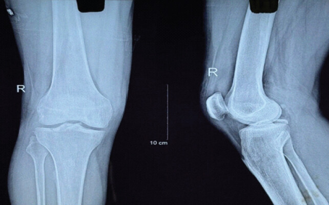
lain radiographs confirmed the PCL tibial avulsion fracture (Fig. 1).
-
RI was reviewed preoperatively to define the avulsion fracture and to exclude associated ligamentous, meniscal, or chondral injury requiring an additional procedure. Detailed lesion-specific MRI grading and standardized tabulation of minor associated findings were not part of the study dataset.
-
he joint was irrigated with normal saline to clear the hemarthrosis and loose fragments.
Fracture reduction and fixation
-
Dagnostic arthroscopy: A 30° 4-mm arthroscope was introduced to assess the PCL avulsion and the remaining intra-articular structures
-
Fagment preparation and reduction: The fracture bed was cleared and the fragment was mobilized and anatomically reduced with a probe; temporary K-wire stabilization was used when required
-
Sture passage: A 45° suture lasso was introduced through the posteromedial portal and passed through or around the PCL fibers close to the tibial insertion. A shuttle wire was used to pass a non-absorbable FiberTape loop
-
Sngle-tunnel creation: A PCL tibial guide set at approximately 55° was positioned at the center of the avulsed fragment/PCL tibial footprint. Through a small anteromedial tibial incision, one 4-mm transosseous tunnel was drilled from the anterior/anteromedial tibial cortex to the center of the fragment bed under arthroscopic and fluoroscopic guidance. No second tibial tunnel or crisscross transosseous construct was used
-
Fnal fixation: Both FiberTape limbs were shuttled through the same single tibial tunnel. With the fragment held in anatomical reduction, the limbs were tensioned and secured over the anterior tibial cortex, and reduction was confirmed arthroscopically and fluoroscopically. The operative records available for this study did not document a more precise external surface landmark or the specific cortical fixation device; this limits exact technical reproducibility and is acknowledged below as a study limitation.
-
Technique clarification: The procedure analyzed in this manuscript was a single-tunnel pull-through construct. Any prior double-tunnel/crisscross wording has been removed because it did not correspond to the technique evaluated in this cohort.
Post-operative management
-
The knee was supported in a PCL brace at approximately 30° of flexion for 3 weeks
-
Weeks 1–3: Isometric quadriceps exercises, straight-leg raises, and non-weight-bearing
-
Weeks 4–6: Hinged brace, gradual weight-bearing, and progressive range of motion
-
Full weight-bearing was permitted by 8 weeks according to clinical progress
-
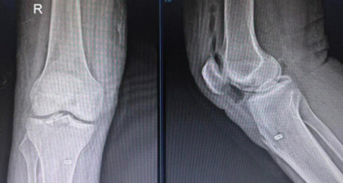
Follow-up radiographs at 3 months were used by the treating team to assess fracture union and overall alignment (Fig. 2). No prospectively defined quantitative threshold for residual displacement, articular congruity, or fragment step-off was recorded.
Outcome measures and statistical analysis
Lysholm scores were recorded preoperatively and at 6 weeks, 3 months, and 6 months and constituted the primary functional outcome measure. International Knee Documentation Committee (IKDC), Knee Injury and Osteoarthritis Outcome Score (KOOS), and Tegner Activity Scale scores were not collected. Radiological union, return to pre-injury activity, extension lag, clinically apparent residual instability, fixation failure, and revision surgery were also recorded. Return to pre-injury activity was based on patient report and treating-team clinical assessment rather than a prespecified validated activity scale. Posterior stability was assessed clinically; stress radiographs, KT-1000/KT-2000 measurements, or other instrumented posterior laxity testing were not performed. Radiological union was recorded according to the treating team’s follow-up radiographic assessment; no prospectively standardized quantitative union or residual-displacement criterion was documented. Continuous data are presented as mean ± standard deviation and categorical data as number and percentage. Serial Lysholm scores were compared with the Friedman test; a two-sided P < 0.05 was considered statistically significant. No fracture-type subgroup analysis was undertaken due to the small and uneven subgroup sizes.
Results
The study included 24 patients, comprising 16 males and eight females, with a mean age of 28 years (range 18–60 years). Ten injuries were sports related and 14 followed motorcycle accidents (Table 1).
Patient demographics and fracture characteristics
| Parameter | Value |
|---|---|
| Total patients | 24 |
| Male patients | 16 |
| Female patients | 8 |
| Mean age | 28 years (range 18–60) |
| Injury mechanism | Sports: 10; motorcycle accidents: 14 |
| Fracture type | Type II: 14; Type III: 8; Type IV: 2 |
Fracture distribution was Type II in 14 patients, Type III in eight patients, and Type IV in two patients (Table 1). Outcomes were not compared statistically by fracture type because the subgroup sizes, particularly Type IV (n = 2), were insufficient for meaningful inferential analysis.
The mean Lysholm score improved from 41.6 ± 3.2 preoperatively to 81.2 ± 5.6 at 6 weeks, 90.7 ± 3.5 at 3 months, and 94.9 ± 2.3 at 6 months. The overall change was statistically significant (Friedman P < 0.001) (Tables 2 and 3).
Functional and radiological outcomes
| Outcome | Pre-operative | 6 weeks | 3 months | 6 months | P-value |
|---|---|---|---|---|---|
| Lysholm score | 41.6±3.2 | 81.2±5.6 | 90.7±3.5 | 94.9±2.3 | <0.001 |
| Fracture union (radiographic) | — | — | 24/24 (100%) | 24/24 (100%) | — |
| Return to pre-injury activity (patient-reported) | — | — | — | 22/24 (~92%) within 4–6 months; no validated activity scale | — |
Patient-level Lysholm scores over follow-up
| Patient No. | Pre-operative | 6 weeks | 3 months | 6 months |
|---|---|---|---|---|
| 1 | 40 | 83 | 91 | 94 |
| 2 | 45 | 82 | 92 | 96 |
| 3 | 42 | 83 | 96 | 97 |
| 4 | 45 | 82 | 85 | 91 |
| 5 | 41 | 74 | 87 | 94 |
| 6 | 43 | 83 | 89 | 94 |
| 7 | 30 | 72 | 85 | 92 |
| 8 | 42 | 83 | 89 | 94 |
| 9 | 42 | 82 | 95 | 96 |
| 10 | 44 | 74 | 90 | 95 |
| 11 | 40 | 83 | 88 | 94 |
| 12 | 44 | 92 | 95 | 98 |
| 13 | 46 | 83 | 88 | 94 |
| 14 | 42 | 84 | 89 | 94 |
| 15 | 43 | 83 | 96 | 98 |
| 16 | 40 | 72 | 85 | 90 |
| 17 | 42 | 83 | 92 | 96 |
| 18 | 39 | 82 | 94 | 98 |
| 19 | 42 | 94 | 92 | 97 |
| 20 | 38 | 73 | 88 | 91 |
| 21 | 44 | 82 | 92 | 96 |
| 22 | 40 | 83 | 93 | 95 |
| 23 | 41 | 74 | 90 | 96 |
| 24 | 43 | 83 | 96 | 98 |
| Mean±SD | 41.58±3.17 | 81.21±5.58 | 90.71±3.53 | 94.92±2.30 |
SD: Standard deviation
All 24 fractures were recorded as united by 3 months on follow-up radiographs. Quantitative residual displacement, articular congruity, and fragment step-off were not measured. Twenty-two patients (approximately 92%) reported return to their pre-injury activity level within 4–6 months; this endpoint was not quantified with a validated activity scale (Table 2).
One patient developed a mild post-operative extension lag that improved with intensive physiotherapy. No non-union, fixation failure, or revision surgery was observed during the available 6-month follow-up. No clinically significant residual posterior instability was documented on routine clinical assessment; however, instrumented or stress-radiographic posterior translation was not measured, so subclinical residual laxity cannot be excluded.
Discussion
A single-tunnel arthroscopic suture technique may be useful when an avulsed PCL fragment is small or comminuted and screw or pin fixation is technically less suitable [8]. The approach permits transosseous suture retrieval and avoids intra-articular hardware. In the present study, however, there was no screw-fixation, suture-anchor, or double-tunnel comparator; accordingly, the findings support feasibility and short-term outcome reporting only and cannot establish superiority, equivalence, or a lower complication rate relative to other fixation methods.
Arthroscopic reduction and internal fixation is recommended for displaced Type III fractures and should be considered for irreducible or unstable Type II injuries [9]. In this cohort, serial Lysholm scores improved significantly and all fractures were recorded as united by 3 months. Most patients also reported return to their pre-injury activity level. These findings are broadly consistent with reports of favorable short-term recovery after suture fixation [10, 11], but direct comparison is limited by differences in fracture patterns, outcome definitions, follow-up duration, and fixation constructs.
Shankar et al. reported a mean post-operative Lysholm score of 94.8 after arthroscopic pull-through suture fixation, while Lamoria et al. reported a mean score of 96.3 [12, 13]. The final mean Lysholm score of 94.9 in this cohort is numerically comparable to those reports; nevertheless, the Lysholm score was the only validated patient-reported outcome used here, and the absence of IKDC, KOOS, Tegner scores, and objective laxity measurements limits a comprehensive assessment of knee function and activity restoration.
One patient developed a transient extension lag that responded to physiotherapy. Mild range-of-motion deficits have been described after PCL surgery [14]. Avoiding prolonged immobilization and following a structured rehabilitation program are important to minimize stiffness. Restoration of native knee biomechanics may also reduce residual instability [15].
Residual laxity, non-union, re-injury, arthrofibrosis, tunnel-related problems, and post-traumatic osteoarthritis remain important concerns after PCL avulsion fixation. The present 6-month follow-up is adequate only for the early union and short-term functional recovery and is not sufficient to determine the frequency of delayed instability, reinjury, or degenerative change [16].
The favorable early findings should be interpreted in the context of several important methodological limitations, detailed below.
Limitations
-
Small sample size: Only 24 patients were included, which limits statistical power, precision of complication estimates, and generalizability to the broader population of patients with PCL tibial avulsion fractures
-
sngle-center setting: All patients were treated at one institution; local surgical expertise, patient characteristics, rehabilitation protocols, and perioperative practices may differ from those at other centers
-
No control or comparison group: The study did not compare the single-tunnel technique with screw fixation, suture-anchor fixation, or double-tunnel fixation; therefore, superiority, equivalence, or comparative safety cannot be established
-
Observational design: Although data collection was prospective, treatment was not randomized and allocation was not controlled, leaving the study susceptible to selection bias and limiting causal conclusions regarding efficacy
-
Short follow-up: Outcomes were reported only through 6 months. This duration is insufficient to evaluate long-term posterior stability, recurrent injury, late tunnel-related problems, post-traumatic osteoarthritis, or durability of return to activity
-
Lack of objective posterior stability testing: Posterior tibial translation was not quantified with stress radiographs, KT-1000/KT-2000 devices, or another instrumented method. Consequently, clinically silent residual laxity cannot be excluded
-
Restricted functional outcome assessment: The Lysholm Knee Score was the sole validated patient-reported outcome measure. IKDC, KOOS, and Tegner Activity Scale data were not available, limiting multidimensional assessment of symptoms, function, and activity level
-
Return-to-activity endpoint: Return to the pre-injury activity level was based on patient report and clinical assessment rather than prospectively defined criteria or a validated activity scale; the reported 22/24 return rate should therefore be interpreted cautiously
-
Heterogeneous fracture patterns: The cohort included Type II, III, and IV fractures. The small and uneven subgroup sizes precluded meaningful fracture-type-specific analysis of reduction, union, functional outcome, or complications
-
Associated injuries: MRI was used to exclude concomitant lesions requiring additional treatment, but detailed standardized MRI grading of meniscal, chondral, and other ligamentous findings was not collected, limiting characterization of potentially minor associated pathology
-
Limited radiological characterization: Union was recorded on routine follow-up radiographs, but prospectively standardized criteria for union, residual fragment displacement, articular congruity, or step-off were not specified or quantitatively measured
-
Technical reproducibility: The analyzed procedure used one transosseous tibial tunnel and not a double-tunnel/crisscross construct. Nevertheless, the available operative records did not document a more precise external tibial entry landmark or the exact cortical fixation device, which should be standardized and reported in future technical studies.
-
Surgical learning curve: The number of operating surgeons, individual arthroscopic experience, and potential change in outcomes with increasing experience were not analyzed; the influence of surgeon expertise on reproducibility is therefore unknown.
-
Complication ascertainment: Only one mild extension lag was observed and no fixation failure or revision surgery occurred during the available follow-up, but the small cohort and short observation period may fail to detect uncommon or delayed complications such as recurrent instability, arthrofibrosis, tunnel-related problems, or degenerative changes.
-
Scope of inference: The study demonstrates feasibility, radiographic union, and favorable short-term Lysholm score improvement in this cohort. It does not provide evidence that the single-tunnel technique is clinically superior to established alternatives. Larger multicenter comparative studies with objective stability testing, standardized imaging and activity outcomes, fracture-type subgroup analyses, and longer follow-up are required.
Conclusion
Within the limitations of this small, single-center prospective observational study, arthroscopic PCL tibial avulsion fixation using a single-tunnel suture pull-through technique was feasible, all 24 fractures were recorded as united by 3 months, and Lysholm scores improved through 6 months. These results represent short-term descriptive outcomes and do not establish superiority or equivalence to screw, suture-anchor, or double-tunnel fixation. Multicenter comparative studies with larger samples, objective posterior stability measurements, standardized radiological and activity criteria, and longer follow-up are needed before definitive comparative conclusions can be made.
Clinical Message
For displaced PCL tibial avulsion fractures, single-tunnel suture pull-through fixation is a hardware-sparing arthroscopic option that showed favorable short-term union and Lysholm score improvement in this 24-patient cohort. The technique should be considered a feasible option rather than a proven superior method until comparative studies with objective stability assessment and longer follow-up are available.
Conflict of Interest:
Nil
Source of Support:
Nil
Consent:
The authors confirm that informed consent was obtained from the patient for publication of this article
How to Cite this Article
Patel PJ, Rajpardhi HA, Modi LP, Goti KH, Gandhi NN, Visani HP. Arthroscopic Posterior Cruciate Ligament Tibial Spine Avulsion Fixation Using a Suture Pull-Through Single-Tunnel Technique. Journal of Orthopaedic Case Reports 2026 October;16(10): 408-414.
References
- Benfaris DM, Aldosari ZA, AlNahari AS, Awwad MW, Alhuqbani MN, Alomar AZ. Tibial spine avulsion injuries in children and adolescents: A narrative review of anatomy, management strategies, and return-to-sport considerations. Healthcare (Basel) 2026;14:1967. [Google Scholar] | [PubMed]
- Cengiz B, Karaoglu S. Case report of concomitant avulsion fractures of the medial meniscus and posterior cruciate ligament. Medicine (Baltimore) 2021;100:e28273. [Google Scholar] | [PubMed]
- Sharma A. Outcomes of arthroscopic fixation methods for tibial eminence fractures: A comparative series. J Clin Orthop Trauma 2026;76:103410. [Google Scholar] | [PubMed]
- Salvato D. Proposing the term “bipolar anterior cruciate ligament injury”. J ISAKOS 2023;8:404-11. [Google Scholar] | [PubMed]
- Haichour I, Boubcheur M, Benalia K, Khatab Z, Abdeljaouad N, Yacoubi H. Imaging and management of posterior cruciate ligament avulsion fracture pseudoarthrosis: A case report. Radiol Case Rep 2026;21:278-85. [Google Scholar] | [PubMed]
- Dung NQ. Clinical evaluation of arthroscopic pull-out suture fixation for tibial spine avulsion fractures. Orthop Rev (Pavia) 2026;18:159605. [Google Scholar] | [PubMed]
- Singhi PK. Modern updates on arthroscopic reconstruction techniques for knee sports injuries. J Arthrosc Surg Sports Med 2026;7:S67-74. [Google Scholar] | [PubMed]
- Dhariwal HV. Management of chronic displaced posterior cruciate ligament avulsion fractures via an open posteromedial approach. J Orthop Case Rep 2024;14:170-5. [Google Scholar] | [PubMed]
- Mousa H, Williams D, Aresti N. Arthroscopic reduction internal fixation for displaced radial head fractures: A systematic review of the outcomes and complications. JSES Rev Rep Tech 2024;4:710-4. [Google Scholar] | [PubMed]
- Pu JS, Zheng L, Jian CC. Clinical efficacy of suture bridge versus hollow screw fixation for PCL tibial avulsion fractures: A comparative study. BMC Surg 2025;25:180. [Google Scholar] | [PubMed]
- Thamyongkit S, Komonsuwan P, Jitaree B, Aroonjarattham P, Rattanapan N, Somtua C. Biomechanical comparison of suture and screw fixation in PCL avulsion with variable bone quality. Sci Rep 2025;15:36243. [Google Scholar] | [PubMed]
- Shankar R. Complication rates and functional outcomes in arthroscopic fracture fixations. Cureus 2025;17:e79902. [Google Scholar] | [PubMed]
- Lamoria R. Functional outcomes of arthroscopic suture fixation versus screw fixation for tibial spine fractures. J Clin Orthop Trauma 2020;11:S362-7. [Google Scholar] | [PubMed]
- Krishna ML, Mittal R, Chauhan N. Posterior cruciate ligament avulsion with posterior tibial rim fracture: A case report. J Orthop Case Rep 2023;13:75-9. [Google Scholar] | [PubMed]
- Patel SK. Functional outcome analysis in arthroscopic fixation of PCL avulsion fractures. Int J Res Orthop 2026;12:641-5. [Google Scholar] | [PubMed]
- Gopinatth V. Midterm to long-term clinical and radiological outcomes after tibial eminence fixation. Orthop J Sports Med 2023;11. 23259671231188383[1. [Google Scholar] | [PubMed]
© 2026 Journal of Orthopaedic Case Reports - Published by Indian Orthopaedic Research Group
About the Authors
-

Parth Jayeshkumar Patel
-

Harshlaxen Ashvinsinh Rajpardhi
-

Lomesh Pravinbhai Modi
-

Krishna Himmatbhai Goti
-

Naushil Nileshkumar Gandhi
-

Hayati Bharatbhai Visani
Related Articles in Journal of Orthopaedic Case Reports
 September 1, 2026 Arthroscopic ACL Tibial Spine Avulsion Fixation Using Suture Pull-Through Double-Tunnel Technique
 October 1, 2026 All Arthroscopic Decompression and Bone Grafting of Cyst in the Talus
 October 1, 2026 Arthroscopic Piecemeal Excision of a Large Intra-articular Infrapatellar Osteochondroma of the Knee: A Case Report
 September 1, 2026 Fixed Flexion Deformity due to Displaced Hoffa Fracture: A Case Report