


Introduction
Intra-articular osteochondromas are rare benign lesions of the knee, distinct from the more common extra-articular osteochondromas that typically arise from the metaphysis and grow away from the joint. The reported prevalence of intra-articular osteochondromas is approximately 1 in 1 million [1]. When located within the infrapatellar fat pad (Hoffa’s fat pad), these lesions present a unique diagnostic and surgical challenge due to their proximity to the patellar tendon and their potential to cause significant mechanical symptoms.
We report a case of a large intra-articular infrapatellar osteochondroma in a middle-aged female, which caused considerable discomfort during ambulation and a significant block to the range of motion (ROM) of the knee. The lesion was removed using a minimally invasive arthroscopic and mini-open technique, which is described in detail. We also review the literature to identify similar cases, their management, and the possible etiologies of such lesions. The differential diagnoses include Trevor’s disease (dysplasia epiphysealis hemimelica), synovial osteochondromatosis, extraskeletal para-articular osteochondroma, hereditary multiple exostoses (HME), and chondrosarcoma (Table 1).
Differential diagnosis of intra-articular and para-articular ossified masses of the knee
| Differential diagnosis | Key clinical features | Distinguishing characteristics |
|---|---|---|
| Intra-articular osteochondroma | Solitary, ossified mass within joint space; mechanical symptoms | Arises within joint capsule; covered by synovial tissue; benign cartilage cap |
| Trevor’s disease (Dysplasia epiphysealis hemimelica) | Developmental disorder; usually presents in childhood/young adults; male predominance | Asymmetrical cartilage overgrowth of the epiphysis; often involves multiple joints |
| Extraskeletal para-articular osteochondroma | Mass adjacent to joint; mechanical symptoms | Develops outside joint capsule; lacks synovial covering; no bony continuity with skeleton |
| Synovial osteochondromatosis | Multiple loose bodies; joint swelling and stiffness | Multiple cartilaginous nodules within synovium; metaplastic origin |
| Chondrosarcoma | Painful, enlarging mass; older age group | Aggressive features on imaging (e.g., cortical destruction); cellular atypia on histopathology |
Case Report
A 46-year-old Filipino female presented with a 6-month history of progressive pain and swelling in the left knee. The pain was of gradual onset, dull in character, and of moderate intensity, occasionally radiating to the left lower limb. Symptoms were aggravated by activity, knee movement, and weight-bearing, and were partially relieved by rest and analgesic medication. The patient also reported swelling, stiffness, intermittent locking, and occasional giving-way episodes, resulting in significant functional limitation including difficulty walking, climbing stairs, and performing daily activities. There was no history of trauma or previous knee surgery. No relevant past medical history or comorbidities were noted.
On physical examination, there was visible fullness over the anterior aspect of the left knee, particularly in the infrapatellar region. The swelling was firm to hard in consistency, fixed to the underlying structures, and non-fluctuant. The overlying skin was normal with no signs of inflammation. Knee ROM was restricted, with an extension lag of 20° and flexion limited to 110°. A painful click was elicited during terminal extension. Joint line tenderness was absent, and ligamentous stability tests were unremarkable. Meniscal tests were negative.
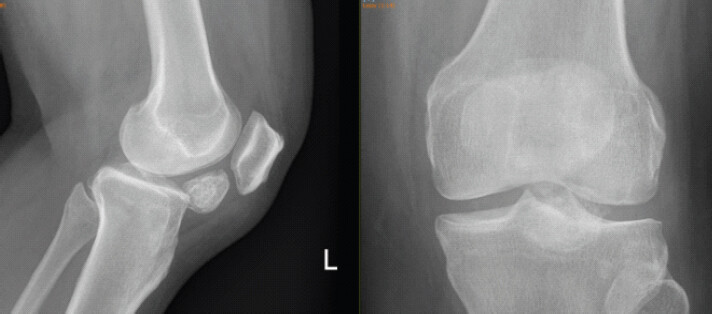
Plain radiographs (Fig. 1) of the left knee demonstrated a well-defined ossified mass in the infrapatellar region within the joint space, appearing similar in size to the patella.
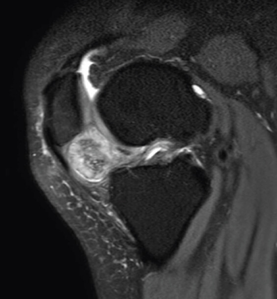
A preoperative non-contrast magnetic resonance imaging (MRI) (Figs. 2 and 3) of the left knee identified a 2.6 × 2.7 × 2.2 cm corticated, T1- and T2-hypointense mass in the infrapatellar fat pad, featuring a thin T2-hyperintense and T1-isointense rim consistent with a cartilaginous surface. Associated smaller foci, edematous changes, and synovial thickening suggested juxta-articular chondromas, with secondary findings including minimal joint effusion and Grade 1 anterior cruciate ligament signal alteration. Multiple smaller osteocartilaginous loose bodies were also identified within the joint cavity. No meniscal tear was observed. The presence of multiple loose bodies argues against a diagnosis of extraskeletal para-articular osteochondroma.
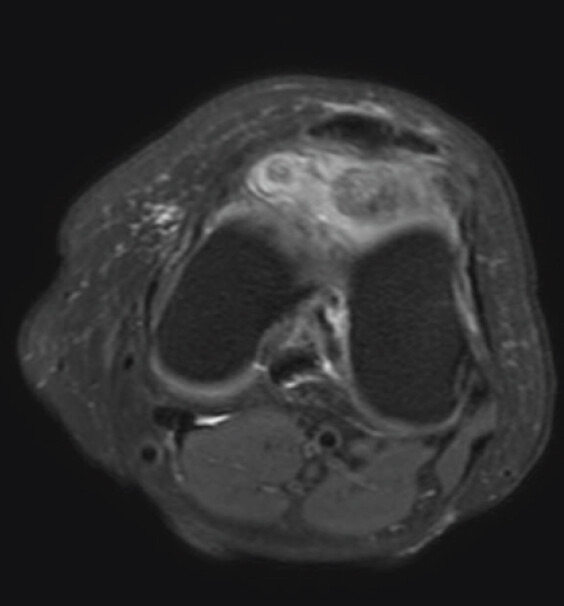
Based on imaging findings, the provisional diagnosis was osteocartilaginous loose bodies with synovial hypertrophy, suggestive of a juxta-articular chondroma. The differential diagnoses considered at this stage included extraskeletal para-articular osteochondroma, synovial osteochondromatosis, Trevor’s disease, and chondrosarcoma. The absence of multiple lesions in the synovium, the patient’s age, and the absence of skeletal dysplasia made synovial osteochondromatosis and Trevor’s disease less likely. The well-defined corticated margins and absence of aggressive features on MRI argued against chondrosarcoma. Intraoperative findings and histopathological examination subsequently confirmed the diagnosis of a large intra-articular infrapatellar osteochondroma.
Surgical technique
The patient underwent arthroscopic excision of the infrapatellar intracapsular osteochondroma under spinal anesthesia. She was positioned supine on the operating table with a pneumatic tourniquet applied to the proximal thigh. The tourniquet was inflated to 250 mmHg. The operative limb was prepared and draped in the usual sterile manner.
Due to the mass effect produced by the infrapatellar lesion, arthroscopic portals were established 5–10 mm more lateral and medial, respectively, relative to the standard anterolateral and anteromedial portals, based on palpation of the soft spot. This modification was necessary to improve visualization and instrument maneuverability within the anterior compartment. A diagnostic arthroscopy was initially performed to evaluate the intra-articular structures.
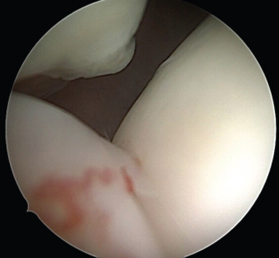
An intracapsular osteochondroma arising from the region deep to the infrapatellar fat pad was identified. The lesion was surrounded by synovial tissue and partially obscured by the hypertrophic fat pad. Careful synovial debridement was performed using an arthroscopic shaver and radiofrequency probe to expose the margins of the tumor (Fig. 4). Throughout the procedure, clear identification and protection of the patellar tendon were maintained, as meticulous delineation of this structure was critical to avoid inadvertent injury during resection.
Once the lesion was adequately exposed and demarcated, the osteochondroma was carefully freed from the surrounding synovium. Arthroscopic shavers and burrs were used to progressively thin the lesion in a controlled piecemeal fashion, reducing its bulk and facilitating safe removal (Fig. 5). The ossified core was particularly dense and required an arthroscopic burr for debulking. After sufficient reduction in size, the residual tumor fragments were retrieved through a small medial parapatellar mini-incision, allowing complete excision of the mass.
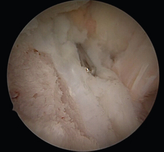
The joint was subsequently inspected arthroscopically to confirm complete removal of the lesion and to ensure that the patellar tendon and surrounding structures remained intact. The joint was thoroughly irrigated to remove debris, hemostasis was achieved, and the arthroscopic portals were closed in standard fashion. Total tourniquet time was 120 min. There were no intraoperative complications.
Surgical tips and pearls
-
Delineate and define the patellar tendon with electrocautery at low power while excising the anterior part of the mass
-
Place portals at the palpable soft spots slightly medial and lateral compared to the standard portals, to accommodate the mass-occupying lesion obstructing standard portal placement
-
Perform a complete arthroscopic knee examination to rule out loose bodies hidden in the gutters and posterior compartment.
Postoperatively, the patient was allowed full weight-bearing and early mobilization. At 6–8 weeks, the extension lag had improved from 20° to 10°, and knee flexion had improved from 110° to approximately 150°. Visual analogue scale score on flexion and attempted terminal extension was 7/10 preoperatively, reducing to 1/10 on flexion and 2/10 on extension at 5-month follow-up.
Pathology
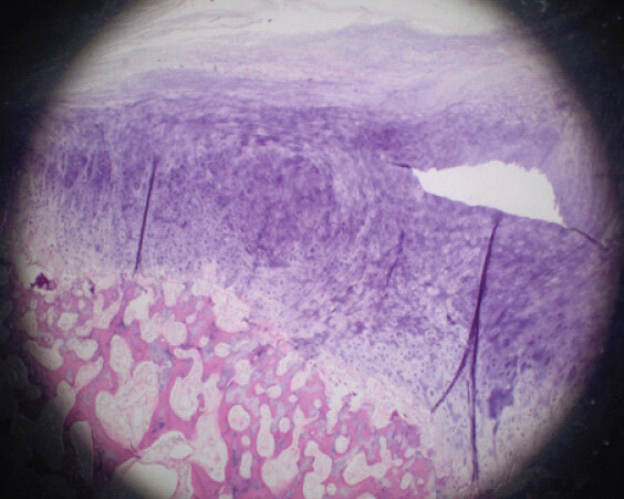
Histopathological examination of the excised specimen revealed sections from the left knee intracapsular mass lesion comprising a hyaline cartilaginous cap of maximum thickness 5 mm, covered by fibro-collagenous tissue with myxoid change. The underlying bone trabeculae were prominent and variably widened, with intervening fatty marrow displaying areas of fat necrosis and calcification. Some tissue fragments appeared infarcted. No features of malignancy were identified (Fig. 6). These findings were consistent with a diagnosis of osteochondroma.
Discussion
Osteochondromas are the most common benign bone tumors, typically arising from the metaphysis of long bones and growing away from the adjacent joint [2]. Intra-articular osteochondromas are considerably rarer, with a reported prevalence of approximately 1 in 1 million [1]. The infrapatellar fat pad (Hoffa’s fat pad) is an exceptionally uncommon location for such lesions, and cases of this size have rarely been described in the literature.
The present case is notable for the size of the lesion (28 × 20 mm), which approximated the dimensions of the native patella, and for the successful application of a minimally invasive arthroscopic piecemeal excision technique. A comprehensive search of the PubMed, MEDLINE, and Google Scholar databases (1990–2024) using the keywords “intra-articular,” “osteochondroma,” “infrapatellar,” and “arthroscopy,” restricted to English-language, full-text publications, revealed a paucity of similar cases. As demonstrated in Table 2, the largest previously reported intra-articular osteochondroma managed arthroscopically measured 2 × 2 cm (Kulkarni et al., 2024 [3]), making the present case, to the best of our knowledge, one of the largest such lesions in the infrapatellar fat pad to be excised arthroscopically in the published literature.
Published cases of intra-articular and para-articular osteochondromas of the knee
| Author (Year) [References] | Age/Sex | Location | Lesion size | Treatment | Outcome and follow-up |
|---|---|---|---|---|---|
| Kesgin et al. (2013) [16] | 27/M | Adjacent to femoral trochlea | Not reported | Arthroscopic resection | Complete relief; return to full activity within 2 weeks; no recurrence at 3 years and 1 year, respectively |
| 24/F | |||||
| Morey et al. (2014) [14] | 16/M | Intercondylar notch of femur | Not reported | Arthroscopic excision (en bloc) | Painless; full ROM (0–140°) at 1-year follow-up |
| Mulcahy and Hoch (2015) [7] | 25/F | Hoffa’s fat pad (extraskeletal para-articular) | 3×5 cm | Open excision | Not reported in abstract |
| Maheshwari et al. (2006) [9] | 35/M | Infrapatellar fat pad (extraskeletal para-articular) | 3.5×2.5 cm | Open excision | Full ROM at 18 months |
| Nishimura et al. (2020) [8] | 35/M | Infrapatellar fat pad (para-articular) | Open simple marginal resection | Pain resolved; full ROM restored; no recurrence at mean 51 months | |
| 38/M | Not reported | ||||
| 55/M | |||||
| Chouliaras et al. (2021) [15] | 32/F | Superolateral aspect of distal femur (intra-articular) | Not reported | Arthroscopic resection (osteotome+burr) | Complete relief; return to full activity at 1 month; no recurrence at 7-year follow-up |
| Kulkarni et al. (2024) [3] | 55/F | Intercondylar notch (intra-articular loose body) | 2×2 cm | Arthroscopic removal | Complete relief; resumed routine activities within 1 month |
| Present case | 46/F | Infrapatellar fat pad (intra-articular) | 28×20 mm (comparable to native patella) | Arthroscopic piecemeal excision+mini-open retrieval | Significant improvement in ROM and pain (VAS 7/10→1–2/10) at 5-month follow-up |
Numbers in brackets correspond to the reference list. ROM: Range of motion, VAS: Visual analogue scale, M: Male, F: Female
The present case represents, to the best of our knowledge, one of the largest intra-articular infrapatellar osteochondromas managed arthroscopically reported in the literature to date.
Several differential diagnoses were considered in this case.
Trevor’s disease
Trevor’s disease (dysplasia epiphysealis hemimelica) is a developmental disorder of the epiphysis that may present as an intra-articular osteochondroma [4]. It is twice as common in males [5] and most cases present in childhood. Trevor’s disease should be considered as a differential diagnosis when a young adult or child presents with an intra-articular mass [6]. Given the patient’s age of 46 years and the absence of skeletal dysplasia, Trevor’s disease was considered unlikely in the present case.
Extraskeletal para-articular osteochondroma
A close differential diagnosis of intra-articular osteochondroma is extraskeletal para-articular osteochondroma, due to the absence of bony continuity with the underlying skeleton [7,8,9]. The presence or absence of synovial tissue surrounding the lesion is a key differentiating feature. True intra-articular osteochondromas, like the present case, arise within the joint capsule and are covered by synovium, whereas para-articular lesions develop outside the joint capsule. Open excision has been described as the treatment of choice for para-articular lesions [7,8]; however, the present case demonstrates that arthroscopic excision is feasible and may offer advantages in terms of reduced morbidity.
Synovial osteochondromatosis
Synovial osteochondromatosis may present as a solitary giant lesion in the intercondylar notch [10]. Although generally benign, malignant transformation has been reported in approximately 5% of cases [11]. The absence of multiple synovial nodules and the histopathological findings in the present case effectively excluded this diagnosis.
HME
HME is an autosomal dominant disorder that primarily arises from the metaphysis of long bones but may also affect the epiphysis [12,13]. The absence of multiple exostoses in the present case ruled out this diagnosis.
Chondrosarcoma
The risk of malignant transformation in solitary osteochondromas is estimated to be <1%. The absence of aggressive features on MRI and the benign histopathological findings confirmed the diagnosis of osteochondroma in the present case.
The surgical management of intra-articular osteochondromas traditionally involved open excision, particularly for large lesions in the infrapatellar region [7,8]. However, arthroscopic excision has become increasingly favored due to its advantages of lower morbidity, faster recovery, and improved cosmetic outcomes [3,14,15,16]. Open excision, while providing excellent exposure, requires a larger arthrotomy, which can lead to increased post-operative pain, a higher risk of arthrofibrosis, prolonged rehabilitation, and a less cosmetically appealing scar. Conversely, the arthroscopic approach minimizes soft-tissue disruption, preserves the extensor mechanism, and allows for a thorough evaluation of the entire joint to identify and remove any concomitant loose bodies or associated intra-articular pathology. While arthroscopic removal of smaller loose bodies is well-established, the piecemeal excision of a mass measuring 28 × 20 mm from the infrapatellar fat pad represents a significant technical challenge. In this case, the piecemeal approach using arthroscopic shavers and burrs allowed for controlled debulking of the tumor, avoiding the need for a large arthrotomy. The mini-open parapatellar incision was solely for the retrieval of the reduced fragments, maintaining the minimally invasive nature of the procedure. The successful outcome, with significant improvement in ROM and pain scores at 5 months, highlights the efficacy of this approach.
The histopathological findings in this case are of paramount importance. The identification of a benign hyaline cartilaginous cap without features of cellular atypia or malignant transformation definitively confirms the diagnosis of a benign osteochondroma and rules out more sinister pathologies such as chondrosarcoma. Although the risk of malignant transformation in solitary osteochondromas is exceedingly low (<1%), and recurrence following complete excision is rare, we recommend a long-term clinical and radiological follow-up for these patients. Routine surveillance, ideally extending beyond 2–5 years, is advisable to monitor for any signs of recurrence or secondary degenerative joint changes, particularly in cases involving large, mass-occupying lesions. A limitation of this report is the relatively short follow-up period of 5 months; longer-term surveillance will be necessary to fully assess the durability of the outcome and to detect any late recurrence or degenerative joint changes.
Conclusion
Large intra-articular osteochondromas arising from the infrapatellar fat pad are exceptionally rare lesions that may cause significant mechanical symptoms and functional impairment. This case demonstrates that even a lesion approximating the size of the native patella can be safely and effectively managed using minimally invasive arthroscopic piecemeal excision, thereby avoiding the morbidity associated with open surgery. Histopathological examination remains essential to confirm the diagnosis and exclude malignancy, while early mobilization following arthroscopic excision facilitates rapid functional recovery. Although the exact etiology in this case remains uncertain, the absence of synovial tissue favors a diagnosis of extraskeletal para-articular osteochondroma. However, the presence of multiple smaller, morphologically similar loose bodies raises the possibility of synovial chondromatosis.
Clinical Message
Orthopedic surgeons should consider intra-articular osteochondroma in the differential diagnosis of a solitary, ossified mass in the infrapatellar region presenting with mechanical knee symptoms. Arthroscopic piecemeal excision, combined with a mini-open retrieval, is a safe, effective, and minimally invasive treatment option for large lesions, offering significant advantages over traditional open excision. The presence or absence of synovial tissue in the histopathology assists in determining the etiology.
Conflict of Interest:
Nil
Source of Support:
Nil
Consent
The authors confirm that informed consent was obtained from the patient for publication of this article
How to Cite this Article
Jetaji N, Sami S, Babu R, Rajoli SR. Arthroscopic Piecemeal Excision of a Large Intra-articular Infrapatellar Osteochondroma of the Knee: Journal of Orthopaedic Case Reports 2026 October;16(10): 113-119.
References
- Araujo CRJrMontandon S, Montandon C, Teixeira KI, Moraes FB, Moreira MA. Best cases from the AFIP: Dysplasia epiphysealis hemimelica of the patella. Radiographics 2006;26:581-6. [Google Scholar] | [PubMed]
- Murphey MD, Choi JJ, Kransdorf MJ, Flemming DJ, Gannon FH. Imaging of osteochondroma: Variants and complications with radiologic-pathologic correlation. Radiographics 2000;20:1407-34. [Google Scholar] | [PubMed]
- Kulkarni S, Patil VS, Naik S, Bhansali Y. Arthroscopic excision of a solitary intra-articular osteochondroma manifesting as a loose body: A presentation of an extremely rare medical condition. Cureus 2024;16:e66083. [Google Scholar] | [PubMed]
- Gökkuş K, Atmaca H, Sagtas E, Saylik M, Aydin AT. Trevor's disease: Up-to-date review of the literature with case series. J Pediatr Orthop B 2017;26:532-45. [Google Scholar] | [PubMed]
- Rosero VM, Kiss S, Terebessy T, Köllö K, Szöke G. Dysplasia epiphysealis hemimelica (Trevor's disease): 7 of our own cases and a review of the literature. Acta Orthop 2007;78:856-61. [Google Scholar] | [PubMed]
- Maheshwari AV, Pitcher JD. Intraarticular osteochondroma of the knee. Indian J Orthop 2015;49:260-1. [Google Scholar] | [PubMed]
- Mulcahy H, Hoch B. An extraskeletal para-articular osteochondroma of Hoffa's fat pad. Radiol Case Rep 2015;5:420. [Google Scholar] | [PubMed]
- Nishimura T, Saku I, Kanda S, Fukushima T, Akiyama T. Para-articular osteochondroma of the infrapatellar fat pad: A report of three cases. Int J Surg Case Rep 2020;69:105-8. [Google Scholar] | [PubMed]
- Maheshwari AV, Jain AK, Dhammi IK. Extraskeletal paraarticular osteochondroma of the knee--a case report and tumor overview. Knee 2006;13:411-4. [Google Scholar] | [PubMed]
- Fornaciari P, Schai PA, Niehaus R, Exner UG. Intra-articular giant synovial osteochondroma: Case reports of the ankle and knee joint. Case Rep Orthop 2015;2015:320139. [Google Scholar] | [PubMed]
- Davis RI, Hamilton A, Biggart JD. Primary synovial chondromatosis: A clinicopathologic review and assessment of malignant potential. Hum Pathol 1998;29:683-8. [Google Scholar] | [PubMed]
- Stieber JR, Dormans JP. Manifestations of hereditary multiple exostoses. J Am Acad Orthop Surg 2005;13:110-20. [Google Scholar] | [PubMed]
- Choudhry A, Gimm G, Kang HW, Shin CH, Yoo WJ, Choi IH. Intra-articular acetabular osteochondroma in patients with multiple hereditary exostoses. J Pediatr Orthop B 2022;31:e90-4. [Google Scholar] | [PubMed]
- Morey VM, Jalan D, Mittal R, Gamangatti S. Intraarticular osteochondroma of the knee. Indian J Orthop 2014;48:332-4. [Google Scholar] | [PubMed]
- Chouliaras V, Sotiriadis I, Flevas DA. Arthroscopic excision of an intra-articular osteochondroma of the knee. Cureus 2021;13:e17154. [Google Scholar] | [PubMed]
- Kesgin E, Çelik C, Karaoğlu S. Arthroscopic resection of osteochondroma of the knee: Two case reports. Eklem Hastalik Cerrahisi 2013;24:112-6. [Google Scholar] | [PubMed]
© 2026 Journal of Orthopaedic Case Reports - Published by Indian Orthopaedic Research Group
About the Authors
-

Nazmuddin Jetaji
-

Sara Sami
-

Rajan Babu
-

Sreekanth Reddy Rajoli
Related Articles in Journal of Orthopaedic Case Reports
 February 1, 2026 A Rare Case of Calcaneal Osteochondroma: Case Report
 October 1, 2026 All Arthroscopic Decompression and Bone Grafting of Cyst in the Talus
 October 1, 2026 Recurrent Ventral Scapular Osteochondroma Presenting with Pseudo-winging in an Adolescent: A Case Report and Follow-up of a Previously Published Case
 October 1, 2026 Arthroscopic Posterior Cruciate Ligament Tibial Spine Avulsion Fixation Using a Suture Pull-Through Single-Tunnel Technique