


Introduction
Femoral neck fractures in younger individuals usually occur following high-energy trauma. The choice of treatment is influenced by several factors, including patient age, functional demands, fracture morphology and bone quality. Nearly one-fifth of these fractures are non-displaced [1]. In patients below 60 years of age, preservation of the native femoral head is preferred to prevent the long-term complications associated with prosthetic replacement. Management options include closed or open reduction with internal fixation, with or without the use of bone grafts. Even with appropriate reduction and stable fixation, the incidence of non-union remains considerable, ranging from 10% to more than 40% in some reports. In elderly patients with osteoporotic bone, outcomes are frequently poor due to impaired blood supply to the femoral head and instability produced by posterior cortical comminution, which eliminates the normal buttress effect against external rotational forces. Internal fixation alone may be inadequate in such situations due to the presence of cavitary defects in the posterior femoral head and neck, predisposing the fracture to secondary displacement. For this reason, adjunctive bone grafting has been advocated to enhance stability [2]. The mechanical stability achieved after fixation is closely related to the extent of posterior comminution and bone loss. This is clinically relevant, as posterior cortical defects have been observed in a substantial proportion of femoral neck fractures up to 70% [3]. For displaced femoral neck fractures in younger patients, surgeons commonly prefer fixation with cannulated screws or a dynamic hip screw (DHS). Rotational instability of fracture fragments is a critical factor contributing to fixation failure in comminuted fractures. Rau et al. reported that DHS fixation alone may be insufficient to control the proximal fragment and can result in loss of reduction and rotational instability during lag screw insertion [4]. Fibular strut grafting provides both biological supports for fracture healing and mechanical reinforcement of the posterior cortex during reconstruction of the femoral neck [5]. Fibular grafting was used because these fractures had posterior comminution, which leads to instability, high risk of non-union and fixation failure. Since routine DHS fixation alone is insufficient in such cases, a fibular strut graft was added to provide structural support to the posterior cortex, improve mechanical stability, and enhance biological healing, especially important for preserving the femoral head in young patients.
In this case series study, we aimed to analyze the functional outcome of femoral neck fractures with posterior comminution managed by DHS fixation combined with fibular strut grafting.
Materials and Methods
Study design
This was a prospective case series study.
Study duration
This study duration was 1 year, from August 2023 to August 2024.
Study department and place
Department of orthopedics, Osmania general hospital, Hyderabad.
Study criteria
-
Inclusion criteria included patients with fractures of the neck of the femur associated with posterior comminution, aged between 20 and 60 years, with a history of trauma within the preceding 3 weeks, without any uncontrolled systemic diseases and who were willing to provide informed consent for participation in the study.
-
Exclusion criteria included patients younger than 20 years or older than 60 years, those who were medically unfit to undergo surgery and patients with associated neurovascular injuries or head injuries.
Study population
Six radiologically confirmed patients of femoral neck fractures with posterior comminution, scheduled for open reduction and internal fixation using a DHS with non-vascularized fibular strut grafting, were included in the study. IEC Number and approval date: ECR/300/Inst/AP/2013/RR-16, and January 20, 2023.
Pre-operative evaluation and operative notes
Pre-operative evaluation was performed using plain radiographs and computed tomography (CT) scans. All procedures were carried out under fluoroscopic guidance on a standard fracture table. Closed reduction was achieved in all cases and was confirmed on anteroposterior (AP) and lateral views using the Garden alignment index and Lowell’s method. The patient was positioned supine and the proximal femur was exposed through a standard lateral approach.
Two guide pins were inserted that is the primary guide pin was placed inferiorly for the lag screw of the DHS and a second guide pin was inserted parallel and proximal to the primary pin using a guide pin placement device. The second guide pin served as a guide for fibular graft insertion and provided temporary stability in case of primary guide pin back-out after reaming, in addition to preventing femoral head rotation during reaming and screw insertion. Fixation was achieved using a DHS (Fig. 1).
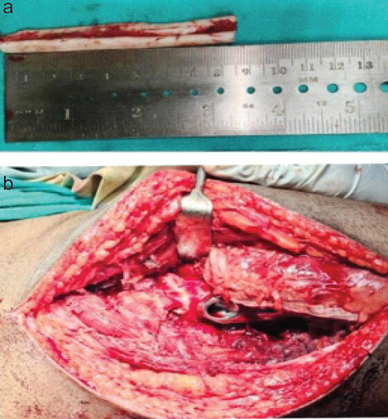
The level for placement of the 135° angle plate was identified using the tip of the lesser trochanter, approximately 2 cm below the vastus lateralis ridge. A fixed-angle guide was positioned midway along the lateral cortex and a 3.2-mm guide pin was inserted toward the apex of the femoral head, parallel to and just inferior to the center of the femoral neck on the AP view and centrally on the lateral view. The tip–apex distance was maintained at 25 mm or less after correcting for magnification. Once satisfactory guide pin placement was confirmed, the appropriate lag screw length and reaming depth were determined. Reaming was performed over the primary guide pin according to the measured length and a lag screw of corresponding size was inserted.
Following fixation of the lag screw and side plate, a channel for the fibular graft was prepared using a proximal femoral nail reamer over the proximal guide pin. A non-vascularized fibular graft was harvested from the ipsilateral leg through a posterolateral approach as described by Nagi and Dhillon [6]. The graft was obtained using an oscillating saw, with the middle third of the fibula harvested while preserving at least 10 cm of distal fibula to maintain ankle stability; therefore, syndesmotic screw fixation was not required in any case. The leading edge of the graft was beveled for approximately 1 cm and impacted over the guide wire into the femoral neck using a graft impactor until a subchondral position approximately 3–5 mm from the joint line was achieved. The wound was closed in layers over a surgical drain.
Post-operative care
All patients were encouraged to begin in-bed exercises from the 1st post-operative day (POD). Surgical drains were removed on POD 2 and routine wound dressings were performed on POD 2, 5, and 10. Suture removal was carried out on POD 10. Non-weight-bearing ambulation was initiated on POD 5. All patients were discharged on the 5thPOD and were followed up regularly at the 2nd, 3rd, 6th, and 12th weeks after surgery. Partial weight-bearing was commenced at the 3rd post-operative week and full-weight-bearing was allowed from the 6th post-operative week. Functional outcomes were assessed in all patients using the Modified Harris hip score (mHHS) at 3 months and 6 months following surgery.
Results
Six patients (Fig. 2,3,4,5,6) diagnosed with femoral neck fractures associated with posterior comminution were included in this study. The patients belonged to the age group of 20–60 years and most injuries were secondary to high-energy mechanisms such as road traffic accidents and falls from height. All individuals presented with acute hip pain, inability to bear weight, and restricted mobility of the affected limb. Clinical examination consistently revealed tenderness around the hip joint, painful restriction of movements, and external rotation deformity.
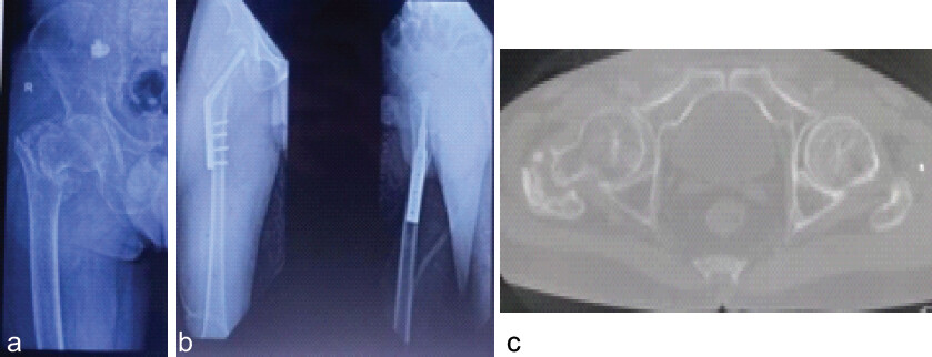
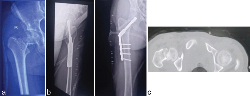
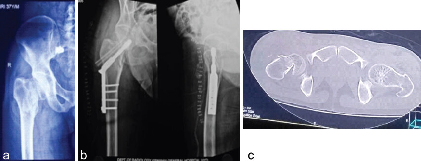
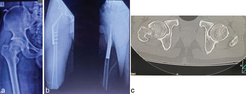
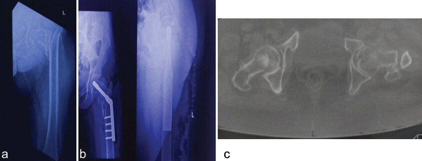
Initial assessment with AP and lateral radiographs of the hip suggested femoral neck fractures in all cases. Further evaluation with CT scans provided detailed visualization of fracture configuration and confirmed posterior cortical comminution, along with varying degrees of displacement and bone loss. Surgical planning was individualized based on fracture morphology, extent of comminution and bone quality. Closed reduction was successfully achieved in all patients and intraoperative fluoroscopy confirmed satisfactory alignment based on the Garden alignment index and appropriate tip–apex distance.
All patients were treated with DHS fixation augmented by a non-vascularized fibular strut graft. The surgical procedures were completed without intraoperative complications. The addition of the fibular graft contributed to improved posterior cortical support and enhanced rotational stability. Postoperatively, patients were managed with a uniform rehabilitation protocol that included early mobilization, progressive weight-bearing, and scheduled follow-up assessments.
In Case 1, the patient sustained a high-energy injury and presented with severe pain and inability to walk. CT imaging demonstrated a displaced femoral neck fracture with a significant posterior cortical defect. Following successful reduction and fixation, the patient achieved an excellent functional outcome at 3 months (mHHS >90), characterized by absence of pain, normal gait, and full return to daily activities. Radiographs indicated progressive fracture healing. At 6 months, the patient maintained excellent functional and radiological status, with no signs of complications such as implant failure, non-union, or avascular necrosis.
Case 2 showed similar presenting complaints and CT imaging revealed posterior comminution with moderate displacement. The patient demonstrated steady post-operative recovery and achieved an excellent functional outcome at 3 months, with minimal pain, satisfactory range of motion, and independent ambulation. Radiological findings were consistent with ongoing healing. This favorable outcome was sustained at the 6-month follow-up.
In Case 3, CT imaging confirmed a displaced fracture with posterior cortical comminution. Post-surgical recovery was uneventful and the patient exhibited rapid functional improvement. At 3 months, the patient was pain-free, ambulatory without support and able to perform routine activities without limitation, corresponding to an excellent mHHS score. Radiographic evaluation suggested good progression toward union, which was maintained at 6 months.
Case 4 presented with moderate pain and limited weight-bearing ability before surgery. Imaging studies confirmed disruption of the posterior cortex. At the 3-month follow-up, the patient achieved a good functional outcome (mHHS 80–89), with mild intermittent pain, near-normal gait, and slight difficulty during stair climbing. Radiological assessment demonstrated satisfactory healing progression. At 6 months, the functional outcome remained stable without any deterioration.
In Case 5, clinical and radiological findings were comparable, with CT confirming posterior comminution. The patient responded well to surgical intervention and rehabilitation. At 3 months, a good functional outcome was noted, with only mild pain and independent ambulation without assistive devices. Radiographs showed satisfactory alignment and early signs of union. This clinical status was maintained at 6 months.
Case 6 presented with more severe symptoms, including persistent pain, limp, and significant functional impairment. CT imaging revealed a displaced femoral neck fracture with extensive posterior comminution and bone loss. Despite adequate reduction and fixation, the patient had a poor functional outcome at 3 months (mHHS 59.5), with ongoing pain, dependence on a walking aid and difficulty performing daily activities. Radiological findings suggested delayed union. At 6 months, the patient exhibited further clinical deterioration, including increased pain and worsening limp, possibly indicating delayed healing or early mechanical failure.
At the 3-month evaluation, functional outcomes were categorized as excellent in three patients, good in two patients and poor in one patient. At 6 months, patients who initially demonstrated good or excellent results maintained their functional status, indicating sustained stability and progression of healing. (Table 1–5).
Interpretation of mHHS
| Modified Harris hip score range | Outcome category |
|---|---|
| <70 | Poor |
| 70–79 | Fair |
| 80–89 | Good |
| 90-100 | Excellent |
mHHS: Modified Harris hip score
Functional outcome at 3-month follow-up
| Outcome category | Number of patients | Percentage |
|---|---|---|
| Excellent | 3 | 50 |
| Good | 2 | 33.3 |
| Poor | 1 | 16.7 |
| Total | 6 | 100 |
Clinical features at 3-month follow-up
| Outcome group | Pain | Gait | Stair climbing | Walking aid | Public transport use |
|---|---|---|---|---|---|
| Excellent (n=3) | No significant pain | Normal | No difficulty | Not required | Able |
| Good (n=2) | Occasional mild pain | Near normal | Slight difficulty | Not required | Able |
| Poor (n=1) | Mild pain | Slight limp | Difficult | Cane most of the time | Unable |
Comparison of functional outcome at 3 and 6 months
| Outcome at 3 months | Outcome at 6 months | Number of patients |
|---|---|---|
| Excellent → Excellent | Maintained | 3 |
| Good → Good | Maintained | 2 |
| Poor → Poor (worsened) | Deteriorated | 1 |
Functional status at 6-month follow-up
| Outcome category | Number of patients | Functional status |
|---|---|---|
| Excellent | 3 | No deterioration in mHHS |
| Good | 2 | No deterioration in mHHS |
| Poor | 1 | Worsening pain and increased limp |
| Total | 6 |
mHHS: Modified Harris hip score
At the 6-month follow-up, the five patients who had good or excellent outcomes at 3 months showed no deterioration in functional status as assessed by mHHS. In contrast, the patient with a poor outcome exhibited further functional decline, with worsening pain and increased limping reflected by a reduced mHHS score.
Discussion
The frequency of femoral neck fractures among young and middle-aged individuals has increased in recent years, largely due to high-energy trauma. These injuries significantly impair quality of life, hip function, and work capacity, thereby imposing a substantial socioeconomic burden worldwide [7]. Previous studies have reported that femoral neck comminution is present in approximately 50–96% of cases, with posterior comminution accounting for the majority, occurring in about 50–82% of patients [8, 9].
Huang et al. observed that the incidence of osteonecrosis of the femoral head following internal fixation was markedly higher in patients with posterior comminution (34.2%) compared with those without such defects. Several investigations have identified cortical deficiency as a key contributor to nonunion in femoral neck fractures. Huang et al. further demonstrated that posterior comminution reduces axial load-bearing capacity, leading to early fracture displacement [10]. Similarly, Rawall et al. reported increased rates of non-union in young patients with significant posterior cortical defects [11]. When fractures with posterior comminution are anatomically reduced and rigidly fixed, contact is often achieved primarily along the anterior fracture surface, leaving the posterior defect unsupported. Ye et al. concluded that anatomical reduction combined with DHS fixation alone may be inadequate to achieve or maintain stable fixation in the presence of posterior comminution [12].
Fibular strut grafting functions both as a structural support and as a biological graft, enhancing fracture union while restoring posterior cortical integrity during reconstruction of the femoral neck and head [13]. Elgeidi et al. reported a union rate of 97% in acute femoral neck fractures treated with DHS fixation supplemented by a fibular strut graft [14]. Lazaro et al. proposed that an optimal fixation construct should provide angular and length stability, resist physiological forces across the fracture site and permit controlled micromotion. They utilized an endosteal fibular allograft as a biological intramedullary strut to reinforce the comminuted femoral neck and improve the mechanical strength of the fixation construct [15]. The limitation of our study is we have followed up only for 6 months and the long-term outcome of the procedure cannot be identified.
Conclusion
Posterior comminution of the femoral neck is a significant factor influencing functional outcome. In this case series, five out of six patients demonstrated favorable functional results when femoral neck fractures with posterior comminution in young adults were managed by closed reduction followed by fixation with a DHS combined with fibular strut grafting. These findings suggest that DHS fixation augmented with a fibular strut graft provides stable fixation and can be considered a reliable treatment option for femoral neck fractures with posterior comminution in young adult patients.
Clinical Message
In femoral neck fractures with posterior comminution, DHS fixation alone may be insufficient due to instability, increasing the risk of non-union and vascular collapse of the femoral head. Augmentation with a fibular strut graft enhances mechanical stability and supports biological healing and bone growth, leading to improved functional outcomes. This combined approach is a reliable option for preserving the femoral head in young and middle-aged patients.
Conflict of Interest:
Nil
Source of Support:
Nil
Consent:
The authors confirm that informed consent was obtained from the patient for publication of this article
How to Cite this Article
Swaroop MS, Rajeek KM, Reddy GR, Reddy YT. Functional Outcome of Femoral Neck Fractures with Posterior Comminution Managed by Dynamic Hip Screw with Fibular Strut Grafting: A Case Series Study. Journal of Orthopaedic Case Reports 2026 October;16(10): 478-484.
References
- Bartonickek J. Pauwels' classification of femoral neck fractures: Correct interpretation of the original. J Orthop Trauma 2001;15:358-60. [Google Scholar] | [PubMed]
- Meyers MH, Moore TM, Harvey JP Jr. Displaced fracture of the femoral neck treated with a muscle-pedicle graft. With emphasis on the treatment of these fractures in young adults. J Bone Joint Surg Am 1975;57:718-20. [Google Scholar] | [PubMed]
- Meyers MH, Harvey JP Jr., Moore TM. The muscle pedicle bone graft in the treatment of displaced fractures of the femoral neck: Indications, operative technique, and results. Orthop Clin North Am 1974;5:779-92. [Google Scholar] | [PubMed]
- Rau FD, Manoli A 2nd, Morawa LG. Treatment of femoral neck fractures with the sliding compression screw. Clin Orthop Relat Res 1982;163:137-40. [Google Scholar] | [PubMed]
- LeCroy CM, Rizzo M, Gunneson EE, Urbaniak JR. Free vascularized fibular bone grafting in the management of femoral neck nonunion in patients younger than fifty years. J Orthop Trauma 2002;16:464-72. [Google Scholar] | [PubMed]
- Nagi ON, Dhillon MS. Management of neglected/ununited fractures of the femoral neck in young adults. Curr Orthop 2003;17:394-402. [Google Scholar] | [PubMed]
- Miyamoto RG, Kaplan KM, Levine BR, Egol KA, Zuckerman JD. Surgical management of hip fractures: An evidence-based review of the literature. I: Femoral neck fractures. J Am Acad Orthop Surg 2008;16:596-607. [Google Scholar] | [PubMed]
- Scheck M. Intracapsular fractures of the femoral neck. Comminution of the posterior neck cortex as a cause of unstable fixation. J Bone Joint Surg Am 1959;41-A:1187-200. [Google Scholar] | [PubMed]
- Collinge CA, Mir H, Reddix R. Fracture morphology of high shear angle “vertical” femoral neck fractures in young adult patients. J Orthop Trauma 2014;28:270-5. [Google Scholar] | [PubMed]
- Huang TW, Hsu WH, Peng KT, Lee CY. Effect of integrity of the posterior cortex in displaced femoral neck fractures on outcome after surgical fixation in young adults. Injury 2011;42:217-22. [Google Scholar] | [PubMed]
- Rawall S, Bali K, Upendra B, Garg B, Yadav CS, Jayaswal A. Displaced femoral neck fractures in the young: Significance of posterior comminution and raised intracapsular pressure. Arch Orthop Trauma Surg 2012;132:73-9. [Google Scholar] | [PubMed]
- Ye Y, Hao J, Mauffrey C, Hammerberg EM, Stahel PF, Hak DJ. Optimizing stability in femoral neck fracture fixation. Orthopedics 2015;38:625-30. [Google Scholar] | [PubMed]
- Jaiswal A, Pruthi KK, Goyal RK, Pathak V, Habib M, Tanwar YS. Evaluation of osteosynthesis with dual fibular bone grafting for neglected femoral neck fractures. J Clin Orthop Trauma 2013;4:58-69. [Google Scholar] | [PubMed]
- Elgeidi A, El Negery A, Abdellatif MS, El Moghazy N. Dynamic hip screw and fibular strut graft for fixation of fresh femoral neck fracture with posterior comminution. Arch Orthop Trauma Surg 2017;137:1363-9. [Google Scholar] | [PubMed]
- Lazaro LE, Birnbaum JF, Farshad-Amacker NA, Helfet DL, Potter HG, Lorich DG. Endosteal biologic augmentation for surgical fixation of displaced femoral neck fractures. J Orthop Trauma 2016;30:81-8. [Google Scholar] | [PubMed]
© 2026 Journal of Orthopaedic Case Reports - Published by Indian Orthopaedic Research Group
About the Authors
-

M S Swaroop
-

K Mohith Rajeek
-

G Ramakrishna Reddy
-

Y Thimma Reddy
Related Articles in Journal of Orthopaedic Case Reports
 October 1, 2026 Dual Mobility Total Hip Arthroplasty for Recurrent Dislocation After Femoral Neck Fracture in an Elderly Patient: A Case Report
 October 1, 2026 Primary Total Hip Arthroplasty for a Displaced Femoral Neck Fracture in an Above-the-Knee Amputee: A Case Report
 September 1, 2026 Application of 3D-printed models in procedural training for dynamic hip screw fixation and intraosseous vascular access: An original article
 August 1, 2026 Posterior Acetabular Wall Fracture Mimicking a Subcapital Femoral Neck Fracture on Plain Radiographs: A Diagnostic Pitfall and the Role of Intraoperative Fluoroscopy: A Case Report