The occurrence of a valgus deformity resulting from an injury to the periosteum can be present and must be taken into account.
Dr. D Gallucci Gerardo, Department of Orthopaedics and Traumatology, “Dr Carlos E. Ottolenghi” of the Hospital Italiano de Buenos Aires Argentina; Juan Domingo Perón 4190, C1199 ABH, Buenos Aires Argentina. E-mail: gerardo.gallucci@hospitalitaliano.org.ar
IntroductionThe aim of this paper is to report a rare case of a child who suffered a simple elbow dislocation (SED) that developed a post-traumatic valgus deformity and a subsequent posterolateral elbow instability.
Case ReportWe report a case of a female patient who suffered a posterolateral SED of her elbow at the age of 12. She was treated with closed reduction and over the years, she developed an asymptomatic valgus deformity. At the age of 16, she suffered a fall trauma while playing field hockey with a re-dislocation of the elbow. Since then, she presented multiple episodes of subluxation. A supracondylar subtractive wedge osteotomy of 20° and double plate osteosynthesis was performed with reconstruction of the ulnar lateral collateral ligament.
ConclusionThe focus of this article must be on the unusual occurrence of this sequence of conditions. SED is rare in children and generally associated with medial epicondyle fractures. The occurrence of a valgus deformity resulting from an injury to the periosteum can be present and must be taken into account. Posterolateral instability is rare in the context of a valgus elbow. Angular correction osteotomy and ligament reconstruction can be associated with good functional and aesthetic results.
KeywordsSimple elbow dislocation, valgus deformity, posterolateral instability, periosteum.
Simple elbow dislocation (SED) is an uncommon injury [1] occurring between 3% and 6% of all pediatric elbow injuries [2, 3]. Non-operative treatment has good outcomes [4] and recurrent dislocation due to residual instability is unusual [5]. Valgus deformity is a rarely encountered complication of traumatic elbow in children [6]. However, it has been reported after elbow dislocation [7]. Combined elbow injuries such as angular deformities associated with recurrent elbow instability are rare. Posterolateral instability (PLI) is associated with varus elbow deformities [8] and the medial ligament complex insufficiency has been reported in cubitus valgus [9]. To the best of our knowledge, there are no reports about the presence of PLI in the context of cubitus valgus.
The aim of this paper is to report a rare case of a child who suffered a SED that developed a post-traumatic valgus deformity and a subsequent posterolateral elbow instability.
A 20-year-old female patient who had suffered a posterolateral SED of her left elbow at the age of 12 was admitted to our service (Fig. 1).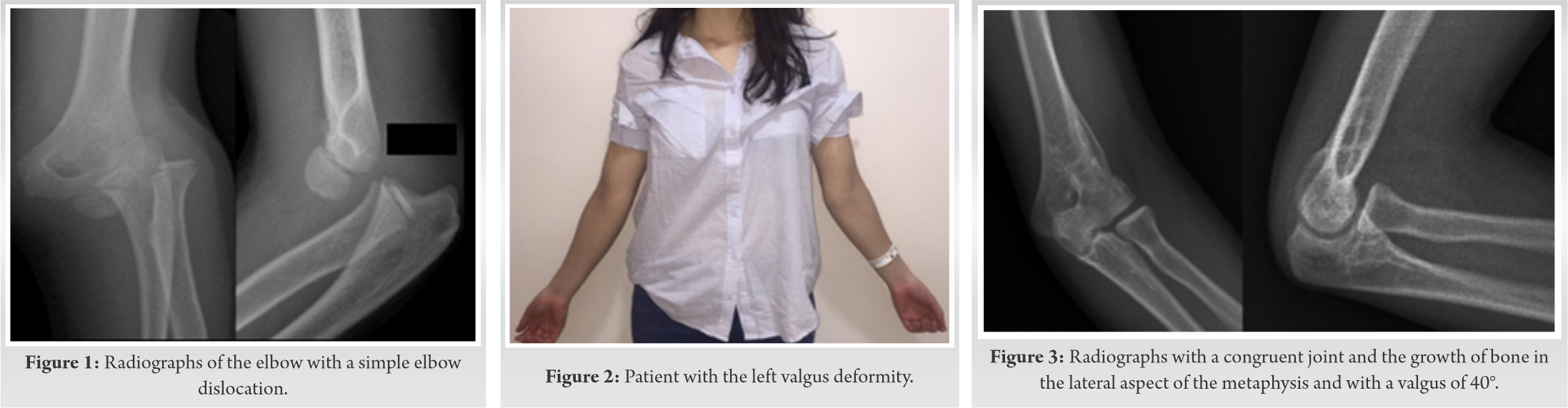 Initially, she was treated with closed reduction and a cast for 3 weeks. Over the years, she developed an asymptomatic valgus deformity. At the age of 16, she suffered a fall trauma while playing field hockey with a re-dislocation of the elbow. Since then, she presented multiple episodes of subluxation. With this clinical situation, the patient was referred to the senior author with pain on the lateral aspect of the elbow, paresthesia in the fourth and fifth finger, and a feeling of instability (Fig. 2). In addition, she was complaining about an aesthetic problem. The range of motion was 135° of flexion and 0° of extension (the same as the contralateral side). Tinel signs at the cubital tunnel and the lateral pivot-shift apprehension tests were positive. Radiographs showed a congruent joint with the growth of bone in the lateral aspect of the metaphysis. For the measurement of the deformity, we used the humerus-elbow-wrist angle in the anteroposterior projection with extended elbow and in full supination. The valgus deformity was 40° versus 20° measured on the contralateral side (Fig. 3). The medial prominence index was 46%. Magnetic resonance imaging (MRI) demonstrated the elongation of the ulnar lateral collateral ligament (ULCL) (Fig. 4). The pain, according to the visual analog scale (VAS), was 5 out of 10. The clinical results were evaluated according to the Mayo elbow performance score (MEPS) and the Quick DASH questionnaire. The scores were 65 and 34, respectively. Under a regional block, a positive Pivot Shift test confirmed the ULCL injury A supracondylar subtractive wedge osteotomy of 20° and double plate osteosynthesis was performed with anterior transposition of the ulnar nerve. Reconstruction of the ULCL was performed with a slice of the triceps tendon. At 16 months post-operative, the patient presented a mobility of 135°–0° with pain of 2, a MEPS of 85, and a DASH of 13. According to the VAS, satisfaction with the procedure was 9 out of 10, and the result was excellent, according to the Oppenheim scale. Radiographs showed the union of the osteotomy with a final valgus of 20° and a medial prominence of 56% (12% of increase over pre-operative) (Fig. 5). She was aesthetically satisfied (Fig. 6).
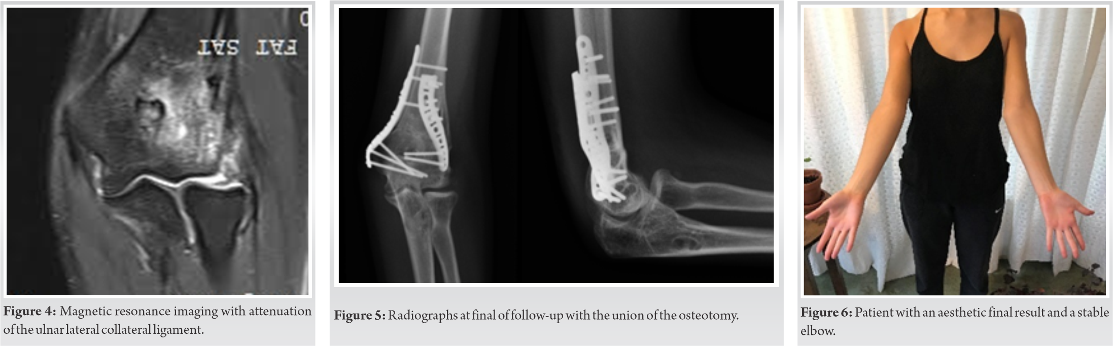
We introduce here a patient with two concomitant unusual conditions, the development of a cubitus valgus deformity after a SED and PLI in the context of a cubitus valgus. SED is uncommon in children over 10 years old. This incidence in the second decade of life is explained by the partial closing of the growth plates around the elbow joint that increases the structural bone strength [2, 3], transmitting force to weaker tissues such as collateral ligaments, causing rupture [1, 3]. The most common displacement is the posterolateral type at roughly 70% from all pediatric dislocations [10, 12]. Radiographs in these cases must be meticulously evaluated because the presence of associated lesions is usually high, being avulsion of the medial epicondyle the most common one [13]. It is uncommon to have recurrent dislocation and instability in SED following closed reduction, since there was no evidence of recurrent instability in cases of complex elbow dislocations. Carlioz and Abols [14] reported a long series of 58 pediatric patients with elbow dislocations and evidenced associated fractures in 75% of the cases. Despite some reported complications, none of them presented instability. It is difficult to know for certain if our patient evolved with a proper ligament healing or if some residual instability remained after the first episode of dislocation. However, since we have not found in the literature reports of instability after a SED, it would be logical to believe that the subsequent instability may not be related to this first episode of dislocation. Another rare condition in our patient is the presence of valgus deformity. It is significantly less frequent than varus and occurs most commonly due to a pseudoarthrosis of the lateral humeral condyle. However, the development of a valgus deformity after a SED has been described. Adaş et al. [7] investigated the functional and radiological outcomes of conservatively treated SED and the subsequent incidence of the development of a cubitus valgus. They reported that 4 out of 11 patients with SED developed a valgus deformity with an average increase of 14.5° of the carrying angle. Although all patients were examined with MRI, no osseous problems were found that could explain this elbow deformity. They suggested that the deformity could be a sequel from medial epicondyle’s growth stimulation after the dislocation.
What is certain is that information is limited or restricted on this topicin the literature. On this context, a possible explanation is that the injury to the periosteum might affect bone growth. Periosteal rupture may cause asymmetrical growth disturbance, angular deformity, and limb discrepancies [15, 16]. Dimitriou et al. [15] reported the results of periosteal dissection in rabbits and its correlation with long bone growth. They concluded that the cross-section of the periosteum increases the longitudinal growth of long bones and that partial section in the proximal tibia leads to a valgus deformation.
Haasbeek et al. [16] reported, in two clinical cases, the angular deformations when tightening the periosteum near the physis. They suggested that when there’s deformation of the bone growth, and there is no physical abnormality, the injury of the peristium as the leading cause should always be considered. PLI has been associated with cubitus varus. Varus deformity causes a repetitive varus torque at the elbow during axial loading and extension resisted as when standing up from a chair. This varus moment can result in chronic attenuation of the ULCL over time [17, 18]. To the best of our knowledge, there is no reported PLI in the context of a valgus elbow deformity. In general, valgus deformity produces the attenuation of the medial collateral ligament, but in this case, the patient experienced trauma while playing field hockey and suffered a posterolateral dislocation. We consider that this trauma occurred in a stable elbow because, as we previously mentioned, residual instability in children is rare and has not been reported.
The multiple occurrences of repeated traumas in this patient led to the development of PLI, which was present at the time of the consultation.
ULCL reconstruction is associated with excellent results [19]. Because of the severe valgus presented in this patient, with ulnar nerve symptoms and PLI, the surgical plan was to perform a corrective osteotomy in conjunction with ligament reconstruction and subcutaneous transposition of the ulnar nerve.
The focus of this article must be on the unusual occurrence of this sequence of conditions. SED is rare in children and generally associated with medial epicondyle fractures. The occurrence of a valgus deformity resulting from an injury to the periosteum can be present and must be taken into account. PLI is rare in the context of a valgus elbow. Angular correction osteotomy and ligament reconstruction can be associated with good functional and esthetic results.
Valgus deformity after SED is rare in children. The injury of the periosteum can develop a valgus deformity. PLI is rare in the context of a valgus elbow.
References
- 1.Subasi M, Isik M, Bulut M, Cebesoy O, Uludag A, Karakurt L. Clinical and functional outcomes and treatment options for paediatric elbow dislocations: Experiences of three trauma centres. Injury 2015;46:S14-8. [Google Scholar]
- 2.Kozin SH, Abzug JM, Safier S, Herman MJ. Complications of pediatric elbow dislocations and Monteggia fracture-dislocations. Instr Course Lect 2015;64:493-8. [Google Scholar]
- 3.Lieber J, Zundel SM, Luithle T, Fuchs J, Kirschner HJ. Acute traumatic posterior elbow dislocation in children. J Pediatr Orthop B 2012;21:474-81. [Google Scholar]
- 4.Kaziz H, Naouar N, Osman W, Ayeche M. Outcomes of paediatric elbow dislocations. Malays Orthop J 2016;10:44-9. [Google Scholar]
- 5.Atarraf K, Arroud M, Chater L, Afifi MA. Chronic dislocation of the elbow in children: Report of 20 cases. Pan Afr Med J 2014;18:348. [Google Scholar]
- 6.Arnold JA, Nasca RH, Nelson CL. Supracondylar fractures of the humerus: The role of dynamic factors in prevention of deformity. J Bone Joint Surg Am 1977;59:589-95. [Google Scholar]
- 7.Adaş M, Bayraktar MK, Tonbul M, Uzun M, Çakar M, Tekin AÇ, et al. The role of simple elbow dislocations in cubitus valgus development in children. Int Orthop 2014;38:797-802. [Google Scholar]
- 8.Beuerlein MJ, Reid JT, Schemitsch EH, McKee MD. Effect of distal humeral varus deformity on strain in the lateral ulnar collateral ligament and ulnohumeral joint stability. J Bone Joint Surg Am 2004;86:2235-42. [Google Scholar]
- 9.Myeroff C, Brock JL, Huffman GR. Recurrent tardy ulnar collateral ligament insufficiency due to cubitus valgus: Management with concomitant osteotomy and dual cortical button suspension technique. JSES Open Access 2018;2:129-32. [Google Scholar]
- 10.Little KJ. Elbow fractures and dislocations. Orthop Clin North Am 2014;45:327-40. [Google Scholar]
- 11.Stans AA, Lawrence JT. In: Flynn JM, Skaggs DL, Waters PM, editors. Dislocation of the Elbow, Medial Epicondylar Fractures. 8th ed. Philadelphia, PA: Wolters KluwerHealth; 2014. p. 651-700. [Google Scholar]
- 12.Rasool MN. Dislocations of the elbow in children. J Bone Joint Surg Br 2004;86:1050-8. [Google Scholar]
- 13.Murphy RF, Vuillermin C, Naqvi M, Miller PE, Bae DS, Shore BJ. Early outcomes of pediatric elbow dislocation-risk factors associated with morbidity. J Pediatr Orthop 2017;37:440-6. [Google Scholar]
- 14.Carlioz H, Abols Y. Posterior dislocation of the elbow in children. J Pediatr Orthop 1984;4:8-12. [Google Scholar]
- 15.Dimitriou CG, Kapetanos GA, Symeonides PP. The effect of partial periosteal division on growth of the long bones. An experimental study in rabbits. Clin Orthop Relat Res 1988;236:265-9. [Google Scholar]
- 16.Haasbeek JF, Rang MC, Blackburn N. Periosteal tether causing angular growth deformity: Report of two clinical cases and an experimental model. J Pediatr Orthop 1995;15:677-81. [Google Scholar]
- 17.O’Driscoll SW, Spinner RJ, McKee MD, Kibler WB, Hastings H, Morrey BF, et al. Tardy posterolateral rotatory instability of the elbow due to cubitus varus. J Bone Joint Surg Am 2001;83:1358-69. [Google Scholar]
- 18.Arvind C, Hargreaves DG. Tabletop relocation test: A new clinical test for posterolateral rotatory instability of the elbow. J Shoulder Elbow Surg 2006;15:707-8. [Google Scholar]
- 19.Camp CL, Sanchez-Sotelo J, Shields MN, O’Driscoll SW. Lateral ulnar collateral ligament reconstruction for posterolateral rotatory instability of the elbow. Arthrosc Tech 2017;6:1101-5. [Google Scholar]