Avulsion calcaneus fracture fixation with two parallel corticocancellous screws is an excellent method of fracture fixation, but screw pull-out is a major drawback of this method. Oblique placement of one of the screws to nullify tendoachillis forces acting on fracture fragment is an alternative method to prevent the screw pull-out. The author recommends this modified 2 CC screw fracture fixation technique.
Dr Sanket Tanpure, Department of Orthopaedics, Dr. Vithalrao Vikhe Patil Medical College, Ahmednagar, Maharashtra, India. E-mail: sankettanpure55@gmail.com
Introduction: Avulsion tuberosity fracture of the calcaneus is relatively unusual and occurs more frequently in elderly osteoporotic patients. Direct trauma to the heel is a rare cause in young individuals. Failure to perform early open reduction and internal fixation (ORIF) potentially leads to soft-tissue complications due to pressure necrosis of the overlying skin.
Case Presentation: A 29-year-old male patient experienced left heel pain with swelling after the assault as he was hit by an iron rod 8 days prior. Radiographs revealed a Beavis Type 2 calcaneal avulsion fracture. An open reduction with two corticocancellous (CC) screw fixation was intended for the fracture.
Conclusion: The avulsed bone fragment was small causing difficulty in reduction. The result of closed reduction in such a scenario is not promising and usually requires ORIF. We performed a modified surgical procedure of CC screw fixation in which one screw was passed perpendicular to the fracture plane for reduction of the fracture and another screw was passed obliquely to nullify Achilles forces. We believe that this technique of fracture management improves patient outcome and early mobilization.
Keywords: Avulsion, calcaneus fracture, corticocancellous screw fixation, CC screw, tuberosity fracture.
A calcaneal tuberosity avulsion fracture is a rare extra-articular injury that usually occurs due to forceful ankle dorsiflexion against resistance [1, 2]. Direct traumatic tuberosity fracture is a further rare entity [3]. Beavis et al. [4] classify calcaneal tuberosity fractures into three types. Type 1 consists of small cortical avulsion of tuberosity (sleeve fracture). Type 2 consists of beak fracture and Type 3 involves infrabursal avulsion fracture (middle third posterior tuberosity). Fractures of Calcaneus contribute to 2% of all fractures out of which 25–40% are extra-articular in nature [1, 5]. Gastrocnemius-soleus complex contraction results in a posterosuperior tuberosity avulsion fracture. Delayed surgery in case of calcaneal avulsion fractures poses the risk of severe soft-tissue complications like skin necrosis [6]. To prevent skin necrosis, such avulsion or beak fractures (Beavis Type2) need to be reduced as soon as possible [6, 7]. Our patient in this case report is diagnosed with a calcaneal tuberosity avulsion fracture and later on developed soft-tissue problems due to late intervention. This study aims to stress the fact that calcaneal tuberosity avulsion fracture due to direct trauma is a rare entity and such injury needs to be intervened as an emergency so as to prevent further possible soft-tissue complications. Through this report, we also recommend a slightly modified technique in view of corticocancellous (CC) screw trajectory to prevent potential reavulsion injuries and facilitate early rehabilitation.
Patient information
A 29-year-old male patient experienced left heel pain with swelling after the assault as he was hit by an iron rod 8 days prior. There was no history of chronic illness.
Clinical findings
According to Beavis et al. [4] classification, the fracture was Type 2 (Beak fracture) (Fig. 1). A 2 × 2 cm blister was present on the posterior aspect of the heel (Fig. 2). The patient was classed as Stage I by anesthetic profile according to the American Society of Anaesthesiology.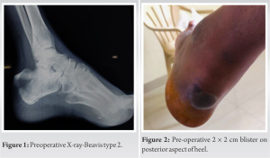
Therapeutic intervention
After spinal anesthesia and tourniquet application to the thigh; the patient was given prone position on OT table. Before starting with the incision; the blister over the fracture fragment was debrided thoroughly. A posterolateral ankle approach skin incision was made to expose the calcaneal tuberosity. After exposure, the displaced fracture fragment was pulled distally and held in place with a ball tip reduction clamp to achieve anatomical reduction. A 6 mm CC screw guide wire was passed perpendicular to the fracture plane under Fluoroscopic guidance (Screw “A”). A second 6 mm CC screw guide wire was passed in an oblique fashion distal to the previous wire (Screw “B”). Both the guide wires were checked for position and trajectory under C-Arm and were measured. Two 6 mm CC screws of measured length were passed over the guide wires along with washers to take plantar calcaneal cortex purchase. Care was taken not to over-penetrate the cortex so as to prevent potential plantar fascia irritation. The second oblique CC screw serves to nullify Achilles tendon force thereby preventing re-avulsion (Fig. 3 and 4). Posterior heel blister was debrided again and covered with cotton gauze (Fig. 5).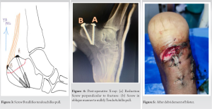
Follow-up and outcome
Post-surgery, the leg was immobilized in a below-knee slab with the foot in equinus position for 2 weeks. At 2-week post-surgery, after removing the sutures and confirming the healthy skin condition; the slab was replaced by a below-knee cast in an equinus position and the patient was encouraged to start partial weight bearing with the help of a walker. 4 weeks after surgery, the cast was removed and ankle range of motion (ROM) was started and he was allowed to walk with complete weight bearing wearing a heel raise. The height of the heel raise was reduced each week and the heel raise was removed completely at 6 weeks. The patient could walk with a single crutch at 4 weeks. The radiological union was appreciated at 3 months without any fracture displacement. There was no restriction or apprehension in ankle ROM at 6 weeks. The skin and blister was healed in 2 weeks. The patient could resume pre-injury day to day activities within 1 month.
Open reduction and internal fixation (ORIF) is usually required for calcaneal avulsions as closed reduction is often unsuccessful for anatomical reduction [8, 9]. If the avulsed bone fragment is large (type 2), Lag screw fixation is the most commonly described technique for fixation. However, lag screw fixation is not effective to nullify tendoachilles pull in small fragments as in Type 1 and Type 3 fractures [10, 11, 12]. Various surgical techniques were proposed in the literature. Tension band wiring or cerclage wiring was the most widely used technique, but there is a risk of wire being cut out through an avulsed fragment or calcaneus [11]. In recent advances, the avulsed fragment of calcaneus tuberosity was fixed with suture anchors [12, 13]. However, there are chances of anchor loosening in osteoporotic patients [10, 14]. There are no such definitive studies in the literature stating that it resists the Achilles tendon forces in early rehabilitation; hence, all previous publish literature advises limited weight bearing at 6–8 weeks post-surgery [10, 12]. However, limitation of weight bearing causes disuse atrophy and decreased ability to walk. Ninomiya et al. [15] proposed a new technique in which Krackow suture was used to fix avulsed fragments along with Achilles tendon so that the fixation of small or thin fragments can be achieved. Banerjee et al. [10] used the Krackow technique in which a non-absorbable suture was passed through the Achilles tendon and brought out through tunnels over calcaneal tuberosity, however, the risk of suture cut out in tunnel limits this technique. Conservative treatment of calcaneal tuberosity avulsion fractures leads to potential complications that include skin necrosis of the heel, Haglund’s deformity, and reduction in plantarflexion power. Posterosuperior heel skin necrosis is a major concern due to underlying fragment pressure. It is of paramount importance to note that the skin overlying the Achilles tendon and calcaneal tuberosity is fragile and has a critical blood supply. Hence, these fractures should be addressed as emergencies [16].
Avulsion calcaneus fracture fixation by two parallel CC screw construct limits the early mobilization and comes with a common complication of screw pull-out. We performed a modified surgical technique of CC screw fixation in which one screw is perpendicular to the fracture plane for reduction of the fracture and another screw in an oblique manner to nullify tendon Achilles forces. We believe this technique of fracture management facilitates early mobilization and improves patient outcomes.
ORIF with two CC screws in a modified manner is a better way to reduce fracture fragments. It provides better stability and early mobilization and reduces the risk of screw pull-out. Early intervention is the key to prevent further soft-tissue complications in such injuries.
References
- 1.Rothberg AS. Avulsion fractures of the oscalcis. J Bone Joint Surg 1939;21:218-20. [Google Scholar]
- 2.Heckman JO. Fractures and dislocations of the foot. In: Rockwood CA, Green DP, Bulchoz RW, Heckman JO, editors. Rockwood and Greens Fracture in Adults. Vol. 2., 4th ed. Philadelphia, PA: Lippincott-Raven; 1996. p. 2332-3. [Google Scholar]
- 3.Biell WC 3rd, Morgan JM, Wagner FW Jr., Gabriel R. Neuropathic calcaneal tuberosity avulsion fractures. Clin Orthop Relat Res 1993;269:8-13. [Google Scholar]
- 4.Beavis RC, Rourke K, Court-Brown C. Avulsion fracture of the calcaneal tuberosity: A case report and literature review. Foot Ankle Int 2008;29:863-66. [Google Scholar]
- 5.Sanders R, Clare M. Campbell’s Operative Orthopedics. Ch. 55., 10th ed. St. Louis: Mosby; 2002. [Google Scholar]
- 6.Protheroe K. Avulsion fractures of the calcaneus. J Bone Joint Surg 1969;51:118-22. [Google Scholar]
- 7.Brunner CF, Weber BG. Special Techniques in Internal Fixation. 1st ed. Berlin: Springer-Verlag; 1982. [Google Scholar]
- 8.Lowy M. Avulsion fractures of the calcaneus. J Bone Joint Surg Br 1969;51:494-7. [Google Scholar]
- 9.Squires B, Allen PE, Livingstone J, Atkins RM. Fractures of the tuberosity of the calcaneus. J Bone Joint Surg Br 2001;83:55-61. [Google Scholar]
- 10.Banerjee R, Chao J, Sadeghi C, Taylor R, Nickisch F. Fractures of the calcaneal tuberosity treated with suture fixation through bone tunnels. J Orthop Trauma 2011;25:685-90. [Google Scholar]
- 11.Khazen GE, Wilson AN, Ashfaq S, Parks BG, Schon LC. Fixation of calcaneal avulsion fractures using screws with and without suture anchors: A biomechanical investigation. Foot Ankle Int 2007;28:1183-6. [Google Scholar]
- 12.Lui TH. Fixation of tendo Achilles avulsion fracture. Foot Ankle Surg 2009;15:58-61. [Google Scholar]
- 13.Robb CA, Davies MB. A new technique for fixation of calcaneal tuberosity avulsion fractures. Foot Ankle Surg 2003;9:221-4. [Google Scholar]
- 14.Gitajn IL, Abousayed M, Toussaint RJ, Vrahas M, Kwon JY. Calcaneal avulsion fractures: A case series of 33 patients describing prognostic factors and outcomes. Foot Ankle Spec 2015;8:10-7. [Google Scholar]
- 15.Ninomiya H, Watanabe M, Kamimura K. Innovative fixation technique for avulsion fractures of the calcaneal tuberosity. J Foot Ankle Surg 2021;60:218-20. [Google Scholar]
- 16.Hess M, Booth B, Laughlin RT. Calcaneal avulsion fracture: Complications from delayed treatment. Am J Emerg Med 2008;26:254.e1-4. [Google Scholar]