Osteochondroma of talar neck can very rarely present late with a large size and a grave clinically picture, however this can be managed successfully by surgical excision of the mass.
Dr. Kamparsh Thakur, Department of Orthopaedics, 166 Military Hospital, Jammu, Jammu and Kashmir, India. E-mail: drkamparsh@gmail.com
Introduction: Osteochondroma is a benign bone tumor of the bone primarily seen in younger age groups. However, late presentation of the same is a rare incidence, as the symptoms develop rapidly due to compression of nearby structures.
Case Report: We present a case of a 55-year-old male patient with a giant osteochondroma originating from the neck of the talus. The patient presented with a huge 100 × 70 × 50 mm swelling over the ankle. The patient underwent an excision of the swelling. Histopathological examination of the swelling confirmed the findings of an osteochondroma. The patient had an uneventful recovery after the excision and resumed his functional activity completely.
Conclusion: Giant osteochondroma around the ankle is an extremely rare entity. Even rarer is a late presentation in the 6th decade onward. However, the management like others involves the excision of the lesion.
Keywords: Giant osteochondroma, neck of talus, excision.
Osteochondromas are a benign tumor of the bone with a cartilaginous cap, which are primarily seen in the adolescent and young people. These form 10–15% of all the benign bone tumors. The most common location for osteochondroma is in the metaphyseal region of long bones [1]Involvement of foot and ankle is rare and is either a solitary presentation or is an occurrence in multiple hereditary exostosis [2]. However, late presentation of osteochondroma is an infrequent finding. We report an unusual case of a giant osteochondroma originating from the neck of talus in a 55-year-old man. We came across only one case with similar presentation in the literature.
A 55-year-old male patient had presented to us in the outpatient department with a gradually progressive swelling over ankle anteriorly, noticeable for the past 15 years. The swelling was globular, bony hard to feel and non-fluctuant in nature. It measured 100 × 70 × 50 mm clinically (Fig. 1).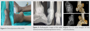 The overlying skin was non-adherent to the underlying swelling, with no local signs of inflammation. Examination revealed dorsiflexion of the ankle beyond neutral to be restricted; however, plantar flexion was preserved. The subtalar movements were unaffected. There was no evidence of compression symptoms of the neurovascular structures in the vicinity. On radiographic evaluation, there was a huge anteromedial bony swelling originating from the neck of the talus and abutting the distal tibia (Fig. 2). Computed tomography scan suggested a benign bony mass originating from the talar neck, likely to be osteochondroma (Fig. 3). In view of the long-standing nature and benign radiographic appearance, the patient was planned for excisional biopsy of the swelling. The patient was operated in supine position, and a single anteromedial incision was given (Fig. 4). The lesion was well-encapsulated and well-marginated from the surrounding soft tissues. It was excised en-mass, following osteotomy from the base and sent for histopathological examination (HPE) (Fig. 5).
 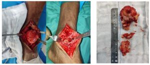 The HPE showed the lesion to be an osteochondroma (Fig. 6). The skin was closed primarily, and the redundant skin was left intact. A well-padded compressive dressing was done for a period of 02 weeks. Post-operative radiographs revealed complete removal of the lesion with a congruent mortise. The patient was full weight-bearing ambulant from post-operative day 1. The dorsiflexion of the ankle improved to 30°. The patient essentially had an uneventful recovery.
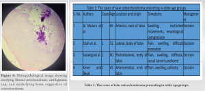
Osteochondroma is the most common benign bone tumor and is generally seen in the metaphyseal region of the long bones forming from enchondral ossification. Ozdemir et al., in their study, reported 196 cases out of the 1786 cases of osteochondroma to be found in foot and ankle region, and most originating from the metatarsals and phalanges [2]. Talus, out of all is an extremely rare site to have this pathology, as only a few are reported as case reports in the literature [3, 4]. Earliest description available of talar neck osteochondroma is by Fuselier et al. in 1984 [5]. As the tumor affects the growing bone, the age of presentation is in adolescent age group. Only a few have reported the presentation in older age group [6, 7]. Similar to us, Al Mutani et al. reported a case in a 65-year-old patient, who had a giant osteochondroma originating from the neck of talus [7]. Other reported cases of talar neck osteochondroma have generally been of younger age group [4, 8]. The case reported by Al Mutani et al. had a presentation like ours in all the ways [7]. Their lesion measured 100 × 90 × 30 mm, whereas ours measured 100 × 70 × 50 mm. The authors had approached the lesion through dual incision, but we could access and excise the osteochondroma using a single anteromedial approach. Overall, both the patients had an uneventful recovery. Late presentation for a talar osteochondroma is a rare phenomenon, as the ankle and surrounding structures are in a closed tight compartment. We did a thorough search of the literature for cases presenting in 5th decade and beyond and came across few cases (Table 1) [6, 7, 9, 10]. The location of osteochondroma in the talus varied for them, and the maximum incidence was of anterior location. Late presentation is also fraught with the increased size and compression symptoms; however, it was realized that pain, swelling, and stiffness were common to all, but compressive neurovascular symptoms were rarely encountered and were mostly accompanied to swelling else anterior. This could be attributed to possible soft-tissue adaptation to the swelling more anteriorly and anatomical morphology of other regions.
Osteochondromas in foot and ankle region are a rare entity and even rarer is involvement of talus. Very few cases of giant osteochondromas of talar neck are seen; however, such cases if encountered can be managed after careful and deliberate planning. Awareness of such an occurrence is essential to the orthopedic surgeons, as these can be tackled at an early stage.
A careful and deliberate planning and a meticulous execution of it can lead to a successful outcome in patients presenting with a large osteochondroma of the talar neck.
References
- 1.Mavrogenis AF, Papagelopoulos PJ, Soucacos PN. Skeletal osteochondromas revisited. Orthopedics 2008;31:1018. [Google Scholar]
- 2.Ozdemir HM, Yildiz Y, Yilmaz C, Saglik Y. Tumors of the foot and ankle: Analysis 196 cases. J Foot Ankle Surg 1997;36:403-8. [Google Scholar]
- 3.Boya H, Özcan Ö, Tokyol Ç. Osteochondroma of the talus: An unusual location. Acta Orthop Traumatol Turc 2014;48:236-9. [Google Scholar]
- 4.Kumar S, Dhammi IK, Jain AK, Shahi P. Osteochondroma of the talus: Three varying cases. BMJ Case Rep 2020;13:e237670. [Google Scholar]
- 5.Fuselier CO, Binning T, Kushner D, Kirchwehm WW, Rice JR, Hetherington V, et al. Solitary osteochondroma of the foot: An in-depth study with case reports. J Foot Surg 1984;23:3-24. [Google Scholar]
- 6.Shah A, Adhikari S, Shah S, Bhandari PS, Uprety S. Osteochondroma of talus: A case report from Nepal. Clin Case Rep 2022;10:e05867. [Google Scholar]
- 7.Al Mutani M, Mahmood A, Chandrasekar CR. Giant osteochondroma of the talar neck. Foot (Edinb) 2013;23:45-9. [Google Scholar]
- 8.Wang C, Ma X, Wang X, Zhang Y, Zhang C, Huang J, et al. Osteochondroma of the talar neck: A case report and literature review. J Foot Ankle Surg 2016;55:338-44. [Google Scholar]
- 9.Keser S, Bayar A. Osteochondroma of the talar neck: A rare cause of callosity of the foot dorsum. J Am Podiatr Med Assoc 2005;95:295-7. [Google Scholar]
- 10.Suranigi S, Rengasamy K, Najimudeen S, Gnanadoss J. Extensive osteochondroma of talus presenting as tarsal tunnel syndrome-report of a case and literature review. Arch Bone J Surg 2016;4:269-72. [Google Scholar]