We should do a thorough clinical examination in all orthopaedic patients due for surgery to rule out ochronosis and be duly prepared for a difficult exposure and difficult surgical procedure in case the patient is positive for ochronosis.
Dr. Arvind, Department of Orthopaedic Surgery, Tata Motors Hospital, Telco, Jamshedpur - 831 004, Jharkhand, India. E-mail: arvindbharadwaj1986@gmail.com
Introduction: Ochronosis is a rare syndrome caused by accumulation of homogentisic acid in connective tissue due to deficiency of enzyme homogentisic acid oxidase. It characterized by blue-black pigmentation connective tissues such as sclera, cartilage of ear, and synovium of joints and it causes destruction of joints cartilage and early arthritis. Urine becomes dark coloured on prolonged standing. Some patient may develop rare cardiac manifestation due to accumulation of homogentisic acid on valves.
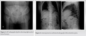
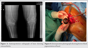
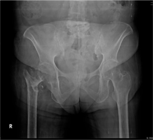
Case Report: A 56-year-old female admitted with neck of femur fracture after history of fall at home. The patient was having chronic back ache and knee pain. Plain radiograph of knee and spine showed severe arthritic changes. Exposure during surgery was difficult due to hard and brittle tendons and capsule of joint. Femur head and acetabulum cartilage appeared dark brown. Dark brown pigmentation of sclera and hands was found on clinical examination postoperatively.
Conclusion: Patients with ochronosis usually develop early osteoarthritis and spondylosis which should be differentiated from other causes of early arthritis such as rheumatoid arthritis and seronegative arthritis. It leads to destruction of joint cartilage and weaking subchondral bone which leads to pathological fracture. And due to stiffness of soft-tissue around joint exposure during surgery can be challenging.
Keywords: Fracture neck of femur, dark brown cartilage, arthritis, pathological fracture, ochronosis.
The term “ochronosis” originates from a Greek word “ochre,” which refers to yellow discoloration [1]. Ochronosis is a rare autosomal recessive disorder occurs due to mutation of HGO gene. It is characteristized by deficiency of enzyme homogentisic acid oxidase which leads to accumulation of homogentisic acid [2]. Patients usually do not develop any symptoms in childhood but later in life homogentisic acid got accumulated and polymerized to form blue-brown pigment which gets deposited in connective tissue, ultimately resulting in brittle and weak tissue [3]. The patient can present with dark pigmentation of sclera, ear cartilage, nasal cartilage and interdigital area in hands, and urine turns dark on exposure to air [3]. This deposition of homogentisic acid with time can weaken the connective tissue in spine and peripheral joints which lead to ochronotic spondylosis and arthropathy [4]. Physical finding may include joint line tenderness, crepitus, decreased, and painful range of motion. Although it is rare cause of early arthritis, still it should be ruled out if the patient only come with pain and swelling around joint as patients with seronegative arthritis may have similar presentation. It also requires high index of clinical suspicion as test for homogentisic acid is not readily available. However, ochronosis usually does not involve small joint of hands, it apparently damages large joint of hip, knee, and spine so it can be easily differentiated [5]. There are some reports which showed decreased bone mineral density in cases of ochronosis due to increased bone resorption as homogentisic acid got deposited in bone matrix and damaged the bone cells [6]. This bone loss could possibly be the reason for common finding of fracture neck of femur and scoliosis in cases of ochronosis. Apart from joint cartilage and bone, it may also involve tendons, muscles, muscles sheaths, fascias, joint capsule and heart valve. Hence, dissection and exposure during surgery can be really challenging. Moreover, anesthetist should be aware of any cardiorespiratory involvement before surgery [7]. We report a case of 56-year-old female with history of fall and chronic back, knee pain which then diagnosed as ochronotic arthropathy.
A 56-year-old female presented to emergency department with history of fall at home 2 weeks back. The patient was managed at home for 2 weeks and brought to hospital when patients’ condition started deteriorating and she developed bed sores. On physical examination, right lower limb was shortened, externally rotated and the patient was not able to do hip flexion. Joint line tenderness and swelling was present on right hip. The patient also complained of chronic back ache and bilateral knee pain from past 10 to 15 years due to which she had difficulty walking without support. On plain radiograph of pelvis, we found fracture neck of femur right side (Fig. 1). 


Ochronosis is a very rare genetic disorder found in few people in millions. The patient usually present in forth decade of life with blackening of urine, dark pigmentation of tissue in sclera, ear, nose, and skin due to accumulation of homogentisic acid [8]. It can be diagnosed by determination of homogentisic acid in urine. No definitive treatment is being used for this disease but there is drug called Nitisinone which has shown some results in treating this disease[9],[10]. Most commonly patient presents with symptoms of arthropathy and it is usually diagnosed at the time of surgery that it is ochronotic arthropathy. Early diagnosis of ochronosis is very crucial to avoid complication such as tendon rupture and cardiorespiratory complications during surgery [7]. In our case, the patient was presented with fracture neck of femur, chronic back, and knee pain which were thought to be age related degenerative changes and treatment plan was made to do bipolar hemiarthroplasty. Diagnosis was made after finding brown-black cartilage of femur head and confirming it with complete history, clinical examinations, and investigations for alkaptonuria. Hence, surgeons and anesthetics both should take history with high suspicion to differentiate other cause of arthropathy from ochronosis to avoid complication during surgery. Arthropathy should also be differentiated from seronegative arthritis so that treatment plan can be made accordingly.
We report this case to show coexistence of scoliosis, knee arthropathy, and fracture neck of femur in patients with ochronosis. This arthropathy should be differentiated preoperatively from other common arthropathy causes and treated accordingly. Common clinical findings of ochronosis such as pigmentation of sclera, ear, and nose cartilage should never be missed while examining patient.
Any patient come with arthropathy and scoliosis should be assessed and investigated for ochronosis. After making diagnosis fracture, the patient should be evaluated for respiratory and cardiac involvement of ochronosis to avoid complications during surgery. Surgical approach can be challenging in such patients as tendons and capsule tissue is brittle and stiff, it can easily be ruptured or avulsed.
References
- 1.Online Etymology Dictionary. Available from: http://www.etymonline.com [Last accessed on 2013 Oct 06]. [Google Scholar]
- 2.Siavashi B, Zehtab MJ, Pendar E. Ochronosis of hip joint; a case report. 2009;2:9337. [Google Scholar]
- 3.Keller JM, Macaulay W, Nercessian OA, Jaffe IA. New developments in ochronosis: Review of the literature. 2005;25:81-5. [Google Scholar]
- 4.Borman P, Bodur H, Cılız D. Ochronotic arthropathy. 2002;21:205-20. [Google Scholar]
- 5.Kihara T, Yasuda M, Watanabe H, Suenaga Y, Shiokawa S, Wada T, et al. Coexistence of ochronosis and arthritis rheumatoid. Clin Rheumatol 1994;13:135-8. [Google Scholar]
- 6.Aliberti G, Pulignano I, Schiappoli A, Minisola S, Romagnoli E, Proietta M. Bone metabolism in ochronotic patients. J Intern Med 2003;254:296-300. [Google Scholar]
- 7.Patel VG. Total knee arthroplasty in ochronosis. Arthroplasty Today 2015;1:77-80. [Google Scholar]
- 8.Zacharia B, Chundarathil J, Ramakrishnan V, Krishnankutty RM, Veluthedath R, Puthezhath K, et al. Black hip, fracture neck of femur and scoliosis: A case of ochronosis. J Inherit Metab Dis 2009;32 Suppl 1:S215-20. [Google Scholar]
- 9.Fisher AA, Davis MW. Alkaptonuric ochronosis with aortic valve and joint replacements and femoral fractures: A case report and literature review. Clin Med Res 2004;2:209-15. [Google Scholar]
- 10.Waschulewski H. Medial femoral neck fractures in ochronotic alkaptonuria. Zentralbl Chir 1969;94:806-10. [Google Scholar]


