
Although a rare entity, osteosarcoma should be considered as a differential diagnosis in chronic pain in adolescents.
Dr. Ratnakar Vecham, Department of Orthopaedics, KIMS Sunshine Hospital, Hyderabad, Telangana, India. E-mail: ratnakarvecham@gmail.com
Abstract
Introduction: Calcaneal osteosarcoma is extremely uncommon, accounting for <1% of all osteosarcomas. They typically exhibit swelling and chronic heel pain and are frequently clinically misdiagnosed as traumatic or inflammatory process.
Case Report: We report a case of a 19-year-old girl with calcaneal osteosarcoma who initially complained of heel pain that was refractory to analgesic medications over a period of 4 months.
Conclusion: The case highlights the importance of early diagnosis and management of osteosarcoma in patients with chronic heel pain and also highlights the importance of considering osteosarcoma as a differential diagnosis in adolescents who present with chronic heel pain, despite the rarity of the condition.
Keywords: Calcaneus, diagnosis, osteosarcoma, pain.
Osteosarcoma is a rare type of bone cancer that typically occurs in adolescents and young adults. It is the most common primary malignant bone tumor in this age group and can occur in any bone but is more commonly found in the long bones of the extremities, such as the femur and tibia. Osteosarcoma is an osteogenic tumor of bone characterized by the formation of neoplastic osteoid tissue. It is the most common primary non-hematopoietic malignant tumor of bone in children and adolescents, second only to chondrosarcoma and Ewing’s sarcoma [1,2].
Calcaneal osteosarcoma is a very uncommon form of osteosarcoma, accounting for <1% of all cases. These tumors commonly present with swelling and persistent heel pain, and they are usually misdiagnosed on clinical examination as traumatic or inflammatory pathology [3]. Diagnostic uncertainty occurs due to the rarity of this entity and the lack of understanding of osteosarcomas in uncommonly affected areas. This typically results in delay in diagnosis and treatment, which may negatively impact the prognosis [1]. We report a case of a 19-year-old girl with calcaneal osteosarcoma who initially complained of heel pain that was refractory to analgesic medications over a period of 4 months. Due to its uncommon nature, and the fact that it may present as chronic heel pain, calcaneal osteosarcoma is often missed.
A 19-year-old girl was seen in the outpatient department with a diffuse, dull aching right heel pain that was the insidious onset and gradually progressive in nature. There was no history of any injury or surgery. The right heel pain was associated with a limp and did not respond to analgesics. There was no diurnal variation of pain. She had been experiencing difficulty in walking and carrying out daily activities for the past 6 months due to this heel pain. A local examination revealed mild swelling but no overt inflammatory signs, such as erythema or the local rise of temperature, and there was no tenderness. Considering her age of presentation, the character of pain, and no signs of inflammation or infections, further investigations were undertaken.

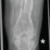
A plain X-ray of the foot showed an ill-defined sclerotic area in the calcaneum with radiating spicules, thinned overlying cortex, and soft-tissue edema over the heel (Fig. 1). Contrast-enhanced magnetic resonance imaging (MRI) of the left ankle confirmed the sclerotic lesion in the calcaneum with extraosseous component and enhancement with contrast. Alkaline phosphatase and lactate dehydrogenase were within the normal range and renal functions were also normal. An ultrasound-guided tru-cut biopsy was undertaken from the lesion, which on microscopy showed abundant osteoid matrix interspersed by pleomorphic cells with elongated oval to spindle hyperchromatic nuclei with increased areas of fibrous tissue, which was the hallmark of osteogenic sarcoma (Fig. 2). Fluorodeoxyglucose positron emission tomography-computed tomography scans (FDG PET-CT) showed no distant metastasis.

Treatment: The patient was given 3 cycles of chemotherapy (cisplatin+adriamycin) at 3-week intervals. After the chemotherapy, a repeat contrast MRI and FDG-PET-CT were undertaken to determine the size and extent of the disease and micrometastasis, which revealed a decrease in the standardized uptake values in the primary lesion and also the extent of the disease.

A limb salvage surgery was undertaken, which involved a posteromedial incision extending 5 cm from above the ankle to the base of first metatarsal along the watershed line (Fig. 3). The biopsy scar was excised, and the neurovascular bundle was isolated and separated. The flexor retinaculum was released, and the lateral plantar vessels had to be sacrificed because of their adherence to the tumor. The medial plantar vessels and nerves were isolated and separated (Fig. 4). The capsule was incised all around the subtalar joint and removed from the navicular and cuneiform bone. The tendon Achilles was resected 1 cm from its insertion, and the plantar fascia was resected 1 cm from its margins. After thorough dissection, a wide en bloc resection of the calcaneum was taken (Fig. 5). The procedure was uneventful. The sample was sent for histopathology, which confirmed osteosarcoma.
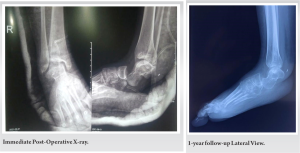
After the surgery, a below-knee splint was applied for 3 months, and the patient was advised non-weight-bearing ambulation. Sutures were removed at 2 weeks, and then three cycles of adjuvant chemotherapy were given. At 3 months of follow-up, partial weight bearing was allowed with elbow crutches and an ankle-foot orthosis. The patient was followed up every 6 weeks. At a 1-year follow-up, a customized silicon heel cup and shoes were given for full weight-bearing ambulation. At the end of 1 year, a PET-CT revealed no evidence of metabolically active disease.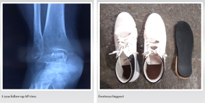
Osteosarcoma most commonly affects the metadiaphysis of long bones and very rarely flat bones or small bones such as the calcaneum. Calcaneal osteosarcoma is an extremely rare form of osteosarcoma, accounting for <1% of all cases [2,4]. This rarity and the lack of understanding of osteosarcomas in uncommonly affected areas make the diagnosis of calcaneal osteosarcoma challenging [5]. In this case, the patient presented with chronic heel pain, which was initially misdiagnosed as an overuse injury. It is important to note that the majority of osteosarcoma cases occur in individuals without any underlying genetic predisposition. However, some rare inherited syndromes have been associated with an increased risk of developing osteosarcomas, such as Werner syndrome, hereditary retinoblastoma, Li-Fraumeni syndrome, and Rothmund–Thompson syndrome [2,6]. In this case, the patient did not have any of these syndromes.
Routine radiographs are often sufficient to make a primary diagnosis of calcaneal osteosarcoma [3]. The characteristic finding of irregular spiculated interrupted periosteal reaction with dense sclerotic bone on radiographic images is a strong indication of calcaneal osteosarcoma as they typically show an ill-defined sclerotic area in the calcaneum with radiating spicules, thinned overlying cortex, and soft-tissue edema over the heel. However, to confirm the diagnosis and determine the local and distant extent of the disease, cross-sectional imaging studies such as contrast-enhanced MRI and FDG PET-CT scans are usually performed. Histopathological examination of a biopsy sample is always necessary to confirm the diagnosis and differentiate it from other lytic sclerotic lesions [7].
With more advanced treatments and improvements in surgical techniques, the survival rate for osteosarcoma has continued to increase. At present, 5-year survival rate for patients with localized osteosarcoma is approximately 60%, and after recurrences or metastases, it is only 20% [8]. It is important to note that early diagnosis and treatment are crucial for improving the prognosis in osteosarcoma. Neoadjuvant chemotherapy, before surgery, reduces the size of the primary tumor and makes it more amenable to surgery thereby increasing the chance of limb-salvage surgery and decreasing the risk of local recurrence. It also allows us to evaluate the response of the tumor to chemotherapy and make decisions about the surgery accordingly. Adjuvant chemotherapy after surgery reduces the risk of micrometastasis and improves the outcome in patients with osteosarcoma. The choice between neoadjuvant and adjuvant chemotherapy depends on the patient’s condition, the size and location of the tumor, and the patient’s overall health. Both approaches have been used in the treatment of osteosarcoma with similar outcomes, but the use of neoadjuvant chemotherapy is becoming increasingly common [9,10].
In this case, the patient was treated with a combination of chemotherapy and limb salvage surgery. Neoadjuvant chemotherapy is used to reduce the size of the tumor before surgery and to prevent micrometastasis. The patient underwent a wide en bloc resection of the calcaneum and received adjuvant chemotherapy after the surgery. The patient was followed up for 1 year and had no recurrence of tumor. She was able to bear weight fully with a customized heel cup.
The case highlights the importance of early diagnosis and management of osteosarcoma in patients with chronic heel pain. The use of neoadjuvant chemotherapy before surgery and adjuvant chemotherapy post-surgery has greatly improved the survival rates in osteosarcoma patients. The patient’s case also highlights the importance of considering osteosarcoma as a differential diagnosis in adolescents who present with chronic heel pain, despite the rarity of the condition.
Prompt clinical and radiological diagnosis with timely management will prevent complications and morbidity in osteosarcoma.
References
- 1.Sarangi PK, Kumar ES, Mohanty J. Calcaneal osteosarcoma: An unusual cause of chronic pediatric heel pain. Oncol J India 2017;1:31. [Google Scholar | PubMed]
- 2.Taslakian B, Issa G, Saab R, Jabbour MN, Khoury NJ. Calcaneal osteosarcoma: A rare cause of heel pain in the paediatric population. BMJ Case Rep 2013;2013:bcr2012008497. [Google Scholar | PubMed]
- 3.Samal BP, Nayak C, Pradhan S, Sahoo TK, Jena AK, Pradhan S. Calcaneal osteosarcoma, a challenge for diagnosis: A rare case report and literature review. Oncol Discov 2015;3:2. [Google Scholar | PubMed]
- 4.Prater S, McKeon B. Osteosarcoma. In: StatPearls. Treasure Island, FL: StatPearls Publishing; 2022, 2023 . [Google Scholar | PubMed]
- 5.Mirabello L, Troisi RJ, Savage SA. Osteosarcoma incidence and survival rates from 1973 to 2004: Data from the surveillance, epidemiology, and end results program. Cancer 2009;115:1531-43. [Google Scholar | PubMed]
- 6.Calvert GT, Randall RL, Jones KB, Cannon-Albright L, Lessnick S, Schiffman JD. At-risk populations for osteosarcoma: The syndromes and beyond. Sarcoma 2012;2012:152382. [Google Scholar | PubMed]
- 7.Pattnaik K, Pradhan P, Kar A, Burma S, Panda S. Unusual histological variants and a rare bone involvement of osteosarcoma in a referral hospital. Oncol J India 2017;1:2-6. [Google Scholar | PubMed]
- 8.Allison DC, Carney SC, Ahlmann ER, Hendifar A, Chawla S, Fedenko A, et al. A meta-analysis of osteosarcoma outcomes in the modern medical era. Sarcoma 2012;2012:704872. [Google Scholar | PubMed]
- 9.Misaghi A, Goldin A, Awad M, Kulidjian AA. Osteosarcoma: A comprehensive review. SICOT J 2018;4:12. [Google Scholar | PubMed]
- 10.Hiraga H, Ozaki T. Adjuvant and neoadjuvant chemotherapy for osteosarcoma: JCOG bone and soft tissue tumor study group. Jpn J Clin Oncol 2021;51:1493-7. [Google Scholar | PubMed]


Related Articles in Journal of Orthopaedic Case Reports
 February 1, 2026 Case Report of Post-Traumatic Monoarticular Tuberculosis of the Knee in a Healthy Young Adult: Diagnostic and Therapeutic Challenges in a Non-Endemic Setting
 January 1, 2026 Case Report: Foreign Body-induced Pseudomonas Arthritis Mimicking Tuberculous Ankle Infection in a Child
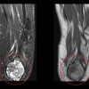 October 1, 2025 Young Female with Calcaneal Chondroblastoma & Secondary Aneurysmal Bone Cyst: Case Report
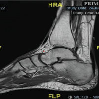 September 1, 2025 Calcaneus and Traumatic Stress Fracture