The surgical technique of a construct that improves fixation for endoprosthesis of the proximal femur is described.
Dr. Hernando Gaitan Lee. Hip and Knee adult reconstructive surgeon. E-mail: gaitanh@javeriana.edu.co
Introduction: Aseptic and septic loosening by mechanical failure is one of the main causes of proximal femur endoprosthesis failure with different clinical consequences such as pain, inflammation, progressive loosening of muscular volume, and strength with functional limitation. We present a case series of four patients with aseptic and septic loosening of femur endoprosthesis that was treated with a novel technique that combines different methods to achieve primary stability.
Case Report: Four patients with an average age of 49.5 years (35–70 years), two women and two men, were referred to the Orthopedic Surgery Department of the San Ignacio University Hospital of Bogota. Two of them with a history of oncological disease, ruling out a tumor relapse through local images and extension examinations; another patient with a history of firearm injury to the left thigh with multiple secondary complications, which required various reconstructive procedures; and the oldest patient with a history of total hip replacement. The main complaint of the four patients was thigh pain not caused by trauma and initial assessment radiographs of all of them showed signs of loosening of the prosthetic material, with septic etiology in two of the patients and aseptic etiology in the other two. Therefore, they were scheduled for revision surgery of prosthetic components. These patients recover functional status and resolved pain in the early post-operative period and during long-term follow-up. Only one of the patients, time after the surgical procedure, required removal of the osteosynthesis material due to persistent bone infectious process.
Conclusion: Septic and aseptic loosening is one of the most common complications of proximal femur endoprosthesis, resulting in significant pain and functional decline in patients. We present a novel surgical technique that allows primary stabilization of the construct that allows early rehabilitation, improvement of functionality, and no signs of new loosening.
Keywords: Proximal femur endoprosthesis, aseptic loosening, septic loosening, mechanical failure, augmentation, condylar plate, polymethylmethacrylate.
Proximal femur replacement has been one of the standard procedures in orthopedic oncology to reconstruct large bone resections of the proximal femur after primary tumors and metastasis [1]. This technique has expanded to non-oncologic indications such as severe femoral bone loss secondary to a failure of primary total hip replacement or multiple revision procedures [2]. Although long-term survival of the implant and functionality is achieved [3], different complications have been described, being aseptic loosening of the femoral stem one of the main ones, which has been reported to reach 93.8% after 10 years of index surgery [4]. We report a case series of four patients, two women and two men, with different medical records, treated with proximal femoral endoprosthesis, who subsequently presented aseptic loosening (2 patients) and septic loosening (2 patients), so they were taken to surgical management with a novel augmentation technique to ensure primary stability of the construct.
This case series includes four patients with an average age of 49.5 years (35–70 years), two women and two men, referred to the Orthopedic Surgery Department of the San Ignacio University Hospital at the Pontificia Universidad Javeriana of Bogota. Two of them with a history of oncological disease (proximal femoral chondrosarcoma and inguino-vulvar myofibroblastic sarcoma), assessed in conjunction with the oncological orthopedic service, ruling out a tumor relapse through local images and extension exams; another patient with a history of firearm injury to the left thigh with multiple secondary complications, which required various reconstructive procedures; and the oldest patient with a history of total hip replacement. The main complaint of the four patients was pain not caused by trauma, and none presented fever or associated systemic inflammatory response symptoms. Initial assessment radiographs of all patients showed signs of loosening of the prosthetic material and with microbiological cultures were established, septic etiology of the loosening in two of the patients and aseptic etiology in the other two patients (Fig. 1). Septic loosening was treated by a two-stage joint revision, once the patient showed no clinical signs of infection, C-reactive protein and arthrocentesis where negative, que second stage was performed. As a preliminary study, an arthrocentesis was performed in the four patients and active periprosthetic joint infection was ruled out, so they were scheduled for revision surgery of prosthetic components. Outpatient clinical control was carried out in an average time of 2 years (8 months–4 years), beginning at 2, 4, and 8 weeks after surgery. The patients present complete resolution of pain; surgical wounds were completely healed without local signs of infection. Femur radiographs showed the implant in adequate position, without any sign of loosening or failure. However, one of the patients (history of firearm injury) required the removal of the osteosynthesis material due to the persistence of the bone infectious process 1 year after the surgical procedure.
Surgical technique
The patient is placed in lateral decubitus position with standard skin cleaning and preparation with chlorhexidine and iodide solution and iodide surgical drape. A hip posterolateral extended approach to the femoral diaphysis is performed to expose the entire endoprosthesis. The endoprosthesis is removed and resection of a segment of femoral diaphysis is done. This resection was performed to remove all devitalized bone tissue. Intraoperatively, the vitality and bleeding of the bone is verified, any segment that shows that it is in poor condition must be resected. Next distal femur preparation is performed with femoral reamers. Retrograded cementation and insertion of the new femoral cemented stem of greater diameter; coupling and fixing the rest of the endoprosthesis. Afterward dissection and exposure of the distal femur application of condylar support plate by a MIPO technique or using a provisional fixation with two Kirschner nails. The position is verified under image intensifier. Definitely, fixation was done with 5 distal locked screws and 2 periprosthetic screws on the distal diaphysis of the femur. Application of a cement mantle to achieve an adequate interface between the endoprosthesis and the plate and that the holes of the plate become full with cement is an essential step that allows better stability (Figs. 2 and 3). Finally, proximal fixation of the plate is performed with surgical cables which are tightened and closed. Inpatient management and rehabilitation follow during the first 3 days, allowing immediate mobilization of adjacent joints and weight-bearing. Follow-up was carried out until the 4th post-operative year. No patient required revision or presented radiological signs of loosening.
Aseptic and septic loosening of the femoral stem is one of the major causes of implant failure in proximal femur endoprosthesis [5]. The main symptom is pain, which usually occurs in the middle third of the thigh, leading to progressive functional decline as in our patient. The initial approach must rule out periprosthetic joint infection because it would implicate variations in management options [6]. We present a group of patients in which a novel surgical technique is used to provide primary stability of a proximal femur endoprosthesis is achieved using multiple methods. In the first instance, thicker steam was used to decrease the possibility of movement [7].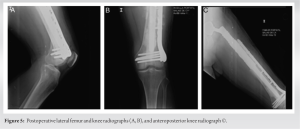 Retrograde cementing technique was performed, which is widely known since Barrack et al. proved in 1992 that it decreases the loosening rate significantly [8]. Recently, in 2019, Phelon et al. reported that cemented femoral endoprosthesis is a versatile option that allows immediate full weight-bearing. Augmenting the construct with a condylar plate fixed with periprosthetic screws and surgical cables is an option that ensures more rigidity in primary stability. Literature shows its use, especially in distal femur periprosthetic fractures [9]. Song et al. reported that fixation of periprosthetic fractures with locking plates provided satisfactory results with a low risk of complications and additional surgeries [10]. In addition, a cement mantle was built between the endoprosthesis to achieve an adequate interface between the endoprosthesis and the plate and avoid metallosis or corrosion between the different metals. Using the following keywords, a literature search was performed on PUBMED and EMBASE: Proximal femur endoprosthesis, aseptic loosening, Augmentation, condylar plate, polymethylmethacrylate. We found no articles reporting this technique in aseptic loosening of proximal femur endoprosthesis. Different therapeutic options have been studied for endoprosthesis aseptic loosening including the use of bone allografts, especially for large femoral bony defects [11], use of wire mesh and cerclage wiring with impaction bone allograft into which the femoral stem is implanted [12], and total femur prosthesis, which provides good functional results but high rates of complications such as infection (13–18%), dislocation (6–10%), and material failure (3–6%) [13, 14]. In the case of periprosthetic infection, the approach must ensure infection control in the first instance to ensure better results. The revision could be performed in one or two stages with reinfection rates of up to 37% [15]. The clinical relevance of our technique is that it allows early mobilization and weight-bearing, allowing rapid rehabilitation and return to the previous functionality. Our series of patients shows good clinical results on the immediate post-operative period, 2 weeks and 2 months and 4 years follow-up, especially with complete resolution of thigh pain and no radiological signs of development of new loosening of the endoprosthesis (Fig. 4 and 5).
Septic and aseptic loosening is one of the most common complications of proximal femur endoprosthesis, resulting in significant pain and functional decline in patients. We present a novel surgical technique that allows primary stabilization of the construct that allows early rehabilitation, improvement of functionality, and no signs of new loosening in a period of 4-year follow-up.
One of the main objectives of orthopedic surgery is to restore the functionality and quality of life of patients. In this paper, we present a novel surgical technique to treat septic and aseptic loosening of proximal femur endoprosthesis that allows to recover functionality in the short, medium, and long term.
References
- 1.Farid Y, Lin PP, Lewis VO, Yasko AW. Endoprosthetic and allograft-prosthetic composite reconstruction of the proximal femur for bone neoplasms. Clin Orthop Relat Res 2006;442:223-9. [Google Scholar]
- 2.Viste A, Perry KI, Taunton MJ, Hanssen AD, Abdel MP. Proximal femoral replacement in contemporary revision total hip arthroplasty for severe femoral bone loss: A review of outcomes. Bone Joint J 2017;99-B:325-9. [Google Scholar]
- 3.Bernthal NM, Schwartz AJ, Oakes DA, Kabo JM, Eckardt JJ. How long do endoprosthetic reconstructions for proximal femoral tumors last? Clin Orthop Relat Res 2010;468:2867-74. [Google Scholar]
- 4.Liang H, Guo W. Reconstruction in orthopaedic oncology: Frontier and horizon. Ann Joint 2020;5:19. [Google Scholar]
- 5.Henderson ER, Groundland JS, Pala E, Dennis JA, Wooten R, Cheong D, et al. Failure mode classification for tumor endoprostheses: Retrospective review of five institutions and a literature review. J Bone Joint Surg Am 2011;93:418-29. [Google Scholar]
- 6.Shehadeh A, Noveau J, Malawer M, Henshaw R. Late complications and survival of endoprosthetic reconstruction after resection of bone tumors. Clin Orthop Relat Res 2010;468:2885-95. [Google Scholar]
- 7.Savino AW, Andersson GB, Andriacchi TP, Hampton S, Galante JO. The influence of femoral stem thickness and implantation technique on the strength of the bone cement bond. Acta Orthop Scand 1982;53:23-7. [Google Scholar]
- 8.Barrack RL, Mulroy RD Jr., Harris WH. Improved cementing techniques and femoral component loosening in young patients with hip arthroplasty. A 12-year radiographic review. J Bone Joint Surg Br 1992;74:385-9. [Google Scholar]
- 9.Tomás T, Nachtnebl L, Otiepka P. Distal femoral periprosthetic fractures: Classification and therapy. Acta Chir Orthop Traumatol Cech 2010;77:194-202. [Google Scholar]
- 10.Song SJ, Kim KI, Song WJ, Kim DK, Bae DK. Treatment of distal femur fractures with locking plates: Comparison of periprosthetic fractures above total knee arthroplasty and non-periprosthetic fractures. Acta Orthop Belg 2014;80:380-90. [Google Scholar]
- 11.Rogers BA, Sternheim A, Backstein D, Safir O, Gross AE. Proximal femoral allograft for major segmental femoral bone loss: A systematic literature review. Adv Orthop 2011;2011:257572. [Google Scholar]
- 12.Schoof B, Jakobs O, Gehrke T, Gebauer M. Proximal femoral reconstruction after aseptic loosening following proximal femoral replacement for ewing sarcoma: A case report with one-year follow-up. Hip Int 2014;24:103-7. [Google Scholar]
- 13.Friesecke C, Plutat J, Block A. Revision arthroplasty with use of a total femur prosthesis. J Bone Joint Surg Am 2005;87:2693-701. [Google Scholar]
- 14.Medellin MR, Fujiwara T, Clark R, Stevenson JD, Parry M, Jeys L. Mechanisms of failure and survival of total femoral endoprosthetic replacements. Bone Joint J 2019;101-B:522-8. [Google Scholar]
- 15.Funovics PT, Hipfl C, Hofstaetter JG, Puchner S, Kotz RI, Dominkus M. Management of septic complications following modular endoprosthetic reconstruction of the proximal femur. Int Orthop 2011;35:1437-44. [Google Scholar]