Counsel patients who present with isolated digital dactylitis of premature growth arrest and potential functional ramifications.
Dr. Pranai Buddhdev, Broomfield Hospital, Chelmsford, Essex, UK. E-mail: p.buddhdev@nhs.net
Introduction: Following trauma, premature growth arrest is a common outcome when the injury affects the pediatric growth plate. Dactylitis describes global inflammation affecting one or more digits in the hand or foot. It occurs in various seronegative arthropathies and septic arthritis. Physeal fusion following dactylitis is uncommon and is not described in the current literature.
Case Presentation: We report the case of a 12-year-old boy, whose minor non-penetrating injury resulted in circumferential edema of his left third upper limb digit, typical of dactylitis. No evidence of infection was found during clinical examination or blood work. Significant stiffness of the digit remained over the course of a few months with spontaneous resolution following functional hand therapy. The child presented to pediatric orthopedics with cessation of longitudinal growth. Evidence of premature physeal fusion of the involved phalanges was confirmed on radiographs.
Conclusion: Growth arrest following dactylitis has not previously been reported. Clinicians managing this condition should be aware of this rare complication. We recommend that inflammation is treated promptly, and patients are monitored clinically and radiologically to address any potential functional deficit.
Keywords: Dactylitis, pediatric orthopedics, orthopedics, rheumatology, growth arrest, premature growth arrest.
Dactylitis is defined as global inflammation of a digit, affecting the digital flexor tendon sheaths, and occasionally causing synovitis of the joints involved. Published literature reports that up to 52% of cases of dactylitis involve the joints themselves, with radiographic evidence of bony erosion in 50% [1]. Dactylitis results in a characteristic sausage-like appearance of the digit. It is most associated with infection, peripheral spondyloarthritis (including psoriatic arthritis [PsA]), sickle cell disease, osteoid sarcoma, sarcoidosis, and microcrystalline deposition [2]. Physeal fusion occurs naturally during puberty, signifying skeletal maturity and determining our adult bone size. In premature growth arrest, longitudinal and/or appositional growth is interrupted following insult to the growth plates before skeletal maturity [3]. Common etiologies include trauma, infection, irradiation, ischemia, and iatrogenic injury, with the most commonly reported cause of premature growth arrest being skeletal trauma around the physis [3]. We report the first case of premature growth arrest following dactylitis in a 12-year-old boy affecting the left middle finger.
A prepubescent 12-year-old boy of normal stature and without significant medical history was referred to the pediatric orthopedic clinic after presenting to his family doctor with a chronically painful and swollen left middle finger. A minor non-penetrating injury caused the development of dactylitis within 72 h, with a classic presentation of circumferential edema and limited movement persisting 2 weeks following the injury. Initial radiographic investigations identified soft-tissue swelling but no associated fracture, bony pathology, evidence of a penetrating foreign body, or osteomyelitis (Fig. 1). Routine biochemical and hematological investigations showed no evidence of infection or raised inflammatory markers.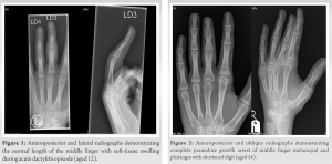
Significant stiffness of the isolated affected digit persisted for 12 months, requiring functional hand therapy to restore function and return to normal activity.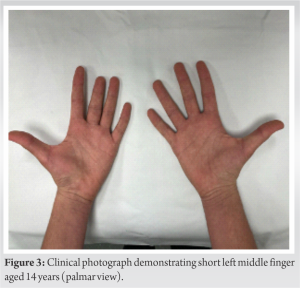
Despite the patient remaining asymptomatic, it was observed that interstitial growth of the affected finger had ceased (Fig. 2); presenting clinically with a shortened digit and radiological evidence of complete growth arrest of all the physes of phalanges with loss of cortical definition and a loss of joint space in the PIPJ and distal metacarpals. The length of phalanges of the affected middle finger remained unchanged from the year; he presented with dactylitis, but its diameter had increased slightly, causing it to be clinically shortened compared to his other fingers by approximately 1 cm (Figs. 3 and 4). At the final follow-up, the patient presented with pain-free full range of movement and normal power and pincer grips (Fig. 5). Imaging confirmed the absence of ongoing active inflammation in both joints and soft-tissues following pediatric rheumatology review.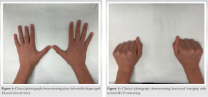
Dactylitis is more commonly recorded in the literature as a complication of a disease state rather than a condition causing complications itself. Causes of dactylitis include infection, inflammatory arthropathies and spondyloarthropathies (especially Juvenile idiopathic arthritis and PsA), gout, sarcoidosis, and sickle cell disease. Underlying infections that predispose to dactylitis include chronic infections such as Mycobacterium tuberculosis, Treponema pallidum, or Mycobacterium leprae [4]. Spina ventosa (tuberculous dactylitis of the bones of the hands and feet) is uncommon in patients after age 6 [5]. Dactylitis is also commonly associated with penetrating wounds, foreign bodies, and interventions such as metalwork implantation. This is particularly true of external fixation and the use of percutaneous devices such as K-wires, which are commonly complicated by pin-site infections [6, 7]. Trauma to a flexor crease is frequently associated with pyogenic flexor tenosynovitis, which presents similarly to dactylitis, but without bony involvement [8]. Joints affected by arthritis usually demonstrate advanced growth (pannus formation) due to angiogenesis and hyper-vascularization as a direct result of inflammatory cascades [9]. In PsA, osteoclast action at the bone-tendon junction causes characteristic bone erosions and enthesitis, and this inflammatory process can also cause dactylitis. The link between chronic inflammation, osteogenesis, and bone remodeling could be due to the interactions of VEGF and its receptors. Following fractures, hematoma formation occurs, leading to an inflammatory cascade and VEGF secretion with subsequent angiogenesis [10]. This inflammatory process is a requirement for secondary healing [11]. However, VEGF receptors are also located on the surface of osteoblasts and osteoclasts, thus it is plausible that VEGF at the growth plate is required for conversion of fibrous to bony repair tissue and bone bridge formation through direct (intramembranous ossification) and indirect (endochondral ossification) bone formation mechanisms [12]. This may cause undesirable conversion in chronic inflammation such as chronic dactylitis, leading to premature epiphyseal fusion.
We provide evidence for post-dactylitis growth arrest in a prepubescent patient. Children who present with dactylitis need to be formally assessed in pediatric rheumatology to ensure accurate diagnosis and prompt treatment of inflammation. Our paper has shown that follow-up is vital even in the absence of ongoing inflammation due to the potential for premature growth arrest and functional deficit which requires hand therapy.
Resolution of chronic dactylitis in children is significant as failure to treat or otherwise resolve the issue can lead to premature growth arrest of the digit, which could cause functional deficits as well as psychosocial issues, for example, bullying by peers. Parents and patients should also be made aware of the possibility of growth arrest.
References
- 1.Kaeley SG, Eder L, Aydin SZ, Gutierrez M, Bakewell C. Dactylitis: A hallmark of psoriatic arthritis. Semin Arthritis Rheum 2018;48:263-73. [Google Scholar]
- 2.Dabash S, Prabhakar G, Potter E, Thabet AM, Abdelgawad A, Heinrich S. Management of growth arrest: Current practice and future directions. J Clin Orthop Trauma 2018;9 Suppl 1:S58-66. [Google Scholar]
- 3.Brockbank JE, Stein M, Schentag CT, Gladman DD. Dactylitis in psoriatic arthritis: A marker for disease severity? Ann Rheum Dis 2005;64:188-90. [Google Scholar]
- 4.Woodworth MH, Marquez C, Chambers H, Luetkemeyer A. Disabling dactylitis and tenosynovitis due to Mycobacterium haemophilum in a patient with human immunodeficiency virus/acquired immune deficiency syndrome. Open Forum Infect Dis 2017;4:ofx165. [Google Scholar]
- 5.Abebe W, Abebe B, Molla K, Alemayehu T. Tuberculous dactylitis: An uncommon presentation of skeletal tuberculosis. Ethiop J Health Sci 2016;26:301-3. [Google Scholar]
- 6.Kofman KE, Buckley T, McGrouther DA. Complications of transcutaneous metal devices. Eur J Plast Surg 2012;35:673-82. [Google Scholar]
- 7.Kazmers NH, Fragomen AT, Rozbruch SR. Prevention of pin site infection in external fixation: A review of the literature. Strategies Trauma Limb Reconstr 2016;11:75-85. [Google Scholar]
- 8.Clark DC. Common acute hand infections. Am Fam Physician 2003;68:2167-76. [Google Scholar]
- 9.Schell H, Duda GN, Peters A, Tsitsilonis S, Johnson KA, Schmidt-Bleek K. The haematoma and its role in bone healing. J Exp Orthop 2017;4:5. [Google Scholar]
- 10.Marsell R, Einhorn TA. The biology of fracture healing. Injury 2011;42:551-5. [Google Scholar]
- 11.Chung R, Xian CJ. Recent research on the growth plate: Mechanisms for growth plate injury repair and potential cell-based therapies for regeneration. J Mol Endocrinol 2014;53:T45-61. [Google Scholar]
- 12.Strunk J, Heinemann E, Lange U. A new approach to study angiogenesis in rheumatoid arthritis by power Doppler sonography and serum vascular endothelial growth factor level measurement. Arthritis Res Ther 2004;6 Suppl 1:103. [Google Scholar]