

Multiple high-dose intra-articular corticosteroid injections may be associated with an accelerated risk of developing rapidly destructive osteoarthritis, even in younger male patients not typically considered high-risk.
Dr. Sharath Raj, Department of Orthopedic Surgery, Postgraduate Institute of Medical Education and Research, Chandigarh, India. E-mail: sharathraj.pgi@gmail.com
Abstract
Introduction: Rapidly destructive osteoarthritis (RDO) is a rare and severe form of joint degeneration with an unclear aetiology. Recently, intra-articular corticosteroid injections, a common treatment for osteoarthritis, have been implicated as a potential iatrogenic cause. This case is important as it documents this phenomenon in an atypical patient demographic, strengthening the evidence for this serious adverse event.
Case Report: A 55-year-old male with a history of mild-to-moderate left hip osteoarthritis received two fluoroscopically guided, high-dose intra-articular injections of 80 mg Kenalog 6 months apart. Following the second injection, he experienced a rapid and severe worsening of pain and functional limitation. Radiographs taken 6 months post-injection revealed complete joint space loss and significant femoral head collapse, consistent with RDO. The patient subsequently underwent a successful total hip arthroplasty.
Conclusion: This case strengthens the association between multiple, high-dose intra-articular steroid injections and the subsequent development of RDO. The clinical implication is that clinicians must be aware of and counsel patients on this potential severe adverse event, particularly when considering repeated high-dose injections as a management strategy for hip osteoarthritis.
Keywords: Rapidly destructive osteoarthritis, hip osteoarthritis, intra-articular injections, glucocorticoids, total hip arthroplasty, adverse effect.

Rapidly destructive osteoarthritis (RDO), first described in 1957, is a rare and poorly understood subset of degenerative hip disease characterised by swift joint deterioration [1]. In 1970, a formal definition was established, requiring a joint space reduction of at least 50%, or >2 mm of femoral head bone loss, over a 1-year period in the absence of other identifiable causes of destructive arthropathy [2]. Pathological features include flattening of the femoral head, subchondral bone damage, and a notable absence of significant osteophyte formation. The condition typically affects women in their seventh or eighth decade of life and presents unilaterally in 80–90% of cases. This typical patient profile makes the presentation in younger or male patients particularly noteworthy [3,4,5]. While the underlying pathophysiology remains debated, factors such as subchondral insufficiency fractures and labral inversion have been proposed. More recently, attention has turned toward a potential iatrogenic cause: intra-articular glucocorticoid injections [6,7,8,9]. These injections are a cornerstone of non-surgical management for symptomatic hip osteoarthritis, providing effective short-term pain relief and improved function. However, a growing number of reports suggest a link between these injections and the subsequent rapid collapse of the joint [10]. This report presents the case of a 55-year-old male who developed RDO of the hip after receiving multiple high-dose glucocorticoid injections. This case contributes to the body of evidence on this serious complication and highlights the need for caution and thorough patient counselling. The patient was informed that data concerning his case would be submitted for publication, and he provided consent.
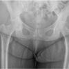
A 55-year-old male with a medical history of gastro-oesophageal reflux disease and hyperlipidaemia initially presented to his primary care physician in 2019 with a 2-year history of left groin pain. Initial radiographs showed mild-to-moderate hip osteoarthritis. After conservative management with non-opioid analgesics and physical therapy failed to provide relief, he was referred to an orthopaedic specialist. Repeat imaging confirmed mild-to-moderate joint space narrowing with subchondral sclerosis and cysts (Fig. 1).
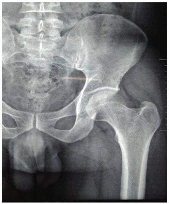
Figure 1: Anteroposterior radiograph of the left hip at initial orthopedic consultation. Images demonstrate mild-to-moderate superior joint space narrowing with subchondral sclerosis and subchondral cysts within the lateral acetabular roof.
After discussing treatment options, the patient opted for non-surgical management and was referred for a fluoroscopically guided intra-articular injection. He received his first injection of 80 mg Kenalog with 3 mL of 0.5% Marcaine into the left hip joint in June 2021. Due to persistent symptoms, a second injection of the same dosage was administered in December 2022 (Fig. 2).
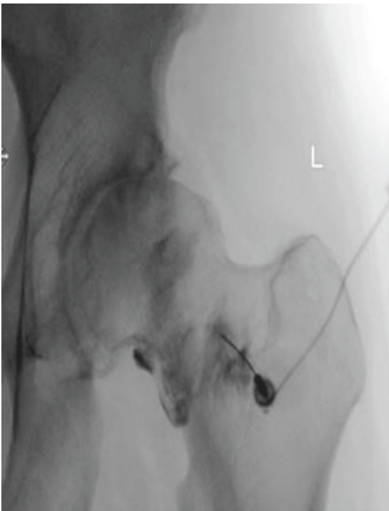
Figure 2: Anteroposterior view of the left hip during a fluoroscopically guided joint injection. Radiograph demonstrates mild-to-moderate superior joint space narrowing with mild sclerosis in the superior acetabulum. No significant osteophytes or erosions are seen.
Following this second injection, the patient’s condition deteriorated dramatically. Within 6 months, he developed severe, debilitating pain that disrupted his sleep and significantly impaired his mobility. On returning to the orthopaedic clinic in 2023, new radiographs revealed severe joint destruction. The images demonstrated near-complete loss of the joint space, flattening and collapse of the femoral head, and a relative lack of osteophytosis, findings highly suggestive of RDO (Fig. 3).
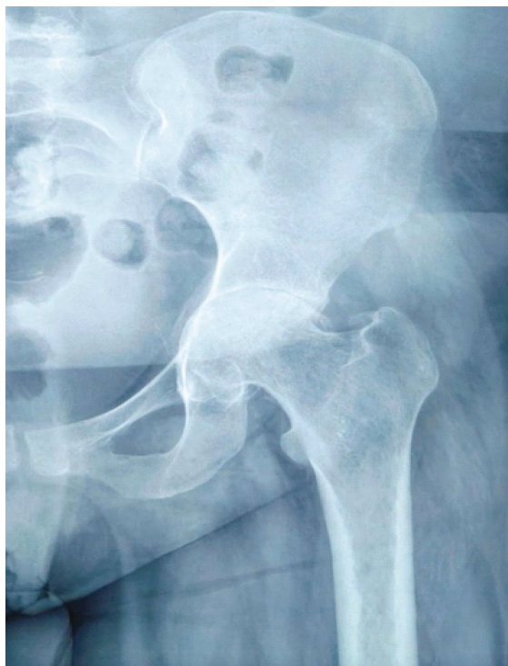
Figure 3: Anteroposterior radiograph of the left hip taken 6 months after the second steroid injection. Images demonstrate severe sclerosis of the weight-bearing aspect of the left femoral head with near-complete loss of joint space, femoral head flattening, and minimal marginal osteophytosis, consistent with rapidly destructive osteoarthritis.
Given the severity of his symptoms and radiographic findings, the patient elected to undergo a left total hip arthroplasty (THA) in September 2023. Intraoperative findings confirmed a flattened femoral head, extensive erosion of articular cartilage, and destruction of the subchondral bone. The procedure was performed without complications, and post-operative radiographs showed excellent implant positioning (Fig. 4).
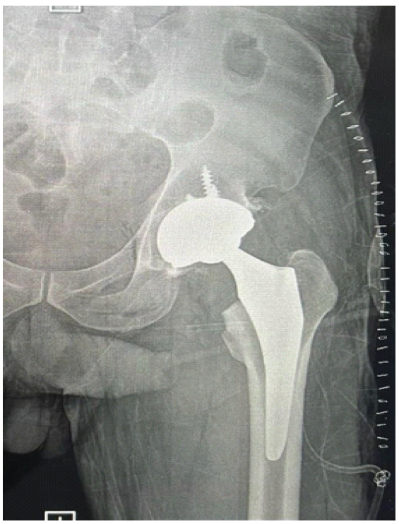
Figure 4: Anteroposterior radiograph of the left hip immediately following total hip arthroplasty. The image demonstrates the prosthesis in proper anatomical alignment with no evidence of periprosthetic fracture or dislocation.
At his 1-year follow-up in September 2024, the patient reported a complete resolution of pain and had returned to his baseline functional status. His surgical incision was well-healed, and follow-up imaging confirmed the prosthesis remained in proper alignment with no complications (Fig. 5).
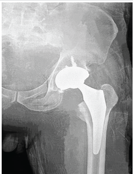
Figure 5: Anteroposterior radiograph of the left hip at 1-year post-operative follow-up. The image demonstrates the left total hip arthroplasty components in stable and proper anatomical alignment with no complicating features.
This case illustrates a potential severe adverse event following a common orthopaedic intervention. The patient’s rapid joint destruction over a 6–12 month period, characterised by femoral head collapse with a notable absence of osteophytes, is the hallmark of RDO. Histologically, RDO is associated with granulomatous foci containing bone and cartilage debris, alongside increased osteoclast activity, suggesting a process of rapid bone breakdown that outpaces normal resorption mechanisms. The radiographic findings in our patient, particularly the lack of osteophytes seen in Fig. 3, align perfectly with this pathological description, strengthening the diagnosis. The central issue raised by this case is the role of intra-articular steroid injections in triggering this destructive process. It has been hypothesised that glucocorticoids may induce chondrolysis, leading to subsequent joint damage. The temporal relationship in this patient is compelling: A stable, albeit osteoarthritic, hip underwent catastrophic failure within months of receiving a second high-dose injection. This aligns with a critical finding from a recent large-scale study by Okike et al., which demonstrated a dose-dependent and frequency-dependent risk. That study found that while the risk of RDO after a single low-dose injection (<40 mg of triamcinolone) was low, the risk escalated significantly with high-dose injections (defined as >80 mg) and with repeated treatments. The patient in this report received two separate 80 mg injections of Kenalog, placing him squarely in the highest-risk category identified in the literature and providing a strong, evidence-based link between his treatment and the adverse outcome. Furthermore, this case is significant because of the patient’s demographic profile. RDO is most commonly reported in elderly women, typically in their 70s or 80s. The development of RDO in a 55-year-old male is an unusual presentation. This deviation from the typical patient profile suggests a powerful pathogenic trigger. It raises the question of whether exposure to high-dose intra-articular steroids can lower the threshold for developing RDO, causing it to manifest in patient populations previously considered to be at low risk. This possibility expands the clinical concern beyond the traditionally susceptible elderly female population. Once RDO is diagnosed, THA is the definitive treatment, as conservative measures rarely halt disease progression. However, THA in this population can be challenging due to significant bone loss, which may lead to longer surgical times and the need for specialised implants. A primary limitation of this report is that it is a single case, which precludes definitive conclusions about causality. However, by documenting this event in detail, it serves as an essential contribution to the cumulative evidence.
This case report documents the development of RDO of the hip in a 55-year-old male following multiple high-dose intra-articular steroid injections. It adds to the growing evidence that this common treatment for osteoarthritis carries a risk of severe, iatrogenic joint destruction. The case is particularly noteworthy due to the atypical patient demographic, suggesting the risk may be broader than previously understood. Healthcare providers must remain vigilant for this potential complication and engage in thorough patient counselling regarding the risks, benefits, and alternatives to high-dose or repeated intra-articular steroid injections.
Clinicians should exercise caution when considering multiple or high-dose (>80 mg) intra-articular steroid injections for hip osteoarthritis, as they may trigger rapidly destructive osteoarthritis. This potential severe adverse event must be discussed with patients as part of the informed consent process.
References
- 1. Lequesne M, De Seze S, Amouroux J. La coxarthrose destructrice rapide. Rev Rhum Mal Osteoartic 1970;37:721-33. [Google Scholar] [PubMed]
- 2. Yamamoto T, Bullough PG. The role of subchondral insufficiency fracture in rapid destruction of the hip joint: A preliminary report. Arthritis Rheum 2000;43:2423-7. [Google Scholar] [PubMed]
- 3. Fukui K, Kaneuji A, Fukushima M, Matsumoto T. Inversion of the acetabular labrum triggers rapidly destructive osteoarthritis of the hip: representative case report and proposed aetiology. J Arthroplasty 2014;29:2468-72. [Google Scholar] [PubMed]
- 4. Bock GW, Garcia A, Weisman MH, Major PA, Lyttle D, Haghighi P, et al. Rapidly destructive hip disease: Clinical and imaging abnormalities. Radiology 1993;186:461-6. [Google Scholar] [PubMed]
- 5. Hu L, Zhang X, Kourkoumelis N, Shang X. The mysteries of rapidly destructive arthrosis of the hip joint: A systemic literature review. Ann Palliat Med 2020;9:1220-9. [Google Scholar] [PubMed]
- 6. Yamamoto T, Schneider R, Iwamoto Y, Bullough PG. Rapid destruction of the femoral head after a single intraarticular injection of corticosteroid into the hip joint. J Rheumatol 2006;33:1701-4. [Google Scholar] [PubMed]
- 7. Tiwari A, Karkhur Y, Keeney JA, Aggarwal A. Rapid destructive osteoarthritis of the hip after intra-articular steroid injection. Arthroplast Today 2018;4:184-6. [Google Scholar] [PubMed]
- 8. Laroche M, Arlet J, Mazieres B. Osteonecrosis of the femoral and humeral heads after intraarticular corticosteroid injections. J Rheumatol 1990;17:549-51. [Google Scholar] [PubMed]
- 9. Hess SR, O’Connell RS, Bednarz CP, Waligora AC 4th, Golladay GJ, Jiranek WA. Association of rapidly destructive osteoarthritis of the hip with intra-articular steroid injections. Arthroplast Today 2018;4:205-9. [Google Scholar] [PubMed]
- 10. Okike K, King RK, Merchant JC, Toney EA, Lee GY, Yoon HC. Rapidly destructive hip disease following intra-articular corticosteroid injection of the hip. J Bone Joint Surg Am 2021;103:2070-9. [Google Scholar] [PubMed]





Related Articles in Journal of Orthopaedic Case Reports
 November 1, 2025 Treatment of Hip Osteoarthritis with Severe Acetabular Bone Defects by Total Hip Arthroplasty Using the Restoration GAP II Reinforcement Ring: A Case Report
 October 1, 2026 Dual Mobility Total Hip Arthroplasty for Recurrent Dislocation After Femoral Neck Fracture in an Elderly Patient: A Case Report
 October 1, 2026 Late Diagnosis of Femoral Shaft Perforation During Total Hip Arthroplasty: Case Report
 October 1, 2026 Real-Time Coagulation Monitoring via Thromboelastography and Platelet Mapping in Total Hip Arthroplasty for a Patient with Factor XI Deficiency: A Case Report