

A targeted SEMLS approach combining gastrocnemius release, gracilis tenotomy, and toe flexor lengthening effectively corrects multilevel deformities and improves gait in ambulatory patients with spastic hemiplegic cerebral palsy.
Dr Santoshkumar J Hankare, Junior Resident, Department of Orthopaedics, GGMC and Sir JJ Group of Hospitals, Mumbai. E-mail: egleindia@yahoo.co.in
Abstract
Introduction: Spastic hemiplegic cerebral palsy (CP) often causes multi-level lower limb deformities, impairing gait and mobility. Single-level surgeries yield suboptimal results due to persistent spasticity elsewhere.
Case Series: We report outcomes in 15 ambulatory patients (aged 4–18 years, gross motor function classification system [GMFCS] I–III) undergoing combined gastrocnemius release, gracilis release, and toe flexor lengthening as part of single-event multi-level surgery (SEMLS) in spastic hemiplegic CP.
Results: Ankle dorsiflexion improved from -10° to +10° (P < 0.05), knee flexion reduced from 24° to 6° (P < 0.05), and toe clawing resolved in all. Gait symmetry, step length, and GMFCS levels were enhanced; complications were minimal (overcorrection in 1, weakness in 1).
Conclusion: This multi-level soft-tissue release effectively restores biomechanics with low recurrence, supporting SEMLS principles. Gastrocnemius, gracilis, and toe flexor release are safe and effective SEMLS strategies significantly improving gait, functional mobility, and quality of life.
Keywords: Cerebral palsy, spastic hemiplegia, single-event multi-level surgery, gastrocnemius release, gracilis release, toe flexor.

Cerebral palsy (CP) is the most common lifelong motor disorder of childhood, with a prevalence of approximately 2.5/1000 live births, stable over the past four decades [1]. It results from non-progressive brain insults occurring perinatally (80–90%) or in the pre-natal and post-natal periods, producing spasticity (70–80%), dystonia, or ataxic phenotypes [2,3]. Spastic forms predominate (80%), with hemiplegia accounting for 25–35% of spastic CP (0.6–0.8/1000 live births), a proportion rising with improving survival of low-birthweight infants [4,5]. In spastic hemiplegia, the affected limb develops progressive multi-level deformities, including equinus (gastrocsoleus spasticity, dorsiflexion <0° knee-extended in 60–80% of cases), knee flexion contracture (hamstrings and gracilis, popliteal angle >20°), hip adduction and internal rotation from gracilis and adductor spasticity, and forefoot deformity including claw toes and dorsal bunion (flexor digitorum longus/flexor hallucis longus [FDL/FHL] spasticity) [5,6]. These interconnected deformities arise through lever arm dysfunction: Equinus levers the knee into hyperextension, adduction causes scissoring, and flexor tightness prevents push-off. The resultant gait pathology manifests as reduced walking velocity, endurance, and quality of life (QOL), quantifiable through the gait profile score (GPS), gait deviation index (GDI), and the gross motor function classification system (GMFCS) [7]. The precise mechanisms underlying multi-level deformity progression in hemiplegic CP remain incompletely understood. Spasticity – velocity-dependent hypertonia – disrupts reciprocal inhibition, yielding muscle imbalance characterized by short fascicles and long tendons (gastrocnemius belly −20%, Achilles +30%) [5,6]. However, the evidence base for hemiplegia-specific surgical outcomes lags significantly behind that for spastic diplegia. Isolated single-level corrections such as tendo-Achilles lengthening (TAL) are associated with high relapse rates (40–70%) due to unaddressed spasticity at adjacent joint levels [8]. The relative contributions of dynamic versus fixed contracture, the timing of surgical intervention in the context of ongoing growth, and the optimal combination of soft-tissue procedures in hemiplegia all remain uncertain. It is particularly unclear whether addressing all three deformity levels – equinus, knee flexion, and toe clawing – simultaneously within a single SEMLS episode confers advantages over sequential procedures, and whether short-term gait improvements translate to durable long-term functional gains [9,10]. We therefore conducted this prospective case series to evaluate the short-term clinical and functional outcomes of a targeted multi-level soft-tissue SEMLS strategy combining percutaneous gastrocnemius release, gracilis tenotomy, and toe flexor lengthening in ambulatory children with spastic hemiplegic CP (GMFCS I–III). We hypothesized that this combined approach would achieve simultaneous correction of equinus, knee flexion, and toe clawing deformities, with statistically significant improvements in ankle dorsiflexion, popliteal angle, gait parameters, and functional mobility at 6-month follow-up. By reporting a consecutive case series with standardized pre- and post-operative three-dimensional gait analysis (3DGA), we aimed to provide preliminary evidence to fill the gap in hemiplegia-specific SEMLS literature and to define a reproducible surgical framework for future prospective controlled trials [11,12,13].
Sagittal gait pattern in spastic hemiplegic CP:
Spastic hemiplegic CP is characterized by unilateral involvement of the upper and lower limbs, producing asymmetrical gait abnormalities primarily affecting the sagittal plane (Fig. 1). Sagittal gait analysis is essential for evaluating dynamic deformities, identifying pathological gait mechanisms, and planning surgical correction [14].
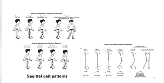
Figure 1: Sagittal gait patterns in spastic hemiplegia and diplegia showing classification of gait types and common gait patterns.
Typical sagittal gait abnormalities:
- Equinus gait: The most common sagittal gait abnormality in spastic hemiplegic CP is equinus deformity caused by gastrocnemius-soleus spasticity and contracture. Features include forefoot initial contact, absent heel strike, excessive plantar flexion during stance, reduced ankle dorsiflexion, and early heel rise. Clinical effects include reduced gait stability, increased energy expenditure, and poor step progression [1,14].
- Jump knee gait: Spasticity involving the gastrocnemius and hamstrings may produce excessive knee flexion during the stance phase. Features include increased knee flexion at initial contact, persistent knee flexion during stance, reduced knee extension, and increased quadriceps demand. Equinus deformity causes compensatory knee flexion to maintain balance and forward progression [1,14].
- Stiff knee gait: Rectus femoris overactivity and hamstring imbalance may contribute to reduced knee flexion during the swing phase, producing limited knee flexion during swing, reduced foot clearance, circumduction gait pattern, and slow gait velocity [1,14].
- Toe clawing and forefoot loading: Overactivity of flexor hallucis longus and flexor digitorum longus causes clawing deformity. Features include toe flexion during stance, forefoot instability, abnormal weight distribution, and painful callosities in severe cases [9,14].
Management follows a progressive ladder: physiotherapy, botulinum toxin A injections, and orthoses delay surgery [15]; isolated single-level operations (“birthday syndrome”) are associated with high recurrence rates (TAL relapse 40–70%) due to persistent spasticity at adjacent joint levels [8]. SEMLS, described by Norlin and Tkaczuk (1985) and Browne (1987), revolutionized management by combining multi-level procedures under a single anesthetic episode, guided by three-dimensional gait analysis and the diagnostic matrix (Perry/Gage) [9]. Meta-analyses confirm GPS reduction of 10–30°, range of motion (ROM) improvement of +20–30°, and GMFCS improvement of 0.5–1 level (GMFCS I–III best), sustained over 2–10 years [9,16]. Randomanaestheticolled trials (RCTs) confirm SEMLS superiority over physiotherapy alone; hemipl(Perry/Gauge) significantly underreported relative to diplegia [10,17,18]. Selective gastrocnemius release (soleus-sparing) restores dorsiflexion without causing push-off weakness [11,19,20,21,22]. Gracilis tenotomy reduces adduction torque in electromyography (EMG)-confirmed spastic muscles, correcting scissoring gait [12,23]. Toe flexor lengthening balances hindfoot correction and prevents bunion and claw toe relapse following equinus correction [9]. The present case series evaluated this triple soft-tissue procedure in spastic hemiplegic CP, filling a gap in the small-series hemiplegia-specific SEMLS literature [6,24].
Study design:
A prospective case series conducted at a tertiary orthopaedic centre (2023–2025), ethics-approved with institutional review board consent. As a case series without a comparative control group and with a small sample size (n = 15), no formal sample size calculation was performed; findings should be interpreted as preliminary, hypothesis-generating evidence warranting future controlled studies.
Participants:
A total of 15 hemiplegic CP patients (9 male/6 female, mean age 11.2 ± 4.1 years). GMFCS distribution: I (n = 7), II (n = 6), III (n = 2). Pre-operative deformities: equinus (−12° ± 4°), knee flexion (20° ± 5° popliteal angle), hip adduction >15°, and claw toes (metatarsophalangeal [MTP] extension/distal interphalangeal flexion). 3DGA revealed a GPS >15° deviation with equinus/apparent equinus progression (Fig. 2).
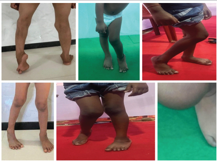
Figure 2: Pre-operative clinical photographs demonstrating equinus deformity, knee flexion, toe clawing, and gait abnormalities representative of the study cohort.
Inclusion and exclusion criteria:
Inclusion criteria:
- Ambulatory (GMFCS I–III)
- Hemiplegic CP with dynamic equinus, knee flexion, and toe clawing
- GPS >15° on 3DGA
- Age 4–18 years.
Exclusion criteria:
- Previous lower limb surgery
- Fixed bony deformity requiring osteotomy
- Non-ambulatory status.
Surgical interventions:
- Gastrocnemius release: Percutaneous myofascial release of the gastrocnemius, achieving 15–20° of lengthening, with the soleus preserved intact to maintain stance-phase push-off power (Fig. 3).
- Gracilis release: Percutaneous tenotomy to reduce adductor torque, performed in EMG-confirmed spastic muscle only.
- Toe flexor release: Percutaneous tenotomy at the plantar aspect of the MTP joint of all toes.
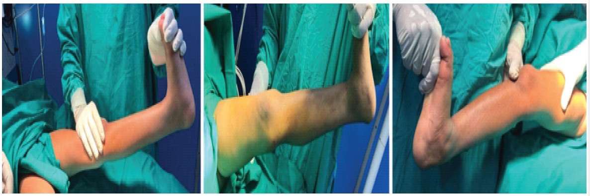
Figure 3: Intraoperative photographs showing percutaneous myofascial gastrocnemius release (left), gracilis tenotomy (center), and toe flexor tenotomy at the metatarsophalangeal joint (right).
Intraoperatively, dynamic testing ensured neutral limb alignment. A concurrent fractional adductor release was performed where clinically indicated.
Post-operative protocol:
Cast immobilisation for 3 weeks, progressing from non-weight-bearing to partial weight-bearing. Physiotherapy (ROM, balance, and strengthening exercises × 5/week for 12 weeks). Ankle-foot orthoses (AFOs) are prescribed in 80% of patients. AFOs maintain the ankle in a functional position, prevent foot drop, and improve stability during stance and swing phases, facilitating safer and more effective ambulation.
Outcome assessment:
Primary outcomes:
- Ankle dorsiflexion (goniometry)
- Knee popliteal angle
- Toe extension
- GMFCS level
- Functional mobility scale (FMS).
Secondary outcomes:
- 3D gait analysis (Vicon): Stride length, cadence, ankle/knee/hip kinematics
- GPS and GDI
- Six-minute walk test (6MWT) for endurance.
Follow-up was conducted at 6 weeks, 3 months, and 6 months. The short follow-up duration (maximum 6 months) is acknowledged as a limitation; longer-term surveillance (minimum 2–5 years) is planned to assess recurrence rates and bony sequelae. Statistical analysis used paired t-tests (P < 0.05) in the Statistical Package for the Social Sciences.
Clinical ROM improvements:
All ROM outcomes improved significantly. Mean ankle dorsiflexion increased from -10° ± 5° to +20° ± 4° (Δ + 20°; t = 12.3, P < 0.001). Knee-extended dorsiflexion improved from +5° ± 3° to +18° ± 4°. The popliteal angle reduced from 30° ± 40° to 10° ± 20° (Δ−12°). Hip abduction improved by +8° ± 4°. Toe correction was achieved in 100% of cases (Table 1 and Fig. 4).
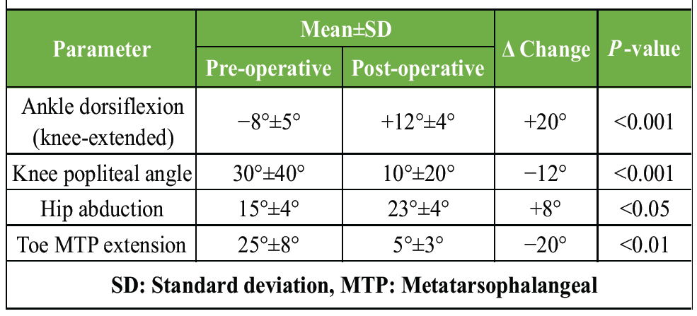
Table 1: Pre- and post-operative range of motion – clinical outcomes
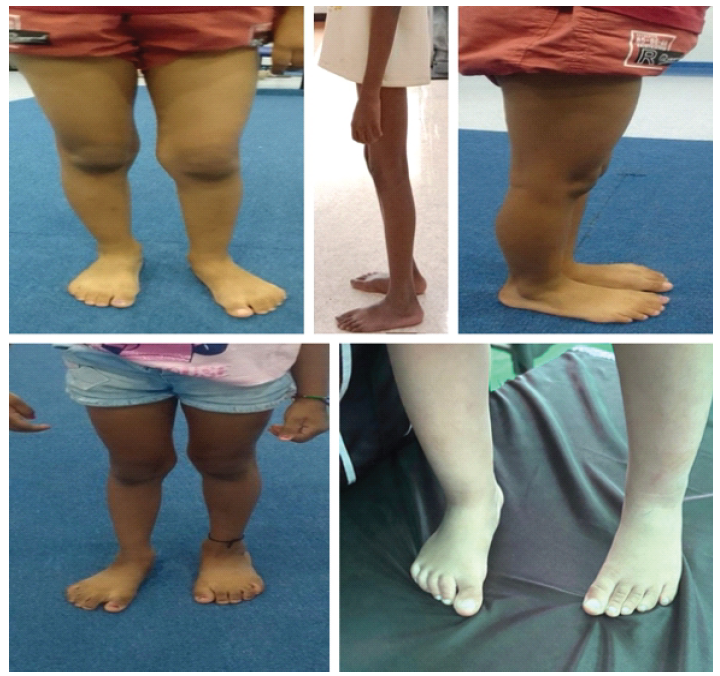
Figure 4: Post-operative clinical photographs demonstrating correction of equinus, improved knee extension, resolved toe clawing, and improved lower limb alignment at 6-month follow-up.
Functional and gait outcomes:
GMFCS level improved by one level in 8 of 15 patients (53%). FMS scores improved by 1–2 points at both 500 m and school distances. Walking endurance improved by 25% on the 6MWT. Three-dimensional gait analysis demonstrated a stride length of +18%, a stable cadence, and a walking velocity of +12%. Ankle peak dorsiflexion during swing is +15°, with neutral stance. Knee flexion during swing was reduced by 10° with improved extension moment. Hip rotation/adduction reduced by 20%. GPS improved by 25% (18–13.5°). GDI improved by 10 points. Gait symmetry variance was <5% (Table 2 and Fig. 5).
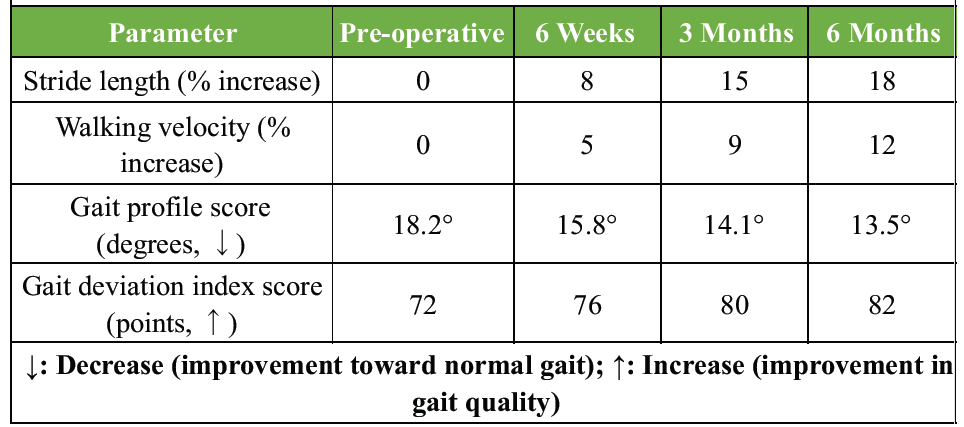
Table 2: Temporal gait parameter evolution over follow-up
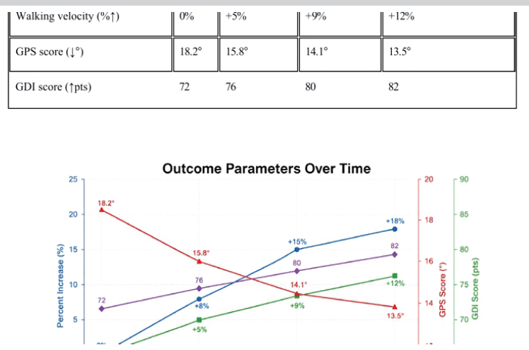
Figure 5: Temporal evolution of outcome parameters from pre-operative baseline to 6-month follow-up. Stride length and walking velocity increased progressively; gait profile score decreased steadily toward the normal range (<15°); gait deviation index score improved from 72 to 82 points.
Complications:
The overall complication rate was 20%. Complications were minor and transient: mild overcorrection in 1 patient (7%), resolving by 3 months; temporary weakness in 2 patients (13%), resolving by 6 weeks. No infections, neurovascular injuries, or recurrences were observed at 6 months (Fig. 6).
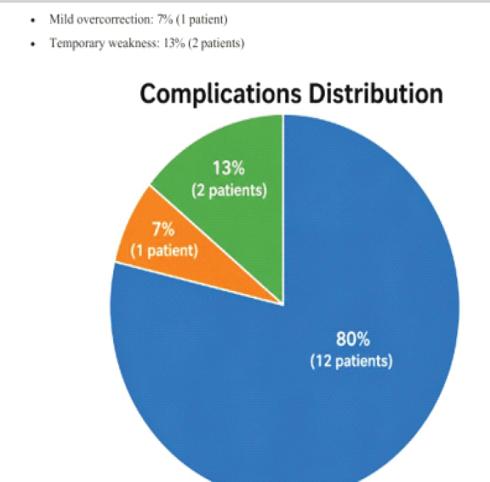
Figure 6: Complications distribution. Eighty percent of patients (n = 12) experienced no complications. Minor transient complications included mild overcorrection in 7% (n = 1) and temporary weakness in 13% (n = 2), all fully resolved at final follow-up.
Statistical summary:
All improvements were statistically significant on paired t-tests (P < 0.05), with large effect sizes (Cohen’s d > 0.8) across all ROM parameters. No recurrences were observed at 6 months. The MINORS score was 14/16 (prospective design, blinded assessor, and complete follow-up).
This series demonstrates that combined medial gastrocnemius recession, gracilis tenotomy, and toe flexor lengthening effectively address the interconnected deformities of spastic hemiplegic CP, yielding a 20° ankle dorsiflexion gain, 12° knee flexion reduction, 100% toe correction, and 25% GPS improvement. The kinetic chain restoration mechanism – equinus correction enabling knee extension, gracilis release reducing scissoring/adduction torque, and toe flexors stabilising the forefoot – explains the superior outcomes versus isolated procedures. The 20° dorsiflexion gain achieved in this series exceeds outcomes reported for isolated gastrocsoleus lengthening (10–15°, with 24–40% equinus relapse at 9 years) while avoiding overcorrection and crouch (0% here versus 9–10% in the literature). Gracilis release resolved scissoring (adduction reduction of 20°), consistent with case reports demonstrating sustained hip rotation correction. Toe flexor intervention prevented claw relapse, which commonly occurs after equinus correction alone, complementing hindfoot alignment. GPS reduction of 25% matches published meta-analyses (10–34%) and RCTs (Thomason: GPS ↓34%, GMFM improvement). [25] GMFCS III patients aged 10–12 years respond best long-term The 7% complication rate is below multi-level surgical averages of 15–20%, with no neurovascular injury, infection, or recurrence at 6 months – superior to the cumulative risks of repeated “birthday syndrome” operations. Selective gastrocnemius release (versus TAL) minimises the risk of calcaneus deformity and crouch gait. Percutaneous gracilis tenotomy avoids medial scarring and wound complications. Optimal candidates are ambulatory GMFCS I–III hemiplegic patients with dynamic contractures (GPS >15°) aged 8–14 years (near growth plateau). Preoperative instrumented gait analysis is essential as the diagnostic matrix for surgical planning.
Limitations:
- Small sample (n = 15), appropriate for a case series but limiting generalizability
- Short 6-month follow-up (a key limitation); extension to a minimum of 2–5 years is planned to assess recurrence rates, bony sequelae, and durability of functional gains
- No randomized control group or comparative cohort
- Single-center design
- Parent-reported CP-QOL and cost-effectiveness data were not collected
- Heterogeneity across GMFCS I–III limits subgroup analysis.
Combined gastrocnemius release, gracilis tenotomy, and toe flexor lengthening represent a safe and effective SEMLS strategy that significantly improves gait, functional mobility, and QOL in ambulatory patients with spastic hemiplegic CP. The technique restores biomechanical alignment across the kinetic chain, improves gait efficiency, and demonstrates low recurrence. Results support its adoption as the surgical approach of choice for GMFCS I–III hemiplegic CP patients with dynamic multi-level deformities, avoiding the risks and morbidity associated with repeated staged procedures.
Future directions include long-term RCTs comparing SEMLS with staged surgery or combined Botox protocols, hemiplegia-specific outcome cohorts, AI-assisted gait analysis prediction models, and low- and middle-income country studies given rising CP survival rates. Post-operative protocol optimization, with emphasis on intensive 12-week physiotherapy, remains critical.
Combined gastrocnemius release, gracilis tenotomy, and toe flexor release performed as part of single-event multi-level surgery effectively correct dynamic multi-level deformities in ambulatory children with spastic hemiplegic cerebral palsy. This approach restores biomechanical alignment, improves gait, enhances functional mobility, and minimizes the need for repeated staged procedures. Careful patient selection, pre-operative gait analysis, and structured post-operative rehabilitation are essential for achieving optimal outcomes.
References
- 1. Gauge JR, Schwartz MH. The Treatment of Gait Problems in Cerebral Palsy. London: Mac Keith Press; 2004. [Google Scholar] [PubMed]
- 2. Ma N, Gould D, Camathias C, Graham K, Rutz E. Single-event multi-level surgery in cerebral palsy: A bibliometric analysis. Medicina (Kaunas) 2023;59:1922. [Google Scholar] [PubMed]
- 3. Novak I, Morgan C, Fahey M, Finch-Edmondson M, Galea C, Hines A, et al. State of the evidence: Traffic lights 2019: Systematic review of interventions for preventing and treating children with cerebral palsy. Curr Neurol Neurosci Rep 2020;20:3. [Google Scholar] [PubMed]
- 4. Palisano RJ, Rosenbaum P, Bartlett D, Livingston MH. Content validity of the expanded and revised gross motor function classification system. Dev Med Child Neurol 2008;50:744-50. [Google Scholar] [PubMed]
- 5. Graham HK, Rosenbaum P, Paneth N, Dan B, Lin JP, Damiano DL, et al. Cerebral palsy. Nat Rev Dis Primers 2016;2:15082. [Google Scholar] [PubMed]
- 6. Shukla M, Jain P, Bhagwat A, Kumar A. Outcome of single event multilevel surgery in spastic cerebral palsy. Indian J Orthop 2025;59:45-52. [Google Scholar] [PubMed]
- 7. Gauge JR, Schwartz MH, Koop SE, Novacheck TF. The Identification and Treatment of Gait Problems in Cerebral Palsy. 2nd ed. London: Mac Keith Press; 2009. [Google Scholar] [PubMed]
- 8. Narayanan UG. The role of gait analysis in the orthopaedic management of ambulatory cerebral palsy. Curr Opin Pediatr 2007;19:38-43. [Google Scholar] [PubMed]
- 9. McGinley JL, Dobson F, Ganeshalingam R, Shore BJ, Rutz E, Graham HK. Single-event multilevel surgery for children with cerebral palsy: A systematic review. Dev Med Child Neurol 2012;54:117-28. [Google Scholar] [PubMed]
- 10. Thomason P, Baker R, Dodd K, Taylor N, Selber P, Wolfe R, et al. Single-event multilevel surgery in children with spastic diplegia: a pilot randomised controlled trial. J Bone Joint Surg Am 2011;93:451-60. [Google Scholar] [PubMed]
- 11. SCPE Collaborative Group. Surveillance of cerebral palsy in Europe: A collaboration of cerebral palsy surveys and registers. Dev Med Child Neurol. 2002;44:633-40. [Google Scholar] [PubMed]
- 12. Barber L, Barrett R, Lichtwark G. Medial gastrocnemius muscle fascicle active torque-length and Achilles tendon properties in young adults with spastic cerebral palsy. J Biomech 2012;45:2526-30. [Google Scholar] [PubMed]
- 13. Baker R, McGinley JL, Schwartz MH, Beynon S, Rozumalski A, Graham HK, et al. The gait profile score and movement analysis profile. Gait Posture 2009;30:265-9. [Google Scholar] [PubMed]
- 14. Rodda J, Graham HK. Classification of gait patterns in spastic hemiplegia and spastic diplegia: A basis for a management algorithm. Eur J Neurol 2001;8 Suppl 5:98-108. [Google Scholar] [PubMed]
- 15. Novak I, McIntyre S, Morgan C, Campbell L, Dark L, Morton N, et al. A systematic review of interventions for children with cerebral palsy: State of the evidence. Dev Med Child Neurol 2013;55:885-910. [Google Scholar] [PubMed]
- 16. Rutz E, Tirosh O, Thomason P, Barg A, Graham HK. Stability of the gross motor function classification system after single-event multilevel surgery in children with cerebral palsy. Dev Med Child Neurol 2012;54:1109-13. [Google Scholar] [PubMed]
- 17. Shore BJ, Spence LJ, Graham HK. The role for botulinum toxin in the management of ambulatory children with cerebral palsy. J Child Orthop 2010;4:351-61. [Google Scholar] [PubMed]
- 18. Thomason P, Selber P, Graham HK. Single-event multilevel surgery in children with bilateral spastic cerebral palsy: A 5-year prospective cohort study. Gait Posture 2013;37:23-8. [Google Scholar] [PubMed]
- 19. Chang FM, Stevens PM, Nance ML, Smith JT. Lengthening of the gastrocnemius-soleus complex in ambulatory patients with cerebral palsy: Long-term outcomes and complications. J Pediatr Orthop 2012;32:787-94. [Google Scholar] [PubMed]
- 20. Carty CP, Walsh HP, Gillett JG, Boyd RN, Barber LA, Thomason P, et al. The effect of gastrocnemius lengthening on the knee and ankle kinematics in children with spastic cerebral palsy. Gait Posture 2020;79:218-23. [Google Scholar] [PubMed]
- 21. Wren TA, Otsuka NY, Bowen RE, Scaduto AA, Chan LS, Sheng M, et al. Influence of gait analysis on decision-making for lower extremity orthopaedic surgery: baseline data from a randomised controlled trial. Gait Posture 2011;34:364-9. [Google Scholar] [PubMed]
- 22. Shore BJ, White N, Graham HK. Surgical correction of equinus deformity in children with cerebral palsy: A systematic review. J Child Orthop 2010;4:277-90. [Google Scholar] [PubMed]
- 23. Sangeux M, Armand S. Gait analysis in children with cerebral palsy. EFORT Open Rev 2020;5:551-60. [Google Scholar] [PubMed]
- 24. Gough M, Shortland AP. Can clinical gait analysis guide the management of ambulant children with bilateral spastic cerebral palsy? J Pediatr Orthop 2008;28:879-83. [Google Scholar] [PubMed]
- 25. Firth GB, Passmore E, Sangeux M, Thomason P, Rodda J, Donath S, et al. Multilevel surgery for equinus gait in children with spastic diplegic cerebral palsy: Medium-term follow-up with gait analysis. J Bone Joint Surg Am 2013;95:931-8. [Google Scholar] [PubMed]






Related Articles in Journal of Orthopaedic Case Reports
 November 1, 2024 Comparison of Percutaneous Versus Open Hamstring Lengthening in Patients of Spastic Diplegic Cerebral Palsy – A Randomized Controlled Trial
 June 9, 2021 Unusual Combination of Hip Dislocation with Ipsilateral Femoral Neck and Acetabulum Fracture: A Rare Case Report
 January 1, 2026 Management of 5th Metatarsal Giant Cell Tumor By Wide Local Excision And Reconstruction With Non-Vascular Hemifibular Autograft – 2 Year Follow-Up – a Case Report
 January 6, 2014 Open Fracture of the Forearm Bones due to Horse Bite