

In case of failure of fixation or revision surgery for acromio-clavicular joint disruptions, it is advisable to go for hybrid reconstruction techniques, focusing on anatomical fixation with augmentation.
Dr. Ranajit Panigrahi, Department of Orthopaedics, Hitech Medical College, Flat 3A, Aurogreen Apartments, Behind Omfed Plant, Chandravihar, Chandrashekharpur, Bhubaneswar - 751017, Odisha, India. E-mail: ranajitpanigrahi@gmail.com
Abstract
Introduction: Biological augmentation of anatomic acromio-clavicular joint (ACJ) reconstruction should be considered in chronic ACJ dislocations because in chronic cases, there is a lack of intrinsic healing of the ligaments. Hybrid techniques that involve the combination of fibertapes or tightropes with tendon grafts have demonstrated native-like biomechanical properties and are thus a suitable mode of reconstruction for chronic ACJ dislocations.
Case Report: We report a case of a 39-year-old right-hand-dominant male who presented to us with failed infected ACJ fixation with a hook plate along with wound dehiscence. The patient presented with pain, deformity, exposed implant, and restricted mobility of the shoulder. The patient was managed in two stages. In the first stage, implant removal was done along with debridement, and infection was controlled with suitable antibiotics. In the second stage, the chronic ACJ dislocation was managed by Mumford’s procedure and hybrid fixation technique using an ACJ fixation device with semitendinosus tendon graft augmentation. Post-operative outcomes showed no signs of infection and considerable improvement in shoulder stability and range of movements.
Conclusion: Anatomical ACJ reduction with suture button devices along with biological augmentation with a tendon graft is a more anatomical and definitive method of fixation of ACJ dislocations, particularly in chronic cases.
Keywords: Acromioclavicular joint, hook plate, hybrid technique, biological augmentation.

Surgical management of chronic acromio-clavicular joint (ACJ) instability has been rarely discussed in literature, because whereas low-grade injuries are often successfully managed conservatively, surgical stabilization of acute high-grade injuries often yields reliable and satisfying results [1]. When conservative management or primary ACJ stabilization fails, chronic instability of the ACJ can develop [2]. For chronic ACJ injuries, surgery is indicated after failed non-operative treatment of 3–6 months [3,4]. Biological augmentation of anatomic ACJ reconstruction should be considered in chronic ACJ dislocations because in chronic cases, there is a lack of intrinsic healing of the ligaments [5]. In addition, hybrid techniques that involve a combination of fibertapes or tightropes with tendon grafts have demonstrated native-like biomechanical properties and are thus a suitable mode of reconstruction for chronic ACJ dislocations [1,6,7]. We share our experience of treating a failed infected ACJ fixation with a hook plate in situ with a hybrid reconstruction technique in combination with distal clavicular resection [8]. Patient’s consent has been obtained for the submission of this case report.
A 39-year-old, right-hand-dominant male presented to our outpatient department after a history of self-fall with a history of pain and restricted mobility of the right shoulder. The patient gave a history of a road traffic accident 1 month prior, was diagnosed as a case of Rockwood type V ACJ dislocation of the right shoulder, and was surgically managed elsewhere with a hook plate. However, the patient continued having pain, swelling, and restricted mobility of the right shoulder with erythema and foul-smelling discharge from the surgical site along with gaping of the lateral end of the surgical stitch line. Now, on local examination of the right shoulder, we find dehiscence of the lateral end of the surgical stitch line from the previous surgery, along with purulent discharge and the implant exposed through the gaping wound. An X-ray of the right shoulder anteroposterior view revealed a dislocated ACJ with a displaced and failed hook plate in situ (Fig. 1).
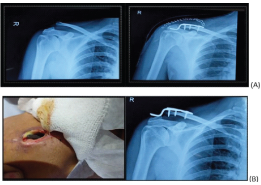
Figure 1: Image series showing clinical picture (a) and the X-ray of the right shoulder antero-posterior view (b) revealed a dislocated acromio-clavicular joint with a displaced and failed hook plate in situ.
Routine pre-surgical blood investigations revealed raised total leukocyte count, raised erythrocyte sedimentation rate (ESR), and C-reactive protein (CRP), indicating definite signs of infection. Management of the case was planned in two stages. In the first stage, implant removal and debridement of the wound were planned. An incision was given in line with the primary surgical scar, soft tissue dissection was carried out, and the failed clavicular hook plate was removed. Swabs were taken, and samples were sent for Gram staining and culture-sensitivity tests. All infected tissue was removed, the margins of the wound debrided, and after thorough lavage, the wound was closed and antiseptic dressing applied. The culture came back positive for Staphylococcus aureus, and the patient was started on intravenous antibiotics (piperacillin+tazobactam and linezolid) as per the antibiotic sensitivity report. After 3 weeks of intravenous antibiotics, the patient was switched to oral antibiotics (oral clindamycin and linezolid) for another 3 weeks (total 6 weeks). At the end of 6 weeks, complete blood count, ESR, and CRP were repeated, and reports were within normal limits. The wound was examined, and the stitch line was found to be dry and healthy without any signs of active infection (Fig. 2).
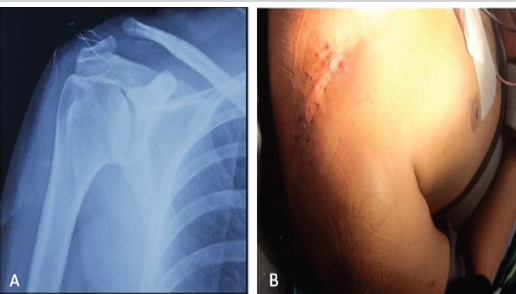
Figure 2: Image series showing the X-ray of the right shoulder antero-posterior view (a) and clinical picture (b) showing the first stage of surgery with the implant removed and stitch line dry and healthy without any sign of infection.
Now, definitive surgery was planned in the second stage. The case was treated as a case of chronic ACJ dislocation and was planned for hybrid reconstruction using an ACJ fixation device along with semi-tendinosus augmentation. The patient was placed in a sloppy beach chair position, and both the affected shoulder and the ipsilateral knee were painted and draped in a sterile manner. Bony landmarks were outlined before surgery. The semitendinosus tendon was harvested from the ipsilateral knee and prepared in a standard manner. After skin incision, soft tissue dissection was carried out till we exposed the dislocated ACJ by dissecting through the clavi-pectoral fascia. After exposing the dislocated ACJ, we tried to reduce the joint, but since it was a chronic dislocation, reduction was not possible, and we proceeded to perform distal clavicle excision or the Mumford procedure. After the distal clavicle resection, the clavicle could be reduced to the acromion, and the ACJ could be relocated (Fig. 3).
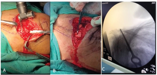
Figure 3: Image series showing after skin and soft tissue dissection and exposing the distal clavicle and acromio-clavicular joint, distal clavicular excision was done using a bone saw (Mumford procedure) to reduce the chronic dislocation (a). Once the excision is completed, dislocated clavicle is attempted to be reduced using bone hook (b). As seen in the C-Arm image, after excision, the dislocated clavicle can be reduced using a bone hook (c).
Now, on the clavicle, we mark two points, 2 cm and 3.5 cm, from the distal end of the clavicle to mark the attachments of the conoid and trapezoid ligaments of the coraco-clavicular complex. After marking, we drill through and through across the clavicle (Fig. 4).
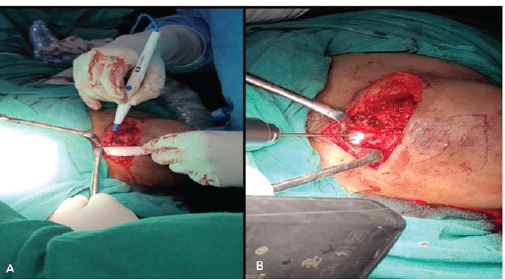
Figure 4: Image series showing the two points 2 cm and 3.5 cm from the distal end of clavicle to mark the attachments of conoid and trapezoid ligaments of the coraco-clavicular complex (a) and drilling through and through across the clavicle (b).
The coracoid is now exposed and drilled through and through for passing the acromio-clavicular (AC) tightrope. Now, with the help of nitinol wire, we pass the AC tightrope device across the coracoid beneath-upward till the AC tightrope button rests on the undersurface of the coracoid (Fig. 5).
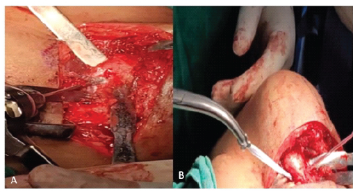
Figure 5: Image series showing exposing the coracoid and then being drilled through and through for passing the acromio-clavicular (AC) joint tightrope. (a) Then, with the help of nitinol wire, we pass the AC tightrope device across the coracoid beneath-upward, till the AC tightrope button rests on the undersurface of the coracoid (b).
Now, from each of the drilled holes in the clavicle, a nitinol wire is passed one after the other, and using the wires, suture arms of the AC tightrope are shuttled across the clavicle at the position of the conoid and trapezoid ligaments of the coraco-clavicular ligament complex. Now using a right-angled forceps, another fiber wire is passed and placed between the two suture arms. Now, the semi-T graft is shuttled across the coracoid and then looped across the clavicle in between the two suture arms of the AC tightrope (Fig. 6).
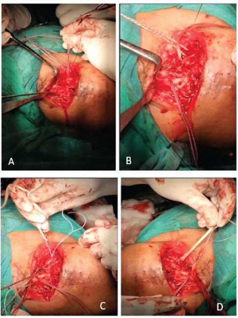
Figure 6: Image series showing passing of nitinol one after another through the drilled hole of the clavicle, (a) and then shuttling the suture arms of the tightrope system across the clavicle at the position of conoid and trapezoid ligament of the coraco-clavicular ligament complex. (b) Also using right-angled forceps, another fiber wire is passed and placed in between the two suture arms, and the Semi-T graft is shuttled across the coracoid (c) and then looped across the clavicle in between the two suture arms (d).
Each of the suture limbs is now passed through an Arthrex dog button, and the sutures are then tied over the dog button on the surface of the clavicle. Now using a bone hook, the ACJ is row-reduced, and in this reduced position, the suture limbs are tightened and knotted over the dog buttons, and the ACJ is stabilized. Now, the reduction is augmented with the semi-T graft looped around the coracoid and brought around the clavicle in between the suture limbs, and the two limbs are knotted and then stabilized with sutures, thus augmenting the ACJ stabilization (Fig. 7).
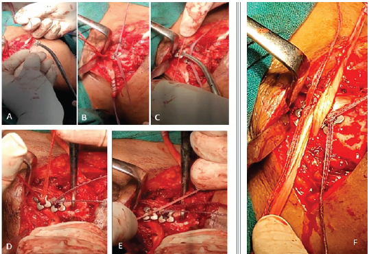
Figure 7: Image series showing each of the suture limbs being passed through Arthrex™ Dog Button (a and b) and the sutures are then tied over the dog-button on the surface of the clavicle (c) and using a bone hook, the acromio-clavicular joint (ACJ) is row reduced and in this reduced position (d), the suture limbs are tightened and knotted over the dog buttons, and the ACJ is stabilized (e). The reduction is augmented with the Semi-T Graft looped around the coracoid and brought around the clavicle in between the suture-limbs, and the two limbs are knotted and then stabilized with sutures (f).
Immediate intraoperative C-arm images and postoperative images showed a reduced and stable ACJ. Patient presented with a healthy and healed suture line, no wound dehiscence, and no sign of infection with improved range of motion of the shoulder (Fig. 8).
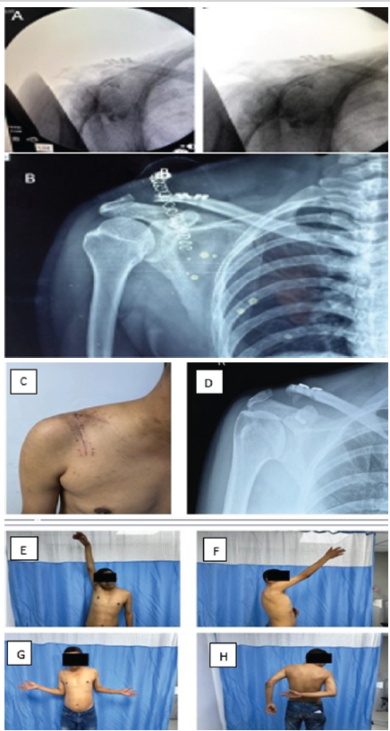
Figure 8: Immediate intra-operative C-ARM images (a) and immediate post-operative images showed a reduced and stable acromio-clavicular joint. (b). Three-month follow-up where the patient presented with healthy and healed suture line, no wound dehiscence, and no sign of infection (c and d) with clinically improved range of motion of the shoulder (e-h).
There exist a vast number of surgical treatment options for ACL injuries, and the current literature has yet to determine an equivocally superior treatment [1,2,3,4]. In chronic ACJ injuries, surgery is indicated after failed treatment for 3–6 months. Anatomic ACJ reconstruction techniques, along with biologic augmentation, should be considered for chronic high-grade instabilities, accounting for the lack of intrinsic healing and scar-forming potential of the ligamentous tissue in the chronic setting [1,7]. This approach has been demonstrated to provide better results regarding anterior and superior translation of the clavicle [9,10]. In addition, recent studies demonstrated native-like biomechanical properties of double graft tendons using hybrid techniques, such as the combination of tight ropes and fibertapes with tendon grafts [1,5,6].
This case report shows that anatomical ACJ double bundle reconstruction with suture button devices along with biological augmentation with a tendon graft is a more anatomical and definitive method of fixation of ACJ dislocations, particularly in chronic cases.
There are a number of techniques described for acute and chronic acromio-clavicular joint fixation. We should choose the proper technique for primary stabilization of acromio-clavicular joint fixation. Hook plates have been used for a long time for acromio-clavicular injuries, but complications and failures have been reported with their use. Hybrid techniques, which involve a combination of fibertapes or tightropes with tendon graft, have demonstrated native-like biomechanical properties and are thus a suitable mode of reconstruction for acromio-clavicular joint dislocations.
References
- 1. Berthold DP, Muench LN, Dyrna F, Mazzocca AD, Garvin P, Voss A, et al. Current concepts in acromioclavicular joint (AC) instability – a proposed treatment algorithm for acute and chronic AC-joint surgery. BMC Musculoskelet Disord 2022;23:1078. [Google Scholar] [PubMed]
- 2. De Groot C, Verstift DE, Heisen J, Van Deurzen DF, Van Den Bekerom MP. Management of acromioclavicular injuries – current concepts. Orthop Res Rev 2023;15:1-12. [Google Scholar] [PubMed]
- 3. Tingle M, Wang T, Hoenecke HR Jr. Current trends in surgical treatment of the acromioclavicular joint injuries in 2023: A review of the literature. JSES Int 2023;8:389-93. [Google Scholar] [PubMed]
- 4. North AS, Wilkinson T. Surgical reconstruction of the acromioclavicular joint: Can we identify the optimal approach? Strategies Trauma Limb Reconstr 2018;13:69-74. [Google Scholar] [PubMed]
- 5. Hollman F, Jomaa MN, Singh N, Pareyón R, Ingoe HM, Whitehouse SL, et al. Combined repair and reconstruction of coracoclavicular and acromioclavicular ligaments for acute and chronic AC joint dislocations: A technical note and prospective case series. J Clin Med 2025;14:1730. [Google Scholar] [PubMed]
- 6. Cerciello S, Corona K, Morris BJ, Proietti L, Mercurio M, Cattaneo S, et al. Hybrid coracoclavicular and acromioclavicular reconstruction in chronic acromioclavicular joint dislocations yields good functional and radiographic results. Knee Surg Sports Traumatol Arthrosc 2022;30:2084-91. [Google Scholar] [PubMed]
- 7. Sircana G, Saccomanno MF, Mocini F, Campana V, Messinese P, Monteleone A, et al. Anatomic reconstruction of the acromioclavicular joint provides the best functional outcomes in the treatment of chronic instability. Knee Surg Sports Traumatol Arthrosc 2021;29:2237-48. [Google Scholar] [PubMed]
- 8. Yang Q, Lu W, You T, Zhang X, Zhang W, Li C. [Effectiveness of arthroscopic distal clavicle resection for symptomatic acromioclavicular joint arthritis]. Zhongguo Xiu Fu Chong Jian Wai Ke Za Zhi 2022;36:698-702. [Google Scholar] [PubMed]
- 9. Tauber M, Valler D, Lichtenberg S, Magosch P, Moroder P, Habermeyer P. Arthroscopic stabilization of chronic acromioclavicular joint dislocations: Triple- versus single-bundle reconstruction. Am J Sports Med 2016;44:482-9. [Google Scholar] [PubMed]
- 10. Mori D, Nishiyama H, Haku S, Funakoshi N, Yamashita F, Kobayashi M. Coracoclavicular and acromioclavicular ligament reconstruction with a double-bundle semitendinosus autograft and cortical buttons for chronic acromioclavicular joint dislocations: Clinical and imaging outcomes. J Shoulder Elbow Surg 2024;33:e507-18. [Google Scholar] [PubMed]




Related Articles in Journal of Orthopaedic Case Reports
 July 10, 2016 Recurrent GCT of Distal Femur Treated with Resection Arthrodesis with Non-Vascularized Bilateral Fibular Graft and A Custom -Made Interlock Nail
 October 10, 2023 Permanent Resection Arthroplasty of the Knee as Limb Salvage Following Recurrent Periprosthetic Infection Complicated with Osteomyelitis
 March 1, 2026 Salvage of a Broken Magnetic Tibia Nail with Plate Fixation Without Nail Removal: A Case Report
 November 23, 2017 Treating a Recalcitrant Non-union of the Radius Using Autogenous Bone, Equine Bone Paste, Equine Demineralized Bone Matrix, Platelet Rich Plasma, and Bone Marrow Aspirate. A Case Report