

Persistent or progressively enlarging plantar heel masses, although rare, may represent atypical lipomatous tumors and require advanced imaging, histopathologic confirmation with MDM2 analysis, and planned excision to prevent poor oncologic outcomes associated with unplanned surgery.
Mr. Sufiyan Ahmed, Podiatric Medical Student at New York College of Podiatric Medicine, New York, New York. E-mail: ahmed.sufiyan2020@gmail.com
Abstract
Introduction: Atypical lipomatous tumor/well-differentiated liposarcoma (ALT/WDL) is the most common liposarcoma subtype, but foot and ankle involvement is exceptionally rare, and plantar heel presentation is particularly uncommon. Such lesions may mimic benign soft-tissue masses, raising the risk of delayed diagnosis or unplanned excision and poorer oncologic outcomes. This case reports a rare sclerosing ALT with prominent myxomatous features arising in the plantar heel, confirmed by diffuse MDM2 immunohistochemical positivity; reports of this specific presentation are extremely limited in the literature. The case underscores the need for a broad differential in persistent or enlarging plantar masses, diagnostic use of magnetic resonance imaging (MRI) and histopathology, and planned excision with negative margins followed by long-term surveillance.
Case Report: A 61-year-old male presented with a slowly enlarging, mildly tender mass on the plantar right heel, measuring approximately 2.7 × 3.4 cm on exam as a firm, non-mobile subcutaneous mass. MRI showed a well-circumscribed, T1-hypointense/T2-hyperintense lesion with inhomogeneous enhancement. Marginal excision confirmed sclerosing ALT with myxomatous features, diffuse nuclear MDM2 IHC positivity, and negative margins. No recurrence was identified at 1-year follow-up.
Conclusion: ALT should be considered in the differential for persistent or progressive plantar soft-tissue masses in middle-aged to older adults. Unplanned excision of foot sarcomas carries significant risk, including recurrence and dedifferentiation. This report is relevant to podiatric surgeons, orthopedic oncologists, and pathologists and, more broadly, to surgical oncology, reinforcing MDM2 IHC, negative-margin excision, and long-term MRI surveillance as essential to the management of ALT in rare anatomic sites.
Keywords: Atypical lipomatous tumor, plantar heel sarcoma, MDM2 immunohistochemistry, well-differentiated liposarcoma, soft-tissue neoplasm.

Liposarcomas comprise three major subtypes: (1) atypical lipomatous tumor/well-differentiated liposarcoma (ALT/WDL) and dedifferentiated liposarcoma (DDL); (2) myxoid/round cell liposarcoma; and (3) pleomorphic liposarcoma [1]. ALT/WDL is the most common, accounting for 40–45% of cases, with an incidence of 0.35/100,000, slight male predominance (1.5:1), and a mean age at diagnosis of approximately 50 years [2,3]. Per World Health Organization (5th edition) nomenclature, ALT and WDL are morphologically and genetically identical, differing only by location [3]. ALT refers to tumors in surgically accessible sites (extremities, abdominal wall, and trunk), whereas WDL denotes identical lesions in deep sites (retroperitoneum, mediastinum, and paratesticular region) [3]. This distinction is prognostically important: extremity ALTs have <2% local recurrence risk versus >40% for retroperitoneal WDL, with up to 40% of recurrent retroperitoneal cases showing dedifferentiation [2]. Both entities are characterized by 12q13–15 amplification (MDM2, CDK4), detectable by immunohistochemistry or fluorescence in situ hybridization (FISH), which is essential to distinguish them from benign lipomatous tumors and other soft-tissue neoplasms [3]. ALT/WDL is rare in the foot and ankle. In one series, only 4 of 401 primary foot soft-tissue tumors were WDL [4], and another 10-year institutional review identified no foot or ankle cases [5]. Unplanned excision of foot sarcomas is associated with worse outcomes, including higher local recurrence, failed limb salvage, and mortality [6]. Although ALT/WDL lacks metastatic potential, incomplete excision may increase dedifferentiation risk; approximately 10% progress to DDL, which carries greater recurrence and metastatic potential [7]. We report a rare case of plantar heel sclerosing ALT with myxomatous features, confirmed by MDM2 immunohistochemistry and treated by marginal excision with negative margins. This case underscores the importance of histopathologic and molecular evaluation of persistent plantar masses and the oncologic risks of unplanned excision in this location.
A 61-year-old male, with no significant past medical history, presented for care complaining of a slowly enlarging lump on the plantar aspect of the right heel. He was 5’10’’ and weighed 166 lbs (body mass index 23.9 kg/m2). Plain radiographs, ordered by a different podiatrist on August 26, 2024, demonstrated a 3 cm soft-tissue density on the plantar heel without calcification. He was referred for treatment in September 2024. On examination, a firm, non-mobile subcutaneous mass (2.7 × 3.4 cm) was palpated in the central plantar heel (Fig. 1).
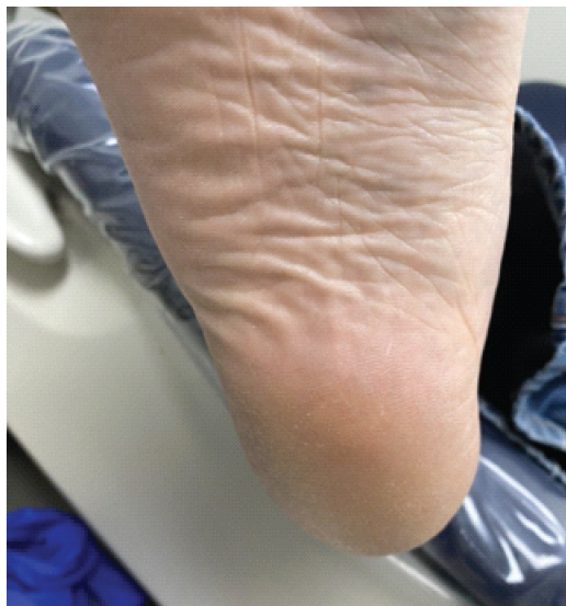
Figure 1: Clinical photograph of plantar heel demonstrating a firm, non-mobile subcutaneous mass in the central heel.
It was mildly tender and did not transilluminate. He had a mild callus present, sensation intact, and palpable pedal pulses. His gait was antalgic with heel offloading, and ankle and subtalar range of motion were full and pain-free. Magnetic resonance imaging (MRI) (October 2024) showed a well-circumscribed subcutaneous lesion (2.7 × 3.4 cm) in the plantar heel within the calcaneal fat pad, subjacent to the calcaneal neck (Figs. 2 and 3).
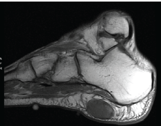
Figure 2: Sagittal T1-weighted magnetic resonance imaging of the right foot demonstrating a T1-hypointense, well-circumscribed subcutaneous lesion within the calcaneal pad, measuring 2.7 × 3.4 cm.
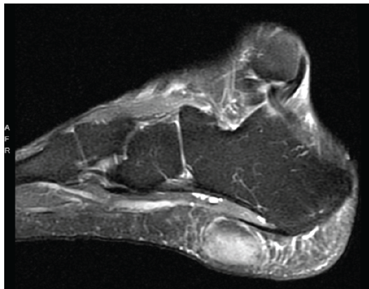
Figure 3: Sagittal T2-weighted magnetic resonance imaging of the right foot showing T2-hyperintensity of the lesion with well-defined margins and no involvement of the plantar aponeurosis or intrinsic musculature.
The lesion was T1 hypointense and T2 hyperintense with heterogeneous post-contrast enhancement, without calcification or surrounding edema. Well-defined margins suggested a non-aggressive process. Differential diagnosis included lipoma, hemangioma, giant cell tumor, desmoid tumor, and neurofibroma. The patient initially declined surgery but returned in April 2025 with enlargement and worsening discomfort. Marginal excision was performed April 3, 2025.
Surgical technique and post-operative care:
The procedure was performed under general anesthesia with local anesthesia (10 mL of a 1:1 mixture of 1% Lidocaine plain and 0.5% Marcaine plain). An ankle tourniquet was inflated to 250 mmHg following exsanguination. A 5-cm horizontal lazy-S incision was made over the central plantar heel. Sharp and blunt dissections were performed until the mass was noted, located 3 cm within the subcutaneous fat. A Freer elevator was used to assess depth. The soft-tissue mass measured 4.2 × 3.2 cm intraoperatively, larger than on the preoperative MRI, consistent with interval growth during the 6-month period between imaging and surgery. The mass was well-encapsulated and glistening, with mixed fibrous-firm and fatty-soft components and no adherent arteriovenous network. It was freed from fibrous adhesions using atraumatic Adson forceps and Stevens scissors, with care taken to preserve capsular integrity. Marginal excision was performed with 1 cm circumferential margins (Fig. 4); the primary specimen and peripheral margin tissues were submitted as separately labeled specimens for pathologic evaluation. Remnants of the soft-tissue capsule noted in the surgical site were also excised and submitted for pathology.
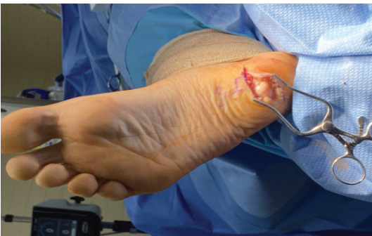
Figure 4: Intraoperative photograph demonstrating marginal excision of the encapsulated plantar calcaneal soft-tissue mass through horizontal lazy-S incisions with approximately 1 cm circumferential margins. Intraoperative size: 4.2 × 3.2 cm.
Dead space was closed in layers using 4-0 polyglactin 910; skin was closed with 3-0 nylon. A dressing and a short Controlled Ankle Motion (CAM) walker were applied postoperatively. The patient remained non-weight-bearing with crutches for 2 weeks. All sutures were removed 2 weeks post-surgery, and the patient reported complete resolution of plantar heel pain. He weight-bearing in the CAM walker at that point and was walking in a sneaker at 3 weeks. At 4 weeks, he walked barefoot without discomfort. Clinical follow-up at 1-year post-excision demonstrated no symptoms and no palpable recurrence. The patient was counseled regarding the long-term recurrence risk associated with ALT/WDL and the approximately 10% risk of dedifferentiation upon recurrence [8]. Annual clinical examination with MRI surveillance has been initiated and is planned for a minimum of 5 years, consistent with published recommendations for extremity ALT [3].
Histopathological findings:
A well-circumscribed, glistening, ovoid mass was submitted for histopathological evaluation on April 3, 2025. Gross pathology (Fig. 5) demonstrated a mucoid mass of 11 g and measuring 3.6 × 2.8 cm with a uniform yellow-white cut surface, represented in three cassettes after additional sections were submitted following initial review.
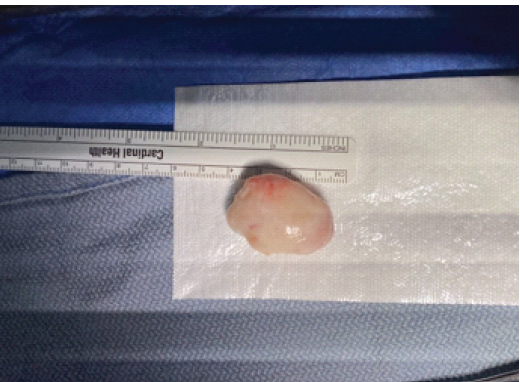
Figure 5: Gross pathology specimen: well-circumscribed, glistening, ovoid mass (3.6 × 2.8 cm; 11 g) with mixed fibrous and fatty cut surfaces.
Peripheral margins consisted of approximately 12 slender mucoid yellow-to-gray-white strips of soft tissue, ranging up to 8 × 2 mm. Microscopic examination confirmed a sclerosing ALT with prominent myxomatous features. Key histologic findings included: Atypical spindle and stromal cells with hyperchromatic, enlarged nuclei within collagenous fibrous septa; scattered lipoblasts; a prominent myxomatous feature was highlighted by Alcian blue staining; and abundant collagen disposition was confirmed by Masson trichrome staining. Cellular pleomorphism was minimal, with rare mitotic activity. Although recurrence may occur, dedifferentiation and metastasis are uncommon (Figs. 6 and 7).
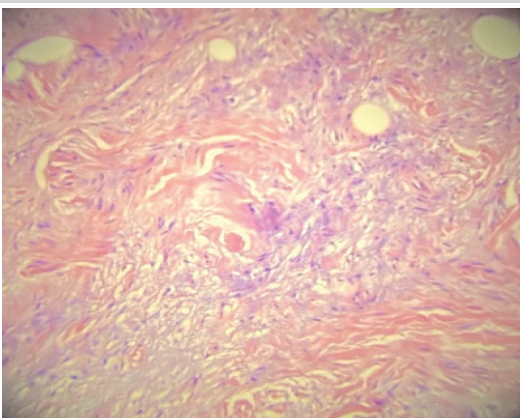
Figure 6: Histological slide of the mass showing small adipocytes in myxomatous stroma with irregular fibrous tissue.
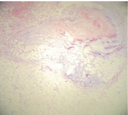
Figure 7: Periphery of surgical pathology image: Gelatinous tissue with adipose tissue separated by thickened fibrous septa.
v Immunohistochemical analysis demonstrated diffuse nuclear MDM2 positivity, consistent with MDM2 overexpression and supporting the diagnosis of ALT/WDL. S100 protein, EMA, desmin, and synaptophysin were all negative. MDM2 was assessed by IHC; FISH for confirmatory MDM2 gene application and CDK4 immunostaining were not performed due to laboratory availability constraints. Peripheral margin specimens were free of tumor. The final pathologic diagnosis was ALT, sclerosing type with myxomatous features.
This case confirms ALT arising in the plantar heel, a rare site for a tumor that typically occurs in the thigh, retroperitoneum, and deep trunk. ALT/WDL is defined by 12q13-15 amplification (MDM2/CDK4) and is distinct from benign lipomas and fibro-osseous lesions like liposclerosing myxofibrous tumors. Diagnosis here relied on diffuse nuclear MDM2 IHC positivity plus characteristic histology (atypical stromal cells, lipoblasts, fibrous septa, and myxomatous change). A prior case report similarly described a dorsal foot ALT/WDL managed with metatarsal ray amputation [8]. Foot location raises the stakes of misdiagnosis: Unplanned excision of foot sarcomas is linked to failed limb salvage, local recurrence, and death [6]. While ALT/WDL itself lacks metastatic potential, incomplete excision may increase the risk of dedifferentiation to DDL, which carries significantly higher recurrence and metastatic potential [7]. In the present case, planned marginal excision with histopathologic guidance was performed, resulting in negative margins and no perioperative complications. The MRI findings in this case reflect the mixed cellularity of the tumor, including its myxomatous matrix and fibrous septa, and differ from the T1-hyperintense, homogeneous signal of a simple lipoma. Burt and Huang describe that ALT/WDL typically demonstrates non-fatty signal components, thick fibrous septa, and inhomogeneous enhancement on MRI, features that reliably distinguish it from benign lipoma [9]. Such features should prompt histopathologic evaluation of plantar soft-tissue masses. The sclerosing variant shows hypocellular collagenous stroma with scattered atypical cells, lipoblasts, and often myxomatous change [7]. MDM2 amplification occurs in >95% of ALT/WDLPS and is absent in lipoma, detectable by IHC or FISH [3]. Diffuse nuclear MDM2 positivity was diagnostic here; CDK4 status was not assessed, a limitation. Surgical resection with negative margins remains the mainstay and is generally curative [10]; radiotherapy and chemotherapy are not indicated due to insensitivity [11]. Systematic reviews support marginal excision for extremity ALT/WDL, demonstrating lower complication rates compared to wide resection, though with a slightly higher local recurrence rate (11.9% vs. 3.3%) [11]; recurrences are usually re-resectable, and metastasis from extremity ALT is unreported in major series [12]. Case series of extremity ALT/WDL confirm that marginal excision is adequate in anatomically constrained sites; Chang et al. reported a series of 45 extremity and trunk wall ALT/WDL cases managed by surgical excision with acceptable local control [13]. Burusapat et al. similarly describe successful surgical management of extremity ALT/WDL, including cases with mixed histologic components [14]. In the present case, planned marginal excision with 1 cm circumferential margins was performed, with the primary specimen and peripheral margin specimens submitted separately for pathologic evaluation, confirming negative margins. Meticulous layered closure to eliminate dead space resulted in an uncomplicated post-operative course, in contrast to reported complications such as aseptic seroma with similar approaches [15]. Extremity ALT carries less than 2% local recurrence risk after complete excision, far lower than retroperitoneal WDL [3], though recurrences warrant re-evaluation given up to 10% dedifferentiation risk [3]. Annual MRI surveillance for at least 5 years is recommended [3]; this patient is enrolled accordingly.
Limitations:
This is a single-patient report. CDK4 IHC and MDM2 FISH were not performed due to laboratory constraints. While the patient has completed 1-year follow-up without clinical recurrence, longer follow-up is needed to draw meaningful conclusions about recurrence risk in this anatomic location. Size discrepancies across modalities (MRI 2.7 × 3.4 cm 6 months preoperatively during a period of reported growth; intraoperative 4.2 × 3.2 cm including reactive tissue; gross pathology) 3.6 × 2.8 cm post-fixation shrinkage) reflect expected measurement variation rather than data inconsistency.
We report a rare plantar heel sclerosing ALT with myxomatous features, confirmed by MDM2 IHC and treated with marginal excision and negative margins. Soft-tissue sarcomas of the foot are rare, and unplanned excision is associated with worse outcomes. Evaluation of persistent plantar masses in older adults should include ALT, with MRI and histopathology when features are atypical. Diagnosis requires integrated histology and MDM2 testing. Marginal excision with long-term MRI surveillance remains standard care.
Persistent or enlarging plantar heel masses should not be presumed benign, as rare entities such as sclerosing atypical lipomatous tumors may require MRI characterization, MDM2-supported histopathologic diagnosis, and planned marginal excision to achieve optimal oncologic outcomes and reduce the risk associated with unplanned surgery.
References
- 1. Coindre JM, Pédeutour F, Aurias A. Well-differentiated and dedifferentiated liposarcomas. Virchows Arch 2009;456:167-79. [Google Scholar] [PubMed]
- 2. Jonczak E, Grossman J, Alessandrino F, Taswell CS, Velez-Torres JM, Trent J. Liposarcoma: A journey into a rare tumor’s epidemiology, diagnosis, pathophysiology, and limitations of current therapies. Cancers (Basel) 2024;16:3858-8. [Google Scholar] [PubMed]
- 3. Yee EJ, Stewart CL, Clay MR, McCarter MM. Lipoma and its doppelganger: The atypical lipomatous tumor/well-differentiated liposarcoma. Surg Clin North Am 2022;102:637-56. [Google Scholar] [PubMed]
- 4. Bakotic BW, Borkowski P. Primary soft-tissue neoplasms of the foot: The clinicopathologic features of 401 cases. J Foot Ankle Surg 2001;40:28-35. [Google Scholar] [PubMed]
- 5. Bird JE, Morse LJ, Feng L, Wang WL, Lin PP, Moon BS, et al. Non-radiographic risk factors differentiating atypical lipomatous tumors from lipomas. Front Oncol 2016;6:197. [Google Scholar] [PubMed]
- 6. Latt LD, Turcotte RE, Isler MH, Wong C. Case series. Soft-tissue sarcoma of the foot. Can J Surg 2010;53:424-31. [Google Scholar] [PubMed]
- 7. Thway K. Well-differentiated liposarcoma and dedifferentiated liposarcoma: An updated review. Semin Diagn Pathol 2019;36:112-21. [Google Scholar] [PubMed]
- 8. Matsuo T, Sugita T, Shimose S, Kubo T, Yasunaga Y, Ochi M. Liposarcoma arising in the foot: A case report. Case Rep Med 2009;2009:630203. [Google Scholar] [PubMed]
- 9. Burt AM, Huang BK. Imaging review of lipomatous musculoskeletal lesions. SICOT J 2017;3:34. [Google Scholar] [PubMed]
- 10. Sbaraglia M, Bellan E, Dei Tos AP. The 2020 WHO classification of soft tissue tumours: News and perspectives. Pathologica 2020;113:70-84. [Google Scholar] [PubMed]
- 11. Lee AT, Thway K, Huang PH, Jones RL. Clinical and molecular spectrum of liposarcoma. J Clin Oncol 2018;36:151-9. [Google Scholar] [PubMed]
- 12. Choi KY, Jost E, Mack L, Bouchard-Fortier A. Surgical management of truncal and extremities atypical lipomatous tumors/well-differentiated liposarcoma: A systematic review of the literature. Am J Surg 2020;219:823-7. [Google Scholar] [PubMed]
- 13. Chang DH, Ma H, Liao WC, Huang MH, Wu PS. Atypical lipomatous tumors of the extremities and trunk wall-the first case series of Chinese population with 45 cases. Ann Plast Surg 2016;76 Suppl 1:S8-12. [Google Scholar] [PubMed]
- 14. Burusapat C, Wongprakob N, Wanichjaroen N, Pruksapong C, Satayasoontorn K. Atypical lipomatous tumor/well-differentiated liposarcoma with intramuscular lipoma-like component of the thigh. Case Rep Surg 2020;2020:8846932. [Google Scholar] [PubMed]
- 15. Uddin A, Flanagan G, Reilly I. Surgical excision of complex lipoma from the foot: A case report. Clin Case Rep 2022;10:e5953. [Google Scholar] [PubMed]





Related Articles in Journal of Orthopaedic Case Reports
 January 1, 2026 A Huge Recurrent Atypical Lipomatous Tumor (Well-Differentiated Liposarcoma) going in to Dedifferentiation 12 years after its First Detection
 October 8, 2012 The Pelvic Digit: A Rare Congenital Anomaly as a Cause of Hip Pain- Abstract
 July 10, 2020 Amputation in Necrotizing Fasciitis – Dilemma or Reality: A Case Report and Literature Review
 December 4, 2020 The Use a Prophylactic Intramedullary Tibial Nail for a Stress Fracture in a High-Level Athlete