

Intraoperative delivery of autologous platelet-rich plasma through cannulated cancellous screws is a safe, low-cost biological adjunct that accelerates radiological union and functional recovery in femoral neck fractures without increasing complication or revision rates.
Dr. Adarsh Baiwal, Department of Orthopaedics, Gandhi Medical College and Associated Hamidia Hospital, Bhopal, Madhya Pradesh, India. E-mail: aadarshbaiwal@gmail.com
Abstract
Aims and Background: Femoral neck fractures remain a major challenge in orthopedic trauma because the tenuous intracapsular blood supply predisposes to non-union and avascular necrosis even after mechanically sound fixation. Internal fixation is the standard head-preserving option in younger adults, yet biological failure persists. This study aimed to determine whether supplementing conventional osteosynthesis with autologous platelet-rich plasma (PRP) accelerates bone healing and improves functional recovery.
Materials and Methods: In this prospective comparative study, 60 patients aged 18–60 years with acute femoral neck fractures were allocated equally to two groups. Group A (n = 30) underwent closed reduction and internal fixation with three cannulated cancellous screws augmented by intraoperative PRP injection through the screw cannulations; Group B (n = 30) received identical screw fixation alone. The groups were comparable in demographics, injury mechanism, and Garden and Pauwels classifications. Outcomes assessed over a minimum 1-year follow-up were time to radiological union, time to full weight-bearing, functional recovery by the Harris Hip Score (HHS), pain by the Visual Analog Scale (VAS), and post-operative complications.
Results: PRP augmentation significantly accelerated healing. Radiological union was achieved at a mean of 3.54 ± 0.32 months in Group A versus 4.03 ± 0.47 months in Group B (P < 0.0001), permitting earlier full weight-bearing (3.58 ± 0.39 vs. 4.19 ± 0.54 months; P < 0.0001). The PRP group reached a higher final mean HHS (91.03 ± 1.38 vs. 88.73 ± 2.18; P < 0.0001). Final VAS scores did not differ significantly between groups (4.43 ± 0.50 vs. 4.70 ± 0.70; P = 0.09). Complication, non-union, and avascular necrosis rates requiring revision were low and comparable between groups.
Conclusion: Autologous PRP is a safe and effective biological adjunct to internal fixation of femoral neck fractures. It shortens the time to union and to full weight-bearing and improves functional hip recovery without increasing surgical complications, and merits consideration as a routine addition to head-preserving osteosynthesis.
Keywords: Femoral neck fracture, platelet-rich plasma, osteosynthesis, fracture healing, Harris hip score.

Femoral neck fractures remain among the most demanding injuries in orthopedic trauma. With rising life expectancy, the global incidence of hip fractures is projected to more than double by 2050, imposing a substantial healthcare burden [1]. The intracapsular location of the femoral neck renders its blood supply vulnerable to disruption at the time of injury, predisposing patients to non-union in approximately 10–30% and to avascular necrosis in 15–35% of cases [2]. These complications frequently necessitate revision procedures such as arthroplasty, which adversely affect functional outcome and quality of life [3].
In younger, physiologically active patients, preservation of the native femoral head by internal fixation with multiple cancellous screws remains the standard of care [4]. However, even when adequate mechanical stability is achieved, fracture healing may be compromised by limited vascularity and insufficient biological stimulation at the fracture site [5].
Platelet-rich plasma (PRP), first described by Whitman et al. in 1997, is an autologous concentrate of platelets that releases growth factors, including platelet-derived growth factor, transforming growth factor-beta, and vascular endothelial growth factor, thereby promoting angiogenesis and osteogenesis [6,7]. By enriching the local biological environment, PRP may stimulate fracture healing and callus formation [7]. Although PRP has yielded encouraging results in long-bone fractures such as the tibial diaphysis [8], its role in femoral neck fractures remains inadequately defined [9].
This study, therefore, aimed to evaluate the effect of intraoperative PRP supplementation of osteosynthesis on fracture healing and functional outcomes in femoral neck fractures.
Study design and patient selection
This prospective comparative study was conducted in the Department of Orthopaedics over 20 months, from July 2024 to March 2026. The study protocol was approved by the Institutional Ethics Committee (Approval No. 16167/MC/IEC/2024), and written informed consent was obtained from every participant before enrolment.
Sixty patients presenting with a femoral neck fracture were recruited on the basis of an a priori sample-size calculation. Assuming a baseline union rate of 60% with standard fixation and an anticipated improvement to 85% with biological augmentation, 30 patients/arm provided 80% power at a two-sided alpha of 0.05, allowing for a projected attrition of 10–15%.
Inclusion criteria were
- Age between 18 and 60 years;
- Acute femoral neck fracture;
- Fracture amenable to acceptable closed reduction and internal fixation with cannulated cancellous screws;
- Willingness to provide informed consent and comply with the follow-up protocol.
Exclusion criteria were
- Fractures requiring open reduction;
- Pathological fractures;
- Active local or systemic infection;
- Pre-existing autoimmune disorders (e.g., rheumatoid arthritis, systemic lupus erythematosus);
- Patients medically unfit for surgery or anesthesia;
- Patients unwilling or unable to complete follow-up.
PRP preparation
Autologous PRP was prepared intraoperatively under strict aseptic conditions. Approximately 50 mL of peripheral venous blood was drawn from each patient (Fig. 1) and processed by a standardized two-step centrifugation technique.
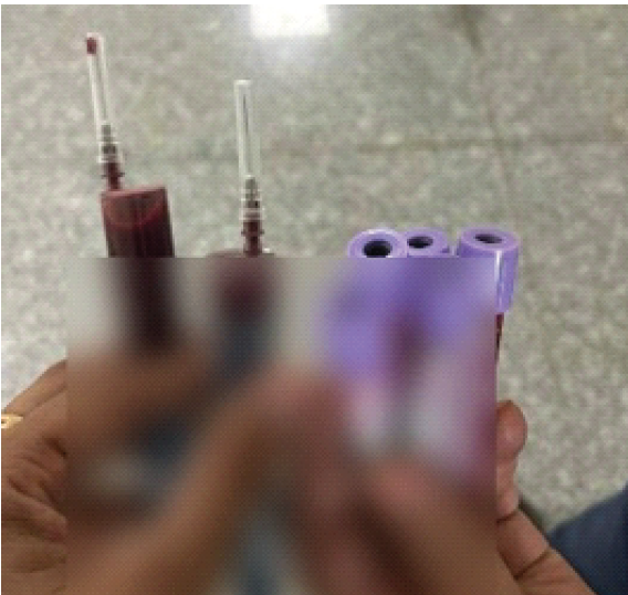
Figure 1: Collection of peripheral venous blood. Approximately 50 mL of peripheral venous blood is withdrawn from the patient and collected for the two-step centrifugation used to prepare autologous platelet-rich plasma. (Printed labels on the collection tubes have been obscured to protect patient confidentiality).
An initial soft spin (1480 rpm for 10 min) separated the sample into red-blood-cell, buffy-coat, and plasma fractions. The plasma and buffy-coat layers were aspirated and subjected to a second hard spin (3400 rpm for 15 min) to yield a concentrated platelet fraction. The final preparation was approximately 5–6 mL of leukocyte-rich PRP with an estimated platelet concentration 4–5 times the baseline peripheral count (Fig. 2). No exogenous activating agent was added before application.
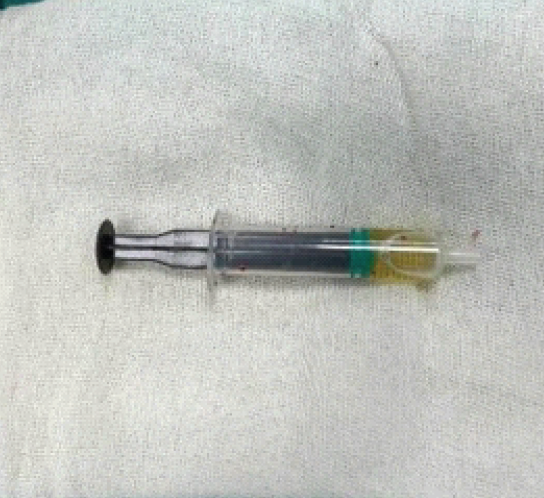
Figure 2: Prepared platelet-rich plasma. Approximately 5–6 mL of leukocyte-rich platelet-rich plasma in the syringe after two-step centrifugation of the patient’s venous blood, ready for intraoperative delivery.
Surgical technique
All procedures were performed with the patient supine on a radiolucent fracture table under regional or general anesthesia. Closed reduction was performed and assessed fluoroscopically; an acceptable reduction was confirmed using the Garden alignment index, targeting angles between 155° and 180° on both anteroposterior and lateral views. Internal fixation was achieved uniformly with three cannulated cancellous screws through a standard lateral approach.
In Group A, after reaming over the three guidewires, the screws were advanced to the fracture site. Each guidewire was then sequentially withdrawn to allow the prepared PRP to be injected directly into the fracture gap through the central cannulation of the screw (Fig. 3).
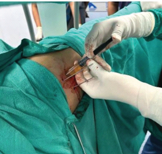
Figure 3: Intraoperative delivery of platelet-rich plasma. Platelet-rich plasma is injected into the fracture gap through the central cannulation of the cannulated cancellous screws after sequential withdrawal of the guidewires.
To prevent gravitational backflow during injection, the limb was internally rotated, and the table was temporarily tilted toward the contralateral side. The guidewires were reinserted to occlude the cannulations, and all three screws were fully advanced and tightened to secure interfragmentary compression (Fig. 4). In Group B, identical fixation was performed without PRP injection.
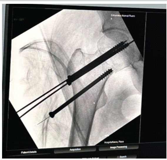
Figure 4: Intraoperative fluoroscopic (C-arm) image. Anteroposterior C-arm view during the procedure, confirming screw position and delivery of platelet-rich plasma to the fracture site.
Post-operative protocol and outcome assessment
The post-operative rehabilitation regimen was standardized for all participants, with the initiation of full weight-bearing individualized to clinical and radiological progress. Follow-up evaluations were performed at 2 weeks and at 1, 3, 6, and 12 months after surgery (Figs. 5 and 6). Radiological union was defined as trabecular continuity across three or more cortices on anteroposterior and lateral views, together with the absence of pain on weight-bearing.
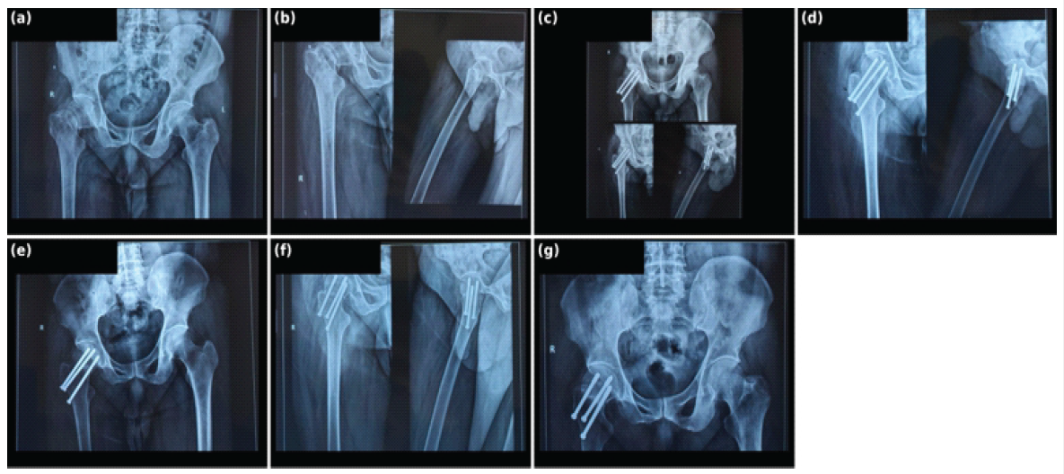
Figure 5: Case illustration, Plate 1 (panels a-g). Serial anteroposterior and lateral hip radiographs of representative patients with femoral neck fractures treated by closed reduction and internal fixation with three cannulated cancellous screws augmented with platelet-rich plasma. Panels (a-g) correspond to follow-up images showing the pre-operative radiograph (a and b), immediate post-operative fixation (c), and progressive early consolidation of the fracture at 2 weeks post-operative (d and e), progressive early consolidation of the fracture at 1 month post-operative (f and g). Anatomical side markers (R/L) are retained; all patient-identifying information has been removed.
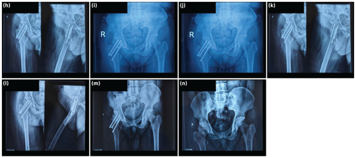
Figure 6: Case illustration, Plate 2 (panels h-n). Continuation of the serial follow-up sequence, demonstrating progressive trabecular union across the fracture line with maintained hardware position through to final follow-up. Panels (h-n) complete the previous Fig. 5 showing 2 months (h and I), 3 months (j and k), 6 months (l and m), and 12 months follow-up (n).
The primary outcome was the time to definitive radiological union, together with monitoring of post-operative complications, including avascular necrosis, non-union, and hardware failure. Secondary outcomes were pain severity, assessed with the Visual Analog Scale (VAS), and hip function, assessed with the Harris Hip Score (HHS) once complete union and rehabilitation were achieved.
Statistical analysis
Data were analyzed with IBM Statistical Package for the Social Sciences Statistics (versions 22–25). The Shapiro–Wilk test assessed the normality of continuous variables. Normally distributed variables were compared with the independent-samples t-test, and non-normally distributed variables with the Mann–Whitney U test. Categorical variables were compared with the Pearson Chi-square test, or the Fisher exact test when an expected cell frequency was below five. P-value below 0.05 was considered statistically significant.
Baseline characteristics
All 60 patients completed the study protocol and the minimum 1-year follow-up, with 30 in the PRP-augmented group (Group A) and 30 in the conventional-fixation group (Group B). The two cohorts were well matched: there were no significant differences in age, sex, side affected, interval from injury to surgery, or mechanism of trauma, and the Garden and Pauwels classifications were evenly distributed between the arms, providing a balanced basis for comparison (Table 1).
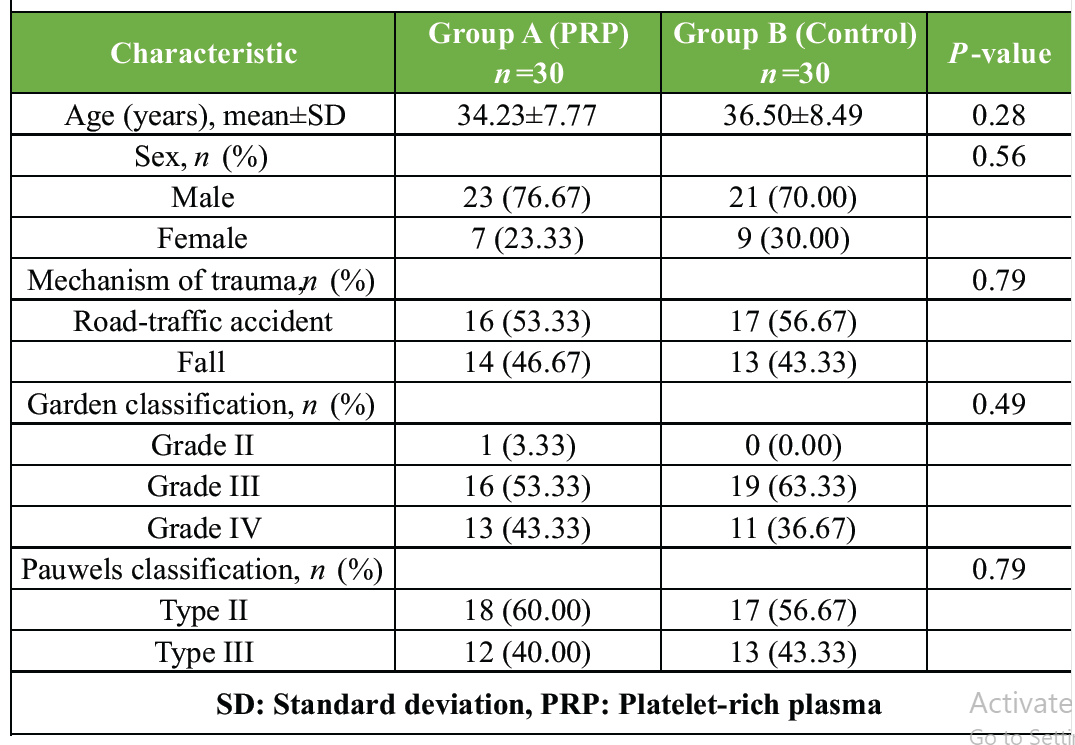
Table 1: Baseline demographic and fracture characteristics of the two groups
Radiological union and weight-bearing
The most marked difference between the two approaches was the speed of healing. Radiological union was achieved significantly faster in the PRP group, at a mean of 3.54 ± 0.32 months, compared with 4.03 ± 0.47 months in the control group (P < 0.0001). This translated into earlier mobilization: the PRP group was cleared for full weight-bearing at a mean of 3.58 ± 0.39 months, versus 4.19 ± 0.54 months in the control group (P < 0.0001) (Table 2).
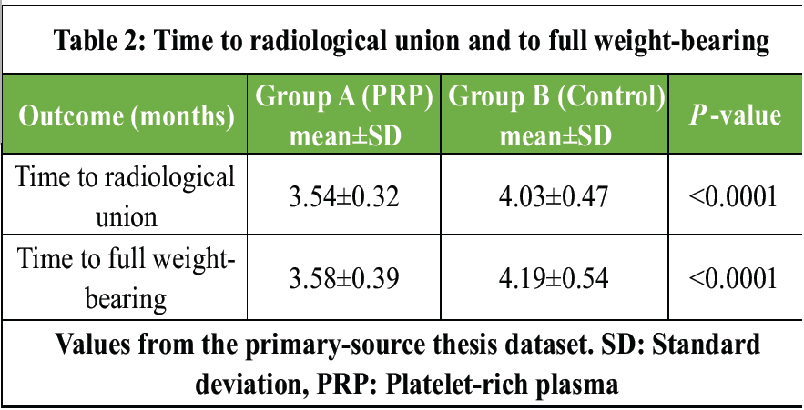
Table 2: Time to radiological union and to full weight-bearing
Functional outcome and pain
Both groups improved substantially in hip function after fixation, but biological augmentation conferred a distinct advantage. The PRP group achieved consistently higher HHSs and reached an excellent final mean of 91.03 ± 1.38, significantly outperforming the control group’s 88.73 ± 2.18 (P < 0.0001). Pain, measured by the VAS, declined steadily in both groups; although PRP patients tended to report marginally lower scores during recovery, the final VAS scores did not differ significantly (4.43 ± 0.50 vs. 4.70 ± 0.70; P = 0.09), indicating that conventional fixation is equally effective for long-term pain relief (Table 3).
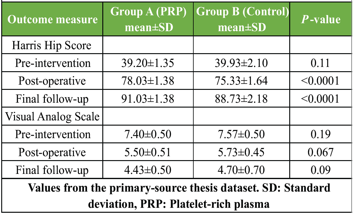
Table 3: Harris hip score and Visual Analog Scale over time
Complications and revision
Most patients avoided severe adverse events, with 27 of 30 (90.0%) in the PRP group and 24 of 30 (80.0%) in the control group requiring no further intervention (P = 0.50). For avascular necrosis, one patient (3.33%) in each group underwent total hip replacement, and one control patient was managed conservatively. For non-union, the PRP group required one total hip replacement and one conservatively managed case, whereas the control group required two total hip replacements and two subtrochanteric osteotomies. Conservative management was reserved for patients who declined revision surgery. Overall, the need for secondary procedures was comparable between the arms (P = 0.44) (Table 4).
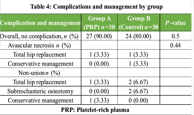
Table 4: Complications and management by group
The principal aim of this study was to determine whether supplementing conventional osteosynthesis with PRP could offset the persistent biological limitations of femoral neck fractures, namely poor vascularity, delayed union, and avascular necrosis. Our findings show that intraoperative PRP delivered through the screw cannulations significantly accelerated union and functional recovery while maintaining a safety profile comparable to that of fixation alone.
The two cohorts were evenly matched at baseline, ensuring that the observed differences reflect the intervention rather than confounding. Men predominated in both groups, and most patients were in their 30s, with no significant differences in age or sex. This demographic profile mirrors the cohorts reported by Ren et al. and Sun et al., who likewise described well-balanced age and sex distributions [9,10]. Injury severity, graded by the Garden and Pauwels systems, was also comparable between arms; our distribution of Garden Grade III and IV fractures is consistent with that of Samy [11].
The most striking clinical advantage was the accelerated healing timeline. PRP-augmented patients achieved definitive union at a mean of 3.54 months versus 4.03 months in controls, and consequently began full weight-bearing significantly earlier. These findings corroborate Ren et al., who similarly reported a significantly shorter time to union with PRP [9]. Whereas Samy found only marginal, non-significant differences in healing between Garden grades, our direct comparison of biologically augmented and conventional fixation demonstrated a highly significant acceleration of bone consolidation [11].
Both groups improved significantly in function and pain, yet the PRP group achieved significantly higher final HHSs, indicating superior functional recovery. The literature is divided on this point: Ren et al. found no significant difference in final HHS between augmented and control groups [9], and Sun et al. reported the opposite trend, with better long-term scores in their non-PRP cohort at 12 and 24 months [10]. Regarding pain, our finding of no significant difference in final VAS scores accords with both Ren et al. and Samy [9,11], whereas Sun et al. observed superior long-term pain relief in their non-PRP cohort [10]. Taken together, these observations suggest that while mechanical stability reliably determines long-term pain relief, the functional benefit of PRP may vary with patient population and preparation protocol.
Mitigating severe biological failure, such as non-union and avascular necrosis, is a central goal of orthobiologic therapy. In our series, the rates of severe complications requiring salvage – total hip arthroplasty or subtrochanteric osteotomy – were low and statistically comparable between arms, and the addition of PRP did not elevate the surgical risk. This favorable safety profile is supported by Ren et al., Griffin et al., and Samy, all of whom reported low and comparable rates of avascular necrosis and other severe complications when biologically augmented fixation was compared with standard hardware [9,11,12].
Limitations
Several limitations should be acknowledged. First, the absence of blinding of the operating surgeons and outcome assessors introduces a potential risk of performance and assessment bias. Second, as a single-center study at a tertiary institution, the generalizability of the findings may be limited across settings with differing demographics and surgical expertise. Third, all procedures were performed by a small number of surgeons, so despite standardized protocols, surgeon-dependent variation may have influenced outcomes. Finally, the modest sample size limits power to detect differences in infrequent complications such as avascular necrosis, and the 1-year follow-up may be insufficient to capture the full long-term course of femoral neck fractures.
Supplementing conventional osteosynthesis with intraoperative PRP is a safe and highly effective biological adjunct for femoral neck fractures. Compared with fixation alone, it significantly accelerates radiological union, enabling earlier full weight-bearing, and produces superior functional recovery as reflected by higher HHSs, without any increase in complications or revision requirements. While mechanical stability reliably governs long-term pain relief in both approaches, PRP offers a distinct therapeutic advantage by enhancing the local healing environment.
In physiologically active patients with displaced femoral neck fractures, anatomical reduction and stable screw fixation are necessary but often insufficient to overcome the biological deficits of the intracapsular environment. Delivering autologous PRP through the screw cannulations at the time of fixation is a practical, low-cost, and biologically plausible way to concentrate the patient’s own healing factors at the fracture site, accelerating consolidation and functional recovery and warranting consideration as a routine adjunct to head-preserving osteosynthesis.
References
- 1. Gullberg B, Johnell O, Kanis JA. World-wide projections for hip fracture. Osteoporos Int 1997;7:407-13. [Google Scholar] [PubMed]
- 2. Lu-Yao GL, Keller RB, Littenberg B, Wennberg JE. Outcomes after displaced fractures of the femoral neck. A meta-analysis of one hundred and six published reports. J Bone Joint Surg Am 1994;76:15-25. [Google Scholar] [PubMed]
- 3. Parker MJ, Gurusamy K. Internal fixation versus arthroplasty for intracapsular proximal femoral fractures in adults. Cochrane Database Syst Rev 2006;4:CD001708. [Google Scholar] [PubMed]
- 4. Ly TV, Swiontkowski MF. Treatment of femoral neck fractures in young adults. J Bone Joint Surg Am 2008;90:2254-66. [Google Scholar] [PubMed]
- 5. Hak DJ. The biology of fracture healing in long bones. Injury 1999;30 Suppl 3:C3-6. [Google Scholar] [PubMed]
- 6. Whitman DH, Berry RL, Green DM. Platelet gel: An autologous alternative to fibrin glue with applications in oral and maxillofacial surgery. J Oral Maxillofac Surg 1997;55:1294-9. [Google Scholar] [PubMed]
- 7. Marx RE. Platelet-rich plasma: Evidence to support its use. J Oral Maxillofac Surg 2004;62:489-96. [Google Scholar] [PubMed]
- 8. Malhotra R, Kumar V, Garg B, Chana R, Jain M, Sharma V. Role of autologous platelet-rich plasma in healing of fresh tibial diaphyseal fractures: A randomized control trial. Injury 2011;42:646-9. [Google Scholar] [PubMed]
- 9. Ren W, Wang F, Sun L, Gao W, Ye H, Jia K, et al. Role of autologous platelet-rich plasma in the treatment of femoral neck fractures. Int J Clin Exp Med 2017;10:1610-4. [Google Scholar] [PubMed]
- 10. Sun L, Xu Z, Zhang Z, Zhao J, Sun M, Gao G, et al. Effects of PRP injection combined with intersecting femoral head decompression versus combined with multiple core decompression in the treatment of avascular necrosis of the femoral head: A single-centre retrospective cohort study. BMC Musculoskelet Disord 2025;26:438. [Google Scholar] [PubMed]
- 11. Samy AM. The role of platelet-rich plasma in the management of fracture neck femur: new insights. Int Orthop 2016;40:1019-24. [Google Scholar] [PubMed]
- 12. Griffin XL, Achten J, Parsons N, Costa ML. Platelet-rich therapy in the treatment of patients with hip fractures: A single centre, parallel group, participant-blinded, randomised controlled trial. BMJ Open 2013;3:e002583. [Google Scholar] [PubMed]





Related Articles in Journal of Orthopaedic Case Reports
 December 10, 2022 Subtrochanteric Fracture after Internal Fixation of Femoral Neck Fracture Using the Hansson Pinloc System: A Case Report
 October 1, 2026 Osteogenic Potential of Bone Callus: A Cross-sectional Study on Microscopic Structure and Healing Stages
 October 1, 2026 Comparative Efficacy of Leukocyte-Poor Platelet-Rich Plasma, Leukocyte-Rich Platelet-Rich Plasma, Injectable Platelet-Rich Fibrin, and Corticosteroid Injections in Kellgren–Lawrence Grade 2–3 Knee Osteoarthritis: A Prospective Comparative Study
 October 1, 2026 Dual Mobility Total Hip Arthroplasty for Recurrent Dislocation After Femoral Neck Fracture in an Elderly Patient: A Case Report