

Intra-articular PRP injections provide significant, sustained relief from pain and functional impairment in patients with moderate knee osteoarthritis while demonstrating potential disease-modifying benefits through reduced cartilage degradation and improved MRI parameters.
Dr. Amit Saraf,Professor & Head Department of Orthopaedics, Teerthanker Mahaveer Medical College and Research Centre, Moradabad, Uttar Pradesh, India. E-mail: amitsaraf_75@yahoo.com
Abstract
Introduction: Knee osteoarthritis (KOA) is a prevalent degenerative joint disorder with progressive cartilage loss, chronic pain, and functional limitation. Platelet-rich plasma (PRP), an autologous concentrate with growth factors, has gained attention as a therapeutic modality due to its regenerative and anti-inflammatory properties. This study evaluated clinical, biochemical, and radiological outcomes of intra-articular PRP injections in KOA patients.
Materials and Methods: This randomized controlled trial spanned over 18 months at a tertiary care center. A total of 70 patients with Kellgren-Lawrence grade II and III KOA were randomly allocated into the PRP group (n = 35) and normal saline (NS) group (n = 35). Baseline assessment included Visual Analog Scale (VAS) index, Western Ontario and McMaster Universities Osteoarthritis Index (WOMAC) functional index, serum Coll2-1 levels, and whole-organ magnetic resonance imaging score (WORMS). Patients received ultrasound (USG)-guided intra-articular injections at baseline and 1 month. Follow-up evaluations were performed at 1, 3, 6, and 9 months. Statistical analysis was done using appropriate parametric and non-parametric tests.
Results: Baseline characteristics were comparable between groups. The PRP group demonstrated significant reduction in WOMAC and VAS scores from 1 month onward, with sustained improvement at 3, 6, and 9 months versus the NS group (P < 0.05). Magnetic resonance imaging showed significant reduction in WORMS scores in the PRP group, whereas no significant change was observed in the NS group. In addition, serum Coll2-1 levels significantly decreased in the PRP group at 9 months, indicating reduced cartilage degradation.
Conclusion: Intra-articular PRP injections significantly improved pain, functional outcomes, and structural parameters in patients with moderate KOA. These findings suggest that PRP may serve as an effective therapeutic option with potential disease-modifying benefits in the management of KOA.
Keywords: Knee osteoarthritis, platelet-rich plasma, intra-articular injection, non-steroidal anti-inflammatory drugs.

Osteoarthritis (OA) is a degenerative joint disorder [1]. With a global prevalence of 3.8% and 28.7% in India [2,3], knee OA is projected to affect 642 million individuals by 2050 [4]. At present, no disease-modifying treatments exist. Guidelines recommend education, exercise [5,6], non-steroidal anti-inflammatory drugs (NSAIDs) [7], and intra-articular injections, such as corticosteroids [8] or hyaluronan [9], though these provide limited or short-term relief.
Platelet-rich plasma (PRP), an autologous product rich in growth factors [9,10], has emerged as a promising therapy [11]. Biologically, PRP modulates OA pathways [12], promotes tissue repair [13], and reduces cartilage degradation [14]. However, clinical evidence remains inconsistent due to methodological limitations and a lack of placebo-controlled trials [13,14,15]. Consequently, PRP is absent from standard clinical guidelines. This study evaluates the effect of intra-articular PRP injections in patients with Kellgren-Lawrence (KL) grades 2 and 3 knee OA (KOA).
Study design
The present investigation was a randomized controlled trial.
Study subjects
The present study included participants from >18years of age.
Study place
The present analysis was performed at Teerthanker Mahaveer Medical College and Research Center, in the Department of Orthopaedics.
Study period
The present analysis was carried out over a period of 18 months.
Ethical clearance
Institutional Ethical Committee (IEC) of TMMC & RC, Moradabad (Ref no: TMU/IEC/2024-25/PG/119).
Data collection
All the patients who attended the Orthopedics Department of Teerthanker Mahaveer Medical College and Research Centre who fulfilled the inclusion criteria were enrolled for the study after they gave written and informed consent.
Sample size
The sample size was calculated by using the formula:-
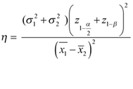
Where,
- η = Total sample size
- σ1 = The standard deviation of the variable in group 1 = 10.27
- σ2 = The standard deviation of the variable in group 2 = 8.95
- = Value of the normal deviate at the considered level of confidence (for a two-sided test) = 2.58 at 99 % confidence interval
- z1-β = Value of the normal deviate at the considered power of the study (for a two-sided test) = 1.29 at 90 % power
- = Mean of the variable group 1 = 70.63
- = Mean of the variable group 2 = 60.37
- According to the calculation, the total sample size is 56 (28 in each group)
- After considering the 25% loss of follow-up final sample size would be 70 (35 in each group).
Inclusion criteria
- Patients over 18 years of age who had been diagnosed with osteoarthritis of the knee according to the American College of Rheumatology (ACR) 2019 criteria were included in this study.
- Among these patients, only those diagnosed with Grade II and Grade III osteoarthritis of the knee joint, as classified by the KL scale, were selected.
Exclusion criteria
The participants were excluded based on the following criteria:
- Any known malignancy
- Gouty arthritis/Inflammatory arthritis, secondary osteoarthritis
- Active knee infection
- Disorders of bleeding
- Uncontrolled diabetes mellitus
- Ligamentous instability of the knee (magnetic resonance imaging [MRI] DIAGNOSED)
- Contraindication to MRI
- Any history of previous intra-articular steroid injection.
Methodology
After obtaining approval from the College Research Committee and IEC, patients were recruited from the outpatient department based on the ACR criteria and KL Grade II and III on standing anteroposterior (AP) and lateral (LAT) X-rays of the knee. Recruitment was conducted after applying the inclusion and exclusion criteria.
Randomization was performed using a computer-generated random sequence, and patients were divided into two groups:
- Group A PRP and
- Group B (normal saline [NS]).
Before administering the first intra-articular injection of PRP or NS, baseline assessments, including the Visual Analog Scale (VAS), Western Ontario and McMaster Universities Osteoarthritis Index (WOMAC), serum Coll2-1NO2 levels, X-ray, and MRI using the whole-organ magnetic resonance imaging score (WORMS), were conducted.
The severity of pain was assessed using the VAS score, which allowed for a subjective evaluation of pain intensity on a standardized scale. To evaluate the functional status of the affected knee, patients were instructed to complete the International Knee Documentation Committee osteoarthritis scale, a validated tool designed to assess knee function in individuals with osteoarthritis.
Plain radiographs of the affected knee were obtained in both AP and LAT views. These X-rays were utilized to classify the severity of KOA based on the KL grading system, a widely accepted radiographic classification method.
In addition to radiographic assessment, a Doppler ultrasonographic examination was performed on the affected knee using a 5–12 MHz linear array transducer (Medison R3). The examination was conducted with the patient in a supine position on an examination bed, with the knee flexed to the maximum extent feasible. The USG evaluation included detailed observations of increased vascularity (indicative of Doppler activity), synovial hypertrophy, cartilage thickness, regularity of cartilage margins, and effusion.
Cartilage degeneration in the osteoarthritic knee was further assessed using an ultrasonographic grading system as described by Saarakkala et al. The patient was positioned supine, and the knee was fully flexed during the procedure. Initially, the intercondylar notch area, encompassing the femoral condyles directly above the patellar bone (referred to as the sulcus), was visualized. Subsequently, the cartilage of the medial and LAT femoral condyles was thoroughly examined by sweeping the USG transducer across the entire cartilage surface from proximal to distal positions. The probe was maintained in a transverse orientation, ensuring that the USG beam remained perpendicular to the femoral surface throughout the assessment.
The cartilage evaluation adhered to a grading system, which included the following categories:
- Grade 0: The cartilage appeared as a monotonous anechoic band with sharply defined hyperechoic anterior and posterior interfaces.
- Grade 1 (Mild degenerative changes): The cartilage exhibited a loss of the normal sharpness of its interfaces and/or an increase in echogenicity. A score of one point was assigned for each observation site where these findings were present, resulting in a maximum of three points if changes were observed in both condyles and the sulcus.
- Grade 2A (Moderate degenerative changes): In addition to the changes noted in Grade 1, clear local thinning of the cartilage, measuring <50% of the normal thickness, was observed.
- Grade 2B: Degenerative changes included local thinning of the cartilage measuring more than 50% but <100%. Two points were assigned at each observation site, with a maximum of six points.
- Grade 3 (Severe degenerative changes): Complete local loss of cartilage tissue (100%) was documented. This grade was assigned three points per observation site, resulting in a maximum of nine points.
The first intra-articular injection of PRP or NS was administered under USG guidance. The injection procedure was carried out with the patient lying supine on the examination table, ensuring optimal comfort and stability. They were precisely advised to avoid any weight-bearing activities or use of the injected leg for 24 h to allow for proper absorption of the injected material and to reduce stress on the joint. The application of ice packs over the injected knee was recommended to alleviate any localized swelling or discomfort. Furthermore, patients were instructed to refrain from the use of NSAIDs during this period, as these medications could interfere with the natural inflammatory response that contributes to the effectiveness of the injection. After 1 month, VAS and WOMAC scores were reassessed, followed by a second USG-guided intra-articular injection of PRP or NS. At 6 months, VAS and WOMAC scores were measured again. At 9 months, VAS, WOMAC, serum Coll2-1NO2 levels, X-ray, and MRI (WORMS) evaluations were conducted to assess the effects of the interventions.
Statistical analysis
All quantitative variables were presented as mean ± standard deviation, providing a measure of central tendency and variability. Qualitative variables were expressed as frequencies and percentages to summarize categorical data effectively. Inferential statistical analyses were conducted using appropriate statistical tests, either parametric or non-parametric, which were selected based on the type of data and the requirements of the study at the time of data analysis.
Baseline parameters were comparable between the PRP and NS groups with respect to age, gender distribution, body mass index (BMI), KL grade, and side involvement, with no statistically significant differences. However, comorbidities were significantly higher in the PRP group. Among laboratory parameters, hemoglobin levels were comparable, whereas total leukocyte count (TLC) and random blood sugar (RBS) showed statistically significant differences between the groups (Table 1).
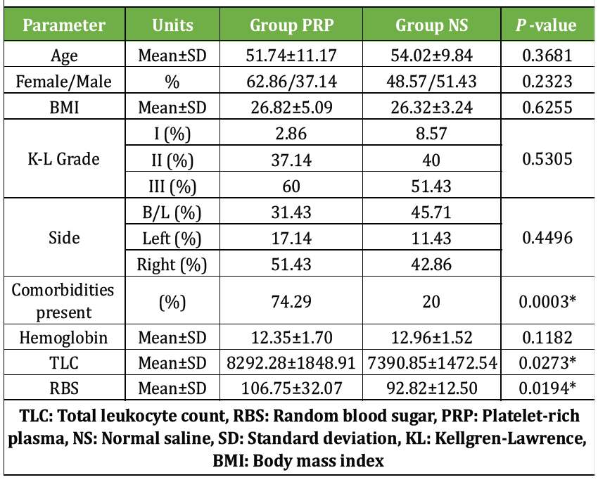
Table 1: Baseline demographic and clinical characteristics of study groups
Baseline WOMAC scores were comparable between the two groups. From 1 month onward, the PRP group showed significantly greater reduction in WOMAC scores compared to the NS group, with sustained improvement observed at 3, 6, and 9 months (Table 2).
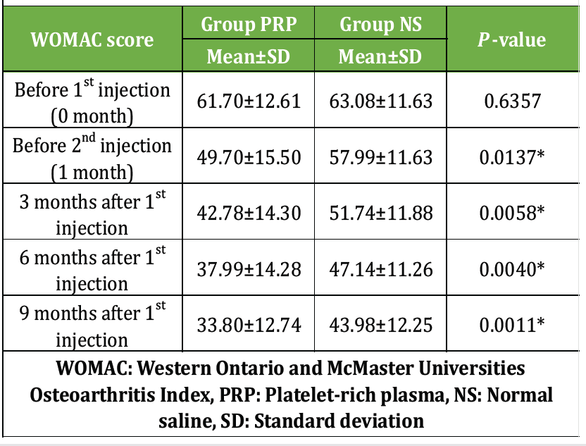
Table 2: Comparison of functional outcome (WOMAC Score)
Baseline VAS scores were comparable between the two groups. From 1 month onward, the PRP group showed significantly greater reduction in pain scores compared to the NS group, with sustained improvement at 3, 6, and 9 months (Table 3).
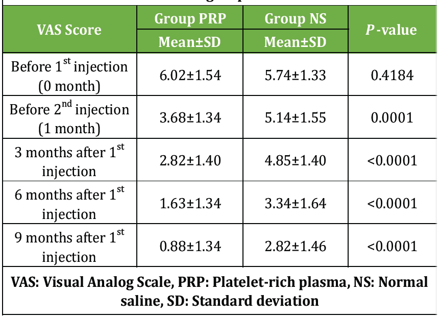
Table 3: Comparison of VAS scores over time between PRP and NS groups
The PRP group showed a significant reduction in WORMS after treatment, whereas the NS group showed no significant change. Intergroup differences were statistically significant both before and after treatment (Table 4).
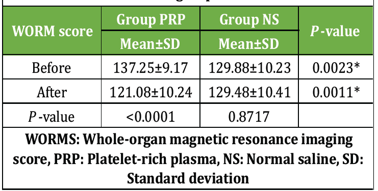
Table 4: Comparison of WORM scores between PRP and NS groups
Baseline serum Coll 2-1 levels were comparable between the two groups. At 9 months, the PRP group showed a significant reduction in levels, whereas the NS group showed higher values, indicating a statistically significant difference between the groups (Table 5).
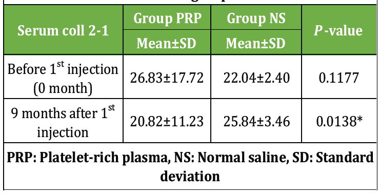
Table 5: Comparison of serum Coll 2-1 levels between PRP and NS groups
Intra-articular PRP has emerged as a promising therapeutic option in the management of KOA [16]. Increasing evidence from randomized controlled trials indicates that PRP is both safe and effective, with outcomes comparable to hyaluronic acid and, in some cases, superior clinical benefits, particularly in younger and more active patients with early-stage disease. However, its therapeutic effect is not permanent and generally declines after 6–9 months, highlighting the need for optimization of treatment protocols [17].
A major limitation in the present literature is the lack of standardization in PRP preparation and administration. Variability in platelet concentration, activation techniques, dosage, and injection frequency may significantly influence outcomes, making it difficult to establish uniform clinical recommendations. Therefore, further high-quality randomized trials are required to define optimal protocols and clarify the role of PRP in OA management.
In the present study, baseline demographic characteristics, including age, gender and BMI, were comparable between the PRP and NS groups, ensuring appropriate matching. Most patients were in the 41–60 years age range, with mean ages of 51.74 ± 11.17 years (PRP) and 54.02 ± 9.84 years (NS), without a significant difference (P = 0.3681). Gender distribution showed a female predominance in the PRP group and a slight male predominance in the NS group, although this was not statistically significant. These findings are consistent with previous studies, which also reported comparable baseline characteristics, supporting internal validity.
In contrast, a significantly higher prevalence of comorbidities was observed in the PRP group (P = 0.0003), unlike earlier studies where comorbid conditions were evenly distributed. This imbalance may act as a potential confounding factor and should be considered while interpreting outcomes.
Radiological severity, assessed using KL grading, was comparable between groups, with the majority of patients classified as Grade II and III OA. This aligns with prior studies and reflects an appropriate patient population for evaluating PRP efficacy, as moderate disease stages are considered optimal for therapeutic intervention. Laterality of knee involvement was also comparable, with a predominance of unilateral and right-sided involvement, consistent with existing literature.
Hematological parameters were largely comparable between groups, although TLC and RBS levels were significantly higher in the PRP group. Despite these differences, all parameters remained within clinically acceptable limits. Notably, literature correlating hematological parameters with PRP outcomes remains limited, indicating an area for future research.
Clinical outcomes demonstrated significant improvement in the PRP group. While baseline WOMAC and VAS scores were comparable, the PRP group showed a statistically significant and sustained reduction in both scores from 1 month onward, persisting through 3, 6, and 9 months. These findings are consistent with previous studies, including Bansal et al., Chu et al., and Huang et al., which reported sustained improvements in pain, stiffness, and function following PRP therapy. The observed peak improvement around 6 months is also in agreement with existing evidence [18,19,20].
Similarly, VAS scores showed a progressive and sustained decline in the PRP group, indicating superior pain relief compared to NS. These findings are supported by studies, such as Subash et al., Spaková et al., and Say et al., all of which demonstrated greater pain reduction with PRP compared to comparator treatments [21,22,23].
Biochemically, the study demonstrated a significant reduction in serum Coll2-1 levels in the PRP group, suggesting decreased cartilage degradation. This observation is in line with previous studies that have reported elevated Coll2-1 levels in advanced OA and their reduction following effective treatment. However, variability in findings across studies highlights the need for further validation of these biomarkers.
Radiological assessment using MRI-based WORMS scoring revealed significant structural improvement in the PRP group, with a marked reduction in scores post-treatment, while the NS group showed no significant change. These findings suggest that PRP may not only provide symptomatic relief but also contribute to structural modification of the joint. Similar improvements have been reported in previous studies, further supporting the potential disease-modifying role of PRP [23,24].
Overall, the findings of the present study are consistent with existing literature and reinforce the role of PRP as an effective modality for improving clinical outcomes and potentially influencing disease progression in KOA.
The present study demonstrates that intra-articular PRP injections provide significant clinical, biochemical, and radiological benefits in patients with KOA (KL Grade II and III). PRP therapy resulted in sustained improvement in pain, stiffness, and functional outcomes, as evidenced by significant reductions in WOMAC and VAS scores over 9 months. In addition, PRP was associated with reduced serum Coll2-1 levels and significant improvement in MRI-based WORMS scores, suggesting a potential disease-modifying effect. In contrast, the NS group showed minimal improvement. These findings support PRP as a safe and effective treatment modality with both symptomatic and structural benefits.
Limitations of the study
Despite its strengths, the present study has certain limitations. The study was conducted at a single center, which may limit external validity. The sample size was relatively modest, reducing statistical power for subgroup analyses. The follow-up period was limited to 9 months, restricting assessment of long-term outcomes and the need for repeat injections. Baseline imbalance in comorbidities, along with significant differences in TLC and RBS values, may have acted as potential confounding factors. The study did not characterize PRP composition (platelet concentration, leukocyte content, or growth factor levels), limiting reproducibility and comparison with other studies. Patients with advanced OA (KL Grade IV) were not included, limiting applicability to severe disease. MRI and WORMS assessments were performed only at baseline and 9 months, precluding evaluation of temporal structural changes. Lifestyle factors, such as exercise, physiotherapy, weight reduction, and activity modification were not standardized or analyzed. Outcomes relied predominantly on patient-reported measures (VAS and WOMAC), and quality-of-life measures and cost-effectiveness were not evaluated.
Intra-articular PRP injections serve as a safe, effective, and non-surgical therapeutic option for patients with Grade II and III knee osteoarthritis. Beyond providing durable symptomatic pain relief and functional restoration, USG-guided PRP exhibits potential disease-modifying capabilities by preserving cartilage integrity and reducing joint degradation markers over a 9-month period.
References
- 1. Michael JW, Schlüter-Brust KU, Eysel P. The epidemiology, etiology, diagnosis, and treatment of osteoarthritis of the knee. Dtsch Arztebl Int 2010;107:152-62. [Google Scholar] [PubMed]
- 2. Bannuru RR, Osani MC, Vaysbrot EE, Arden NK, Bennell K, Bierma-Zeinstra SM, et al. OARSI guidelines for the non-surgical management of knee, hip, and polyarticular osteoarthritis. Osteoarthritis Cartilage 2019;27:1578-89. [Google Scholar] [PubMed]
- 3. Kan H, Chan P, Chiu K, Yan CH, Yeung SS, Ng YL, et al. Non-surgical treatment of knee osteoarthritis. Hong Kong Med J 2019;25:127-34. [Google Scholar] [PubMed]
- 4. Jüni P, Hari R, Rutjes AW, Fischer R, Silletta MG, Reichenbach S, et al. Intra-articular corticosteroid for knee osteoarthritis. Cochrane Database Syst Rev 2015;10:CD005328. [Google Scholar] [PubMed]
- 5. Chandler GN, Jones DT, Wright V, Hartfall SJ. Charcot’s arthropathy following intra-articular hydrocortisone. Br Med J 1959;1:952-3. [Google Scholar] [PubMed]
- 6. Sola M, Dahners L, Weinhold P, Van der Horst AS, Kallianos S, Flood D. The viability of chondrocytes after an in vivo injection of local anaesthetic and/or corticosteroid: A laboratory study using a rat model. Bone Joint J 2015;97-B:933-8. [Google Scholar] [PubMed]
- 7. Vandeweerd JM, Zhao Y, Nisolle JF, Zhang W, Zhihong L, Clegg P, et al. Effect of corticosteroids on articular cartilage: Have animal studies said everything? Fundam Clin Pharmacol 2015;29:427-38. [Google Scholar] [PubMed]
- 8. Latourte A, Rat AC, Omorou A, Ngueyon-Sime W, Eymard F, Sellam J, et al. Do glucocorticoid injections increase the risk of knee osteoarthritis progression over 5 years? Arthritis Rheumatol 2022;74:1343-51. [Google Scholar] [PubMed]
- 9. Everts P, Onishi K, Jayaram P, Lana JF, Mautner K. Platelet-rich plasma: New performance understandings and therapeutic considerations in 2020. Int J Mol Sci 2020;21:7794. [Google Scholar] [PubMed]
- 10. Kingsley CS. Blood coagulation: Evidence of an antagonist to factor VI in platelet-rich human plasma. Nature 1954;173:723-4. [Google Scholar] [PubMed]
- 11. Akeda K, An HS, Okuma M, Miyamoto K, Thonar EJ, Lenz ME, et al. Platelet-rich plasma stimulates porcine articular chondrocyte proliferation and matrix biosynthesis. Osteoarthritis Cartilage 2006;14:1272-80. [Google Scholar] [PubMed]
- 12. Dos Santos RG, Santos GS, Alkass N, Chiesa TL, Azzini GO, da Fonseca LF, et al. The regenerative mechanisms of platelet-rich plasma: A review. Cytokine 2021;144:155560. [Google Scholar] [PubMed]
- 13. Simental-Mendía M, Vilchez-Cavazos F, Álvarez-Villalobos N, Villalobos N, Blázquez-Saldaña J, Peña-Martínez V, et al. Clinical efficacy of platelet-rich plasma in the treatment of lateral epicondylitis: A systematic review and meta-analysis of randomized placebo-controlled clinical trials. Clin Rheumatol 2020;39:2255-65. [Google Scholar] [PubMed]
- 14. Jain SK, Suprashant K, Kumar S, Yadav A, Kearns SR. Comparison of plantar fasciitis injected with platelet-rich plasma vs corticosteroids. Foot Ankle Int 2018;39:780-6. [Google Scholar] [PubMed]
- 15. Tucker JD, Goetz LL, Duncan MB, et al. Randomized, placebo-controlled analysis of the knee synovial environment following platelet-rich plasma treatment for knee osteoarthritis. PM R 2021;13:707-19. [Google Scholar] [PubMed]
- 16. Pourcho AM, Smith J, Wisniewski SJ, Gilman JB, Elmore LW, Sell SA, et al. Intraarticular platelet-rich plasma injection in the treatment of knee osteoarthritis: Review and recommendations. Am J Phys Med Rehabil 2014;93:S108-21. [Google Scholar] [PubMed]
- 17. Ismaiel AH. Comparison between the effect of intra-articular injections of platelet-rich plasma and corticosteroids in advanced knee osteoarthritis. J Med Sci Res 2018;1:278-84. [Google Scholar] [PubMed]
- 18. Bansal H, Leon J, Pont JL, Wilson DA, Bansal A, Agarwal D. Platelet-rich plasma (PRP) in osteoarthritis (OA) knee: Correct dose critical for long term clinical efficacy. Sci Rep 2021;11:3971. [Google Scholar] [PubMed]
- 19. Chu J, Duan W, Yu Z, Wilson DA, Bansal A, Agarwal D, et al. Intra-articular injections of platelet-rich plasma decrease pain and improve functional outcomes than sham saline in patients with knee osteoarthritis. Knee Surg Sports Traumatol Arthrosc 2022;30:4063-71. [Google Scholar] [PubMed]
- 20. Huang HY, Hsu CW, Lin GC, Lin HS, Chou YJ, Liou IH, et al. Comparing efficacy of a single intraarticular injection of platelet-rich plasma (PRP) combined with different hyaluronans for knee osteoarthritis: A randomized-controlled clinical trial. BMC Musculoskelet Disord 2022;23:954. [Google Scholar] [PubMed]
- 21. Subash Y, Kamalakumar K, Lydia M. The role of platelet rich plasma injection in the management of early osteoarthritis of the knee. Int J Res Orthop 2018;4:128-32. [Google Scholar] [PubMed]
- 22. Spaková T, Rosocha J, Lacko M, Harvanová D, Gharaibeh A. Treatment of knee joint osteoarthritis with autologous platelet-rich plasma in comparison with hyaluronic acid. Am J Phys Med Rehabil 2012;91:411-7. [Google Scholar] [PubMed]
- 23. Say F, Gürler D, Yener K, Bülbül M, Malkoc M. Platelet-rich plasma injection is more effective than hyaluronic acid in the treatment of knee osteoarthritis. Acta Chir Orthop Traumatol Cech 2013;80:278-83. [Google Scholar] [PubMed]
- 24. Wakayama T, Saita Y, Uchino S, Kobayashi Y, Nishio H, Fukusato S, et al. MRI-based evaluation of PRP therapy in knee osteoarthritis: WORMS and synovial changes at 6 months. J Clin Med 2025;14:6408. [Google Scholar] [PubMed]






Related Articles in Journal of Orthopaedic Case Reports
 October 1, 2026 Comparative Efficacy of Leukocyte-Poor Platelet-Rich Plasma, Leukocyte-Rich Platelet-Rich Plasma, Injectable Platelet-Rich Fibrin, and Corticosteroid Injections in Kellgren–Lawrence Grade 2–3 Knee Osteoarthritis: A Prospective Comparative Study
 October 1, 2026 Efficacy of Intra-Articular Injection of Platelet-Rich Plasma and Growth Factor Concentrate in Degenerative Meniscal Lesions
 December 1, 2025 Functional Outcome of Intra-articular Platelet-Rich Plasma in the Treatment of Frozen Shoulder
 October 1, 2026 Arthrokinematic and Magnetic Resonance Imaging-Based Outcomes of Customized Lateral Wedge Insoles in Medial Compartment Knee Osteoarthritis: Six-Month Interim Findings of a Prospective Cohort Study