

Adequate canal filling and careful assessment of anterior femoral bowing during reamed short proximal femoral nailing help maintain fracture reduction and reduce the risk of post-operative mechanical failure in intertrochanteric fractures.
Dr. Raj Kumar, Department of Orthopaedics, SGRDIMSR, Sri Guru Ram Das University of Health Sciences, Sri Amritsar - 143501, Punjab, India. E-mail: drraj_ortho@yahoo.com
Abstract
Introduction: Intertrochanteric fractures in elderly individuals require stable fixation to achieve union, maintain alignment, and prevent post-operative collapse. Although proximal femoral nailing (PFN) provides reliable fixation in most cases, loss of reduction may still occur during fracture healing, leading to alteration in the femoral neck-shaft angle (NSA) and horizontal femoral offset, thereby affecting hip biomechanics and gait.
Aims: The aim of the study was to evaluate the early radiological outcomes of reamed short PFN in intertrochanteric fractures among patients aged 50 years and above, with special emphasis on maintenance of reduction and biomechanical predictors of reduction loss.
Materials and Methods: A prospective observational study was conducted on 30 patients aged ≥50 years with intertrochanteric femur fractures treated with reamed short PFN over a period of 18 months. Standardized radiographs were obtained preoperatively, immediately postoperatively, and at 3 months' follow-up. Measurements were performed using Digimizer software. Radiological parameters included femoral NSA, horizontal femoral offset, cortical reduction quality, canal filling ratio (CFR), and anterior femoral bowing angle. Loss of reduction was defined using a composite criterion comprising a ≥5° decrease in NSA, a ≥4 mm increase in femoral offset, or deterioration in the cortical reduction pattern.
Results: Radiological union at 3 months was achieved in 28 patients (93.3%). Loss of reduction was observed in 5 patients (17.9%), whereas 23 patients (82.1%) maintained stable alignment. Mean NSA decreased significantly from 130.04° ± 4.61° immediately postoperatively to 128.64° ± 4.79° at 3 months (P = 0.005). Horizontal femoral offset increased from 38.18 ± 6.07 mm to 38.96 ± 6.03 mm (P = 0.040). Patients with loss of reduction demonstrated significantly lower CFR values in both anteroposterior and lateral planes and greater anterior femoral bowing compared to patients with maintained reduction (P < 0.001).
Conclusion: Reamed short PFN achieved a 93.3% radiological union rate, with maintenance of reduction in 82.1% of patients. Lower CFR and greater anterior femoral bowing were significant predictors of reduction loss.
Keywords: Intertrochanteric fracture, proximal femoral nail, reaming, canal filling ratio, anterior femoral bowing, osteoporosis.

Intertrochanteric fractures constitute a major orthopedic challenge worldwide and predominantly affect the elderly osteoporotic population. These fractures are associated with significant morbidity, mortality, and socioeconomic burden due to prolonged immobilization and functional dependency [1]. Early surgical fixation enabling stable reduction and mobilization remains the cornerstone of management. Cephalomedullary nailing has become the preferred treatment modality for intertrochanteric fractures due to its superior biomechanical properties, including improved load sharing, shorter lever arm, and reduced bending moment compared with extramedullary implants [2,3]. However, the role of medullary canal reaming during proximal femoral nailing (PFN) continues to be debated. Advocates of reaming emphasize that sequential canal preparation permits insertion of a larger diameter nail, thereby improving implant-canal congruence and enhancing axial as well as rotational stability [4]. Adequate canal filling ratio (CFR) reduces distal nail toggling and the “pendulum effect,” both of which are implicated in varus collapse and secondary loss of reduction [5]. Reaming may also contribute biologically by depositing autologous osteogenic debris at the fracture site, potentially enhancing fracture healing [6]. Despite these advantages, concerns regarding reaming in elderly osteoporotic patients persist due to the risks of increased intramedullary pressure, fat embolism, thermal necrosis, cortical perforation, prolonged operative time, and increased blood loss [7,8]. Consequently, short PFN is frequently performed without reaming in this age group due to the relatively wider medullary canal. Nevertheless, elderly patients often exhibit substantial femoral canal widening and increased anterior femoral bowing, factors that may compromise nail fit and predispose to mechanical instability. Literature evaluating the impact of controlled reaming on maintenance of reduction in intertrochanteric fractures remains limited, and most available evidence is extrapolated from femoral shaft fracture studies [9]. Despite widespread use of PFNs, limited evidence exists regarding the optimal CFR required to minimize post-operative reduction loss, particularly in osteoporotic elderly patients undergoing reamed short PFN. The present study was therefore undertaken to evaluate the radiological outcomes of reamed short PFN in patients aged 50 years and above with intertrochanteric fractures, with particular emphasis on maintenance of reduction and biomechanical factors associated with reduction loss.
Study design and setting:
This prospective observational study was conducted in the Department of Orthopaedics at Sri Guru Ram Das Institute of Medical Sciences and Research, Amritsar, from July 2024 to December 2025 after obtaining approval from the Institutional Ethics Committee (SGRD/IEC/2024–343).
Study population:
Thirty patients aged 50 years and above presenting with intertrochanteric femur fractures classified according to the AO/OTA classification and treated with reamed short PFN were included in the study. Subgroup analysis according to AO/OTA fracture classification was not performed due to the limited sample size, although fracture morphology may influence post-operative stability.
Inclusion criteria:
Age ≥50 years, intertrochanteric femur fractures, patients managed with reamed short PFN, and availability for follow-up.
Exclusion criteria:
Pathological fractures, fractures with subtrochanteric extension requiring long nails, previous ipsilateral hip or femoral surgery, and abnormal contralateral hip anatomy. Written informed consent was obtained from all participants before enrolment.
Surgical technique:
All procedures were performed on a radiolucent traction table by two senior orthopedic surgeons using a standardized operative technique. Therefore, the results may not be directly reproducible in centers with different levels of surgical expertise. Closed reduction was attempted initially in all cases. Mini-open or percutaneous reduction techniques were employed where satisfactory alignment could not be achieved by closed methods. Temporary fixation with Kirschner wires was performed outside the intended nail trajectory. A proximal incision of approximately 5 cm was made, and the entry point was established slightly medial to the tip of the greater trochanter under fluoroscopic guidance. Sequential flexible reaming of the medullary canal was then performed while maintaining fracture reduction. Definitive fixation was achieved using a short PFN with either a helical blade or lag screw.
Radiological assessment:
Standardized calibrated anteroposterior (AP) pelvis radiographs with 15° internal rotation and lateral radiographs were obtained preoperatively, immediately postoperatively, and at 1 and 3 months’ follow-up. Radiological measurements were performed using Digimizer software version 5.7.0. The following parameters were assessed:
- Femoral neck-shaft angle (NSA): Angle formed between the longitudinal axes of the femoral neck and shaft
- Horizontal femoral offset: Perpendicular distance from the femoral shaft axis to the center of the femoral head
- Cortical reduction quality: Classified as positive, neutral, or negative cortical support on AP radiographs
- CFR: Ratio of nail diameter to intramedullary canal diameter measured at the diaphyseal level on both AP and lateral views
- Anterior femoral bowing angle: Angle between the proximal femoral axis and the femoral shaft axis at the distal nail tip on lateral radiographs
- Radiological union: Defined as bridging callus formation across at least three cortices on two orthogonal views, accompanied by painless full weight-bearing.
Loss of reduction was defined by the presence of any one of the following:
- ≥5° decrease in NSA
- ≥4 mm increase in horizontal femoral offset
- Deterioration in cortical reduction quality.
Functional outcome measures such as the Harris Hip Score, Oxford Hip Score, and quality-of-life assessments were not included, preventing direct correlation between radiological healing and patient function. Radiological measurements were based on standardized plain radiographs rather than computed tomography (CT) imaging. Although CT provides a more precise three-dimensional assessment, routine post-operative CT was not justified for these emergency trauma patients due to additional radiation exposure, cost, and limited clinical indication.
Statistical analysis:
Statistical analysis was performed using IBM Statistical Package for the Social Sciences Statistics version 26.0 (IBM Corp., Armonk, NY, USA). Quantitative variables were expressed as mean ± standard deviation, whereas categorical variables were presented as frequencies and percentages. A paired t-test was used for intragroup comparisons and an independent Student’s t-test for intergroup comparisons. A P < 0.05 was considered statistically significant.
The study included 30 patients, of whom 17 (56.7%) were aged ≥65 years and 17 (56.7%) were female. Two patients were lost to follow-up, leaving 28 patients available for final radiological assessment (Fig. 1).
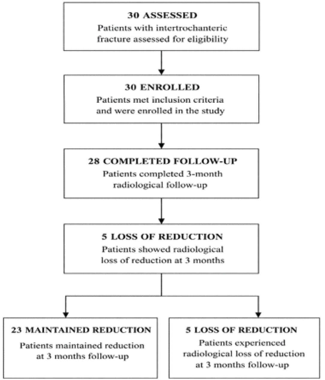
Figure 1: Flow diagram of patient enrolment and follow-up.
Two patients were lost during follow-up, introducing the possibility of attrition bias despite the relatively small proportion of missing data. Radiological union at 3 months was achieved in 28 patients (93.3%). Stable reduction was maintained in 23 patients (82.1%), whereas 5 patients (17.9%) demonstrated significant loss of reduction characterized by varus collapse and increased femoral offset (Table 1).
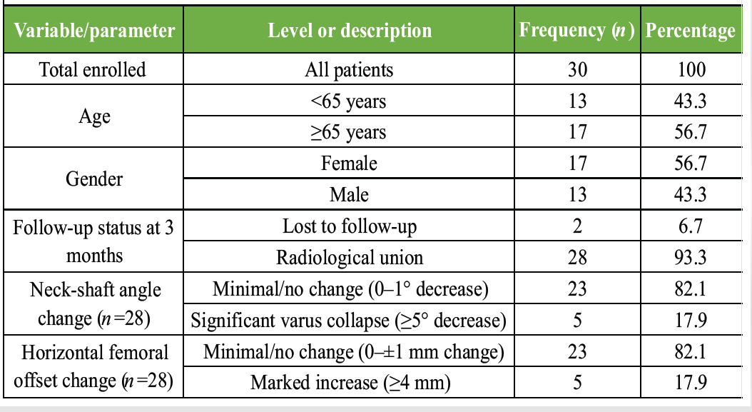
Table 1: Patient demographics and overall radiological outcomes at 3 months
The mean femoral NSA decreased significantly from 130.04° ± 4.61° immediately postoperatively to 128.64° ± 4.79° at 3 months’ follow-up (P = 0.005). Horizontal femoral offset showed a statistically significant increase from 38.18 ± 6.07 mm to 38.96 ± 6.03 mm, P = 0.040 (Table 2).
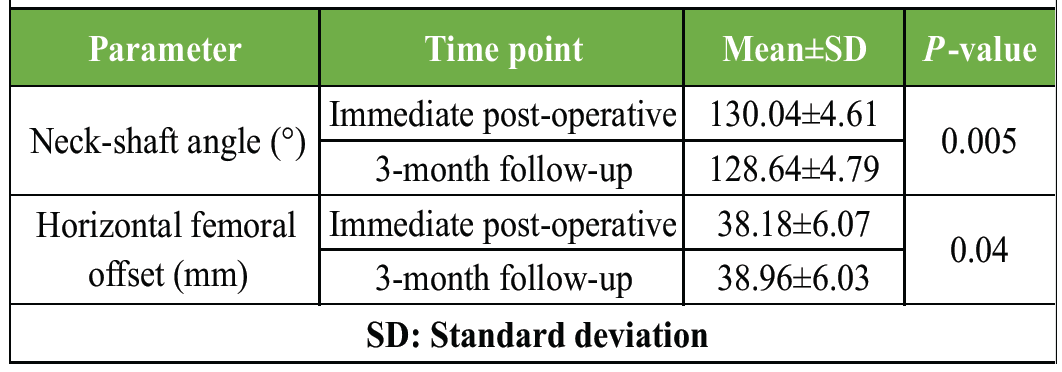
Table 2: Radiological alignment and cortical reduction (immediate post operative versus 3 months)
Patients with loss of reduction exhibited significantly lower CFRs in both AP and lateral planes compared with those maintaining stable reduction. The mean AP CFR was 0.65 ± 0.04 in the loss-of-reduction group versus 0.82 ± 0.04 in the maintained-reduction group (P < 0.001). Similarly, lateral CFR was significantly lower in the loss group (0.55 ± 0.03 vs. 0.74 ± 0.05; P < 0.001). The mean anterior bowing angle was also significantly greater in patients with reduction loss (7.10° ± 0.60° vs. 3.80° ± 0.49°; P < 0.001). Despite these radiological differences, all evaluable patients achieved fracture union at final follow-up (Table 3). The CFR values identified in this study should be considered exploratory and require external validation in larger prospective studies before routine clinical application.
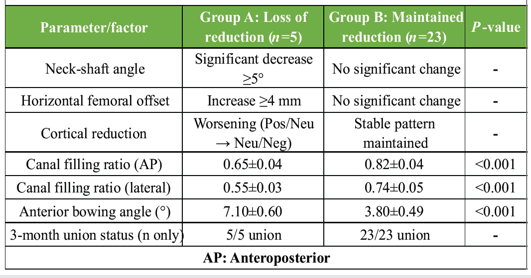
Table 3: Group stratification and associated biomechanical factors
Intertrochanteric fractures in elderly individuals continue to pose significant therapeutic challenges due to osteoporosis, widened femoral canals, and altered femoral anatomy [10]. Cephalomedullary nailing has emerged as the preferred fixation method because it permits stable fixation and early mobilization with favorable biomechanical characteristics [11]. The present study demonstrated a high radiological union rate of 93.3% at 3 months following reamed short PFN. Furthermore, stable reduction was maintained in more than 80% of patients, supporting the effectiveness of controlled reaming in achieving satisfactory implant stability. A major finding of the present study was the strong association between inadequate CFR and reduction loss. Patients demonstrating loss of reduction had significantly lower CFR values in both AP and lateral planes. Poor implant-canal congruence permits nail toggling within the widened osteoporotic femoral canal, thereby predisposing to progressive varus collapse and medialization [12]. Another important observation was the influence of anterior femoral bowing. Increased femoral curvature was significantly associated with reduction loss, likely due to a mismatch between the straight intramedullary implant and the bowed femoral canal. Similar findings have been reported by Xue et al., who highlighted the biomechanical consequences of implant-bone mismatch in Asian femoral morphology [13]. Although reaming has traditionally been avoided in elderly patients due to concerns regarding fat embolism, increased blood loss, and thermal necrosis, no major reaming-related complications were encountered in the present study [7,8]. Controlled and judicious reaming allowed insertion of a larger diameter nail and improved mechanical fixation without increasing perioperative morbidity. Despite mild radiological deterioration in a subset of patients, fracture union was achieved in all cases available for follow-up, suggesting that loss of reduction may primarily represent a mechanical issue rather than impaired biological healing. However, persistent alteration in the NSA and femoral offset may adversely affect long-term hip biomechanics and gait. This study has several limitations. Its single-center design, relatively small sample size, and short follow-up period may limit the generalizability of the findings and preclude assessment of long-term radiological and functional outcomes. As a prospective observational study without randomization or a control group, the possibility of selection bias cannot be excluded. Functional outcome measures, objective bone quality assessment, and detailed perioperative variables were not evaluated, while radiological assessment relied on plain radiographs without CT confirmation. In addition, subgroup analyses by fracture type were limited by sample size, and the standardized surgical technique performed by experienced surgeons may restrict the external applicability of the results. Larger multicenter studies with longer follow-up, comprehensive functional assessment, and advanced imaging are warranted to validate these findings.
Reamed short PFN provides satisfactory early radiological union and maintenance of reduction in elderly patients with intertrochanteric fractures. However, inadequate CFR, particularly in the lateral plane, and increased anterior femoral bowing were associated with post-operative reduction loss. Careful preoperative planning, assessment of femoral morphology, and optimization of implant-canal congruence are essential to minimize mechanical failure and preserve fracture alignment.
Adequate canal filling and assessment of femoral bowing are critical determinants of mechanical stability during short proximal femoral nailing in elderly intertrochanteric fractures. Controlled reaming may improve implant fit and reduce the risk of post-operative loss of reduction.
References
- 1. Mohamed A, Fuad U, Farook U, Nagi A. Management of proximal humerus fractures in elderly patients: A narrative review. Cureus 2025;17:e98292. [Google Scholar] [PubMed]
- 2. Di Martino A, D’Agostino C, Poluzzi R, Brunello M, Geraci G, Traina F, et al. Outcomes and complications of conversion THA after internal fixation of proximal femur fractures: A systematic review. Eur J Trauma Emerg Surg 2025;51:293. [Google Scholar] [PubMed]
- 3. Wittauer M, Henry J, Sánchez-Rosenberg G, Lambers AP, Jones CW, Yates PJ. Evaluation of reduction quality and implant positioning in intertrochanteric fracture fixation: A review of key radiographic parameters. World J Orthop 2025;16:106982. [Google Scholar] [PubMed]
- 4. Turabi R, Frihagen F, McGlasson R, Wyatt D, Trompeter A, Beaupre L, et al. Fragility fracture network position on unrestricted weight-bearing after hip fracture surgery. Geriatr Orthop Surg Rehabil 2025;16:21514593251351136. [Google Scholar] [PubMed]
- 5. Hepgüler S, Cetin A, Değer C, Erkent U. Osteoporotic hip fracture costs in the elderly Turkish population. Acta Orthop Traumatol Turc 2011;45:316-25. [Google Scholar] [PubMed]
- 6. Rodham PL, Giannoudis VP, Kanakaris NK, Giannoudis PV. Biological aspects to enhance fracture healing. EFORT Open Rev 2023;8:264-82. [Google Scholar] [PubMed]
- 7. Mencia MM, Pattron R, Goalan R, Pong CY. Intramedullary reamer failure in orthopaedic surgery: Two case reports and practical recommendations for prevention. Trauma Case Rep 2023;46:100860. [Google Scholar] [PubMed]
- 8. Giannoudis PV, Tzioupis C, Pape HC. Fat embolism: The reaming controversy. Injury 2006;37 Suppl 4:S50-8. [Google Scholar] [PubMed]
- 9. Sullivan MP, Baldwin KD, Donegan DJ, Mehta S, Ahn J. Geriatric fractures about the hip: Divergent patterns in the proximal femur, acetabulum, and pelvis. Orthopedics 2014;37:151-7. [Google Scholar] [PubMed]
- 10. Black LO, Rushkin M, Lancaster K, Cheesman JS, Meeker JE, Yoo JU, et al. Reaming the intramedullary canal during tibial nailing does not affect in vivo intramuscular pH of the anterior tibialis. OTA Int 2023;6:e248. [Google Scholar] [PubMed]
- 11. Chang SM, Song DL, Ma Z, Tao YL, Chen WL, Zhang LZ, et al. Mismatch of the short straight cephalomedullary nail (PFNA-II) with the anterior bow of the femur in an Asian population: J Orthop Trauma 2014;28:17-22. [Google Scholar] [PubMed]
- 12. Guo J, Dong W, Zhou Y, Hu J, Ye P, Chen W, et al. Differences in fixation to young and elderly quadrilateral surfaces with anatomic quadrilateral surface plate (AQSP) based on cortical thickness morphological results. J Orthop Surg Res 2022;17:143. [Google Scholar] [PubMed]
- 13. Xue D, Zheng Q, Li H, Qian S, Zhang B, Pan Z. Reamed and unreamed intramedullary nailing for the treatment of open and closed tibial fractures: A subgroup analysis of randomised trials. Int Orthop 2010;34:1307-13. [Google Scholar] [PubMed]




Related Articles in Journal of Orthopaedic Case Reports
 August 1, 2026 Evaluation of Orthopedic Outcomes of Proximal Femoral Nailing for Intertrochanteric Fractures and Predictive Value of Pre-operative Glycemic Status, Neutrophil-to-Lymphocyte Ratio and Platelet-to-Lymphocyte Ratio: A Prospective Study
 July 1, 2026 Association of Different Levels of Vitamin D Deficiency and Hip Fracture in Indian Population: A Cross-sectional Study
 June 1, 2026 Pseudoaneurysm of the Medial Femoral Circumflex Artery following Surgical Fixation of Trochanteric Femur Fracture: A Case Report
 June 1, 2026 Early Mechanical Dissociation of Helical Blade-Barrel Interface in Proximal Femoral Nail: A Case Report