

Sub-acute Candida septic arthritis of the knee can closely mimic degenerative joint disease with minimal systemic signs; intraoperative suspicion, timely surgical debridement, and targeted antifungal therapy are essential for successful outcomes.
Dr. Aashay Sonkusale, 2605 ZIRCON, Nirmal Lifestyle, LBS Marg, Mulund West, Mulund Colony, Mumbai- 400 080, Maharashtra, India. E-mail: sonkusaleaashay614@gmail.com
Abstract
Introduction: The first mention of Candida vishwanathii dates back to 1959, when it was isolated from the cerebrospinal fluid of a fatal case of meningitis in India. Since its first appearance, information on synovitis of the knee by C. vishwanathii, an opportunistic pathogen with low pathogenicity, is only available from one case report in the world.
Case Report: A 75-year-old male presented to us with right knee pain of 3 months' duration. Examination revealed synovial thickening without a local rise in temperature. Inflammatory markers were not raised. Radiographs suggested osteoarthritis (OA) with periarticular geodes. Initially planned for total knee replacement, intraoperative findings of grossly inflamed synovium prompted deference and complete synovectomy. A 54-year-old male presented similarly with right knee pain and swelling within 1 month of the first case. Radiologic assessment revealed a medial femoral chondral defect. He was planned for a two-stage procedure of biopsy with subsequent chondroplasty. In both cases, microbiological testing reported C. viswanathii with the exact same sensitivity patterns. Both were started on oral fluconazole for 6–8 weeks with complete resolution of symptoms at a 6-month follow-up.
Conclusion: This is only the second case report on C. vishwanathii and describes the indolent pattern of presentation and review of surgical management of subacute septic arthritis.
Keywords: Candida, Vishwanathii, septic, arthritis, synovitis.

The first mention of Candida vishwanathii dates back to 1959, when it was isolated from the cerebrospinal fluid of a fatal case of meningitis in India by eminent chest physician Professor (Dr) Vishwanath and Randhawa [1]. Sandhu et al. suggest that C. vishwanathii is more of an opportunistic pathogen with lower virulence than the more frequently occurring C. albicans. It resonates with both our cases, which present as low-grade knee infections with modestly elevated laboratory infection parameters [2]. The inability to pick up these elusive infections pre-operatively can result in disastrous consequences, especially if they are planned for elective procedures. This case report highlights the importance of intraoperative alertness and dynamic decision-making in the face of non-corroborative findings. It also delineates the clinical presentation, treatment plan with anti-fungals, and a review of literature, which will help in a better understanding of the management of this atypical fungus as to our knowledge, there is only one case report of septic arthritis by this organism. Informed consent was taken from both patients, abiding by the CARE guidelines.
Case 1:
A 75-year-old male presented to the outpatient clinic with chronic pain of left knee, which had aggravated since the last 3 months. He complained of difficulty in climbing stairs and squatting. Clinical examination of the knee revealed synovial thickening with joint line tenderness. Local temperature was not raised. The overlying skin showed no signs of active infection. Significant medical history included uncontrolled diabetes, hypertension since the last 15 years, and a surgical history of 3 angioplasties, spinal fusion in the last 20 years. Laboratory investigations showed a white blood cell (WBC) of 8.36 × 10³/mm3. Erythrocyte sedimentation rate (ESR) of 23 mm in 1st h and C-reactive protein (CRP) of 12 mg/L showed mild elevation. Computed tomography (CT) and magnetic resonance imaging (MRI) scans of the left knee showed a lytic lesion in the lateral femoral condyle (Fig. 1 and 2).
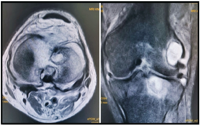
Figure 1: Magnetic resonance imaging of the left knee showing a round cystic lesion with thin sclerotic margins in the lateral femoral condyle, lateral tibial plateau, and intercondylar eminence communicating with the joint. Moderate joint effusion is appreciated.
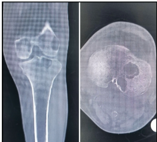
Figure 2: Computed tomography scan of the left knee showing diffuse sub-articular osteopenia. Well-defined lytic lesions seen in the lateral femoral condyle and tibial plateau.
A knee aspiration was done to rule out infection, which revealed a dry tap. We planned for a total knee replacement (TKR), and prior specialist opinions were taken. Intraoperatively, the synovium looked grossly inflamed with fulminant hypertrophy (Fig. 3).
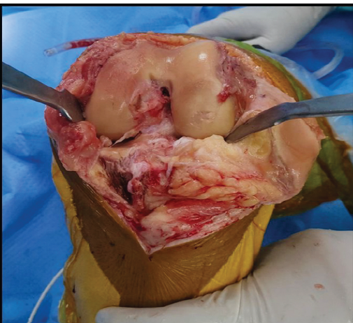
Figure 3: Intraoperative clinical photograph showing unhealthy, inflamed synovium with fulminant hypertrophy not correlating with osteoarthritis.
The clinical picture did not co-relate with OA. An intraoperative decision was taken to defer the TKR. Instead, he was counseled for synovectomy, which was sent for microbiological and histopathological analysis. On the 8th day, C. vishwanathii was detected using the Matrix-assisted Laser Desorption Ionization-Time of Flight (MALDI-TOF) method. He was started on oral fluconazole for a period of 6 weeks. His ESR and CRP were followed up, initially on a weekly basis for the 1st month and subsequently on a monthly basis when they showed a decreasing trend (Fig. 4).
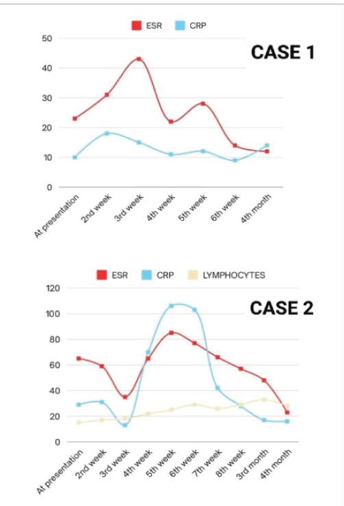
Figure 4: Periodic evaluation of erythrocyte sedimentation rate and C-reactive protein values guiding treatment with fluconazole in both cases.
Antifungal serological testing and therapeutic drug monitoring of fluconazole levels were not performed, as diagnosis was established by culture identification and patients demonstrated a favorable clinical and biochemical response to standard oral dosing in both cases. A 6-month follow-up showed normalized values for ESR, CRP, and serum procalcitonin (0.053 ng/dL). The functional knee range of motion (ROM) improved with Oxford Knee Score (OKS) increasing from 25 to 36 at 18 months follow-up.
Case 2:
A 54-year-old male presented 1 month after the first case, with similar complaints of pain and difficulty in bending of the right knee since the last 3 months. He had no fever and no signs of infection elsewhere in the body. The medical and surgical histories were insignificant. On focused knee examination, the ROM was painful and restricted to 70°. Laboratory investigations showed ESR of 65 mm/1st h, CRP of 29 mg/L along with WBC of 14.6 × 10³/mm3. Radiographic assessment revealed a medial femoral chondral defect with synovitis (Fig. 5).
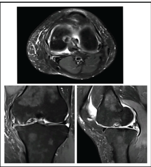
Figure 5: Magnetic resonance imaging of the right knee showing femoral chondral defect in the weight bearing area of the medial condyle.
Patient was planned for a staged procedure- arthroscopic synovial biopsy and subsequent chondroplasty. Routine microscopic examination did not reveal any organism, whereas MALDI-TOF detected C. vishwanathii on the 9th day. As in the previous case, he was started on oral fluconazole for 8 weeks with periodic monitoring of ESR, CRP, and lymphocytes (Fig. 4). ESR and CRP values showed a downward trend from the 8th week on. Patient showed plateauing of ESR and CRP at the 4th month and an improvement in the OKS from 29 to 40 at 20-month follow-up.
Vishwanathii has been demonstrated to be of low virulence in normal mice and highly pathogenic in cortisone-treated immunosuppressed mice [3,4]. The first case in our report can be considered to be at risk for infections in view of his multiple surgeries, medications, and comorbidities. However, the second case is not consistent with this assumption. There are studies that link Candida infections to intra-articular injections [5]. The first case conforms to this etiology, even though the knee aspiration was carried out by an experienced surgeon under all aseptic precautions. The Infectious Disease Clinics of North America states that 3/4 of Candida arthritis involves the knee, which begins as monoarticular arthritis presenting with pain, tenderness, and mild effusion. Patients are typically afebrile with no local erythema and a normal WBC. Unlike bacterial arthritis, candida arthritis manifests as synovitis with subsequent spread to the surrounding bone. This subacute clinical picture requires a high index of suspicion [6]. A careful history of previous surgeries, intravenous drug abuse, and penetrating trauma to the knee can help establish the source of infection in delayed presentations. Candida septic arthritis most commonly involves Candida albicans, while non-albicans species are far less frequently reported. In the largest review of 112 pediatric and adult cases by Gamaletsou et al., the knee was the most commonly affected joint, with most patients presenting as indolent monoarticular arthritis and minimal systemic features [7].
Anti-fungal therapy with the triazole family has a proven record of good synovial and bone penetration [8]. In both our cases, the C. vishwanathii strain was sensitive to fluconazole. Some infectious disease specialists advocate the continuation of anti-fungal therapy in immunosuppressed individuals to prevent relapses. While there is no general consensus on the appropriate duration of anti-fungal therapy, the decision on the duration of therapy is made considering the extent of infection and patient characteristics. ESR and CRP can be used as objective parameters for resolution of infection; however, they need to be cautiously interpreted as they are only moderately elevated to begin with in candidal arthritis [9]. In most of the cases, although medical therapy alone suffices, surgical management needs to be decided on a case-to-case basis. Complete synovectomy was performed in the first case, which led to a smooth downward trend of ESR and CRP. The second case, on the other hand, showed some waxing and waning of ESR and CRP during the antifungal therapy, which could be attributed to incompleteness of debridement in the form of only a biopsy. In the absence of resolution of symptoms, an open or arthroscopic debridement with irrigation would have been warranted. Medical with surgical management has been associated with reduced mortality in such patients. Current literature supports combined surgical debridement and systemic antifungal therapy for improved outcomes in Candida arthritis, which is consistent with the successful clinical and biochemical resolution observed in our patients [10]. Several limitations of this report should be acknowledged. First, the sample size is limited to two cases, which restricts the generalisability of the findings; however, the rarity of C. vishwanathii septic arthritis makes individual case descriptions clinically valuable. Second, antifungal therapeutic drug monitoring and fungal burden quantification were not performed due to resource constraints. Diagnosis relied on MALDI-TOF identification without molecular confirmation, although this method is considered reliable in clinical microbiology practice. A detailed immunological workup beyond routine investigations was not available. Antifungal sensitivity patterns are not available. Finally, the exact portal of entry could not be definitively established, although possible mechanisms such as prior intra-articular intervention and hematogenous seeding were considered.
Candida septic arthritis of the knee may present with subtle clinical features mimicking degenerative joint disease, leading to delayed diagnosis. A high index of suspicion is essential, particularly when intraoperative findings do not correlate with pre-operative assessment. Early surgical debridement combined with targeted antifungal therapy can result in favorable outcomes. This report adds to the limited literature on C. vishwanathii infections and highlights the importance of considering fungal pathogens in atypical presentations of knee synovitis.
Sub-acute fungal arthritis of the knee presents in an inconspicuous, indolent pattern. Orthopedic surgeons should keep a high index of suspicion for persistent knee pain in susceptible individuals, especially when intraoperative findings are discordant with presumed degenerative pathology.
References
- 1. Viswanathan R, Randhawa HS. Candida viswanathii sp. Novo isolated from a case of meningitis. Sci Cul 1959;25:86-7. [Google Scholar] [PubMed]
- 2. Sandhu RS, Randhawa HS, Gupta IM. Pathogenicity of Candida viswanathii for laboratory animals a preliminary study. Sabouraudia 1965;4:37-40. [Google Scholar] [PubMed]
- 3. Bistoni F, Vecchiarelli A, Cenci E, Sbaraglia G, Perito S, Cassone A. A comparison of experimental pathogenicity of Candida species in cyclophosphamide-immunodepressed mice. Sabouraudia 1984;22:409-18. [Google Scholar] [PubMed]
- 4. Sandhu RS, Randhawa HS. On the re-isolation and taxonomic study of Candida viswanathii Viswanathan et Randhawa 1959. Mycopathologia 1962;18:179-83. [Google Scholar] [PubMed]
- 5. Chung SL, Papanikitas J, Johnstone D, McKean D. Septic arthritis caused by Candida albicans: Post intra-articular steroid injection in a native joint. BMJ Case Rep 2020;13:e233841. [Google Scholar] [PubMed]
- 6. Kohli R, Hadley S. Fungal arthritis and osteomyelitis. Infect Dis Clin North Am 2005;19:831-51. [Google Scholar] [PubMed]
- 7. Gamaletsou MN, Rammaert B, Bueno MA, Sipsas NV, Moriyama B, Kontoyiannis DP, et al. Candida arthritis: Analysis of 112 pediatric and adult cases. Open Forum Infect Dis 2015;3:ofv207. [Google Scholar] [PubMed]
- 8. Felton T, Troke PF, Hope WW. Tissue penetration of antifungal agents. Clin Microbiol Rev 2014;27:68-88. [Google Scholar] [PubMed]
- 9. Neofytos D, Avdic E, Magiorakos AP. Clinical safety and tolerability issues in use of triazole derivatives in management of fungal infections. Drug Healthc Patient Saf 2010;2:27-38. [Google Scholar] [PubMed]
- 10. Wong HM, Sim JH, Zhou YP, Chien JM. The first case report of septic arthritis and osteomyelitis of the knee caused by Candida viswanathii. Infect Dis Clin Pract 2020;28:109-1. [Google Scholar] [PubMed]




Related Articles in Journal of Orthopaedic Case Reports
 February 1, 2026 A Rare Case: Septic Arthritis of The Wrist
 July 1, 2026 Post-traumatic Hemosiderotic Synovitis Mimicking Pigmented Villonodular Synovitis in a Non-hemophilic Child: First Reported Case from the United Arab Emirates
 February 1, 2026 Case Report of Post-Traumatic Monoarticular Tuberculosis of the Knee in a Healthy Young Adult: Diagnostic and Therapeutic Challenges in a Non-Endemic Setting
 February 1, 2026 Gouty Arthritis Versus Chondrocalcinosis in a Stiff Knee, A Diagnostic Dilemma – A Case Report