

These cases highlight the diagnostic limitations of conventional methods and the crucial role of molecular genetics in confirming Ewing sarcoma.
Dr. R Prabhakar, Department of Orthopaedics, Government Stanley Medical College, Chennai, Tamil Nadu, India. E-mail: prabhakarortho@gmail.com
Abstract
Background: Ewing sarcoma, along with its variants that come under the Ewing Sarcoma Family of Tumors, deals with significant diagnostic challenges due to overlapping morphological features with other undifferentiated small round cell tumors. This study explores cases where initial diagnostic uncertainty questioned our conventional diagnostic modalities, highlighting the necessity of recent advancements in molecular analysis.
Methods and Results/Case Reports: Two adolescent patients with clinical and radiological suspicion of Ewing sarcoma underwent biopsy and histopathological evaluation. Immunohistochemical panels were utilized. However, these cases demonstrated diagnostic ambiguity, necessitating further molecular testing for EWSR1 gene rearrangements—tests that were either unavailable or not pursued due to the death of the patient during follow-up, and one patient refused to do so and is in palliative chemotherapy. Usually, Ewing sarcoma presents in the first decade. These cases presented with diagnostic ambiguity because of variation in age, pattern of presentation, lymph node involvement, and histopathological variation.
Conclusion: Our experience brings to light the importance of incorporating molecular genetic analysis in the diagnosis of Ewing sarcoma. As the World Health Organization (WHO) classification evolves to accommodate genetically distinct Ewing-like sarcomas, it is critical to incorporate multidisciplinary consultation and the availability of molecular investigation to provide accurate diagnosis and rapid initiation of precise treatment. It is extremely important for orthopedic surgeons to be aware of these diagnostic nuances so that they can guide patients through proper diagnostic and effective treatment.
Keywords: Ewing sarcoma, Ewing sarcoma family of tumors, small round cell tumors, diagnostic challenges, molecular diagnostics, EWSR1 gene rearrangement, immunohistochemistry, histopathology, resource-limited settings, misdiagnosis, genetic analysis, World Health Organization classification, orthopedic oncology.

As orthopedic surgeons, we are only involved in clinical and radiological assessment of the diagnosis of Ewing sarcoma. Histopathological diagnosis, however, belongs to the department of pathology. Once a sample for biopsy is sent out, we are out of it. Moreover, it is important for us to understand the complexity of histopathological examination too. There is always the risk of diagnostic pitfalls, and there is the challenge posed by morphologically similar round cell tumors. This underlines the necessity of embracing immunohistochemistry and molecular genetic analysis, particularly EWSR1 gene rearrangement detection for accurate diagnosis. Through this, we intend to bring into focus the diagnostic problems that we encountered from our initial clinical and radiological diagnosis to our biopsy report and at last the need for the inclusion of molecular analysis. The case reports below serve to highlight the diagnostic dilemma that was posed by three patients with a presenting suspicion of Ewing sarcoma. Case 1, initially incorrectly diagnosed as chronic osteomyelitis, demonstrates the issue of clinical misdirection. Case 2 demonstrates how the exclusive use of immunohistochemistry can be an issue if results were indeterminate and required further advanced molecular analysis that was not available, thereby losing the patient to follow-up. These examples underscore the importance of increased clinical suspicion and incorporation of molecular testing, including EWSR1 rearrangement analysis, into uncertain cases. Diagnosis of Ewing sarcoma needs to be thorough, as is clear from the problems encountered in the series. Although CD99 immunoreactivity is a good start, it is not specific, and hence, additional investigation is needed. Demonstration of EWSR1 gene rearrangements is essential for diagnostic confirmation, as it is centrally located at the core of tumorigenesis [1]. Multidisciplinary teams incorporating clinicians, radiologists, pathologists, and molecular geneticists are necessary for accurate diagnosis and successful management of the patient.
A male patient aged 17 years presented to our outpatient department complaining of long-standing pain and swelling of the right leg that had continued for approximately 1 year before admission. He was previously treated elsewhere for the same conditions and had a diagnosis of chronic osteomyelitis. His symptomatic treatment in the form of anti-inflammatory therapy and curettage with biopsy did not improve much, and he was referred to our hospital. Our primary clinical and radiological diagnosis was suspicion of osteosarcoma, as he belonged in adolescent group (4), and in radiological assessment at our center, a destructive bone lesion was detected, which was reminiscent of a possible malignancy (Fig. 1a). Therefore, the patient was referred for rigorous examination. This involved a series of investigations, including computed tomography (CT) scans, magnetic resonance imaging (MRI), and pertinent laboratory tests. MRI showed a permeative lytic lesion with cortical erosions, periosteal reaction, and an associated soft-tissue component in the proximal right tibia (Fig. 1b). Additional imaging studies, such as CT scans of the chest and abdomen and a whole-body bone scan, were also conducted. A positron emission tomography (PET) scan revealed a metabolically active permeative lytic lesion with a wide zone of transition and intervening patchy sclerotic areas in the proximal one-third shaft of the right tibia, along with a hypodense perilesional soft-tissue component, multifocal areas of cortical erosions with irregular adjacent periosteal reaction involving the proximal one-third shaft of the tibia, metabolically active enlarged right inguinal and right popliteal nodes, and metabolically active lytic lesions in the right iliac bone, right ischium, a few ribs, and vertebrae—skeletal metastasis. Laboratory tests, including hematologic values (hemoglobin, platelets, and white blood cells), calcium (Ca), blood sugar, thyroid function tests, liver function tests, kidney function indicators (blood urea nitrogen, creatinine), erythrocyte sedimentation rate, and parathyroid hormone, were all normal. Viral marker investigations were also negative. After the initial assessment, an open surgical biopsy was done. To complicate matters further, during the procedure, we got discharged from the site of the lesion, which grossly resembled pus (Fig. 1c) [5].
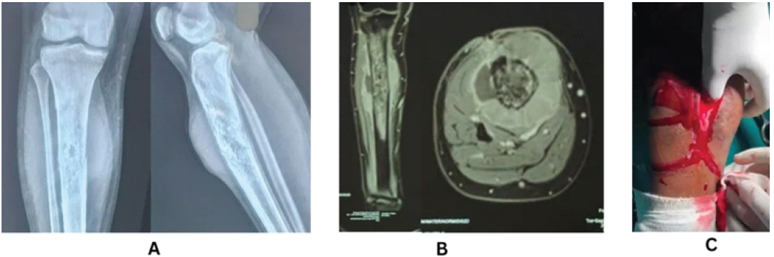
Figure 1: (a) X-ray showing a destructive permeative lesion in the proximal shaft of the right tibia with features suggestive of malignancy, (b) magnetic resonance imaging demonstrating permeative lytic lesion with cortical erosions, periosteal reaction, and associated soft-tissue component in the proximal right tibia, (c) Intraoperative image showing pus-like discharge obtained during open biopsy of the tibial lesion.
Both bone and overlying soft tissue were sent for pathological examination. Microscopic examination of the tissue sample yielded an undifferentiated tumor of small, blue round cells with round to oval hyperchromatic nuclei (Fig. 2a). The histopathologic findings were suggestive of Ewing’s sarcoma. Thus, a suggestion for immunohistochemical (IHC) studies was given. The panel of IHC consisted of markers such as CD99, CD45, pan-CK, S100, WT1, myogenin, vimentin, and desmin. IHC staining showed prominent positivity of the neoplastic cells with CD99, whereas the rest of the markers were negative. Subsequently, the patient was referred to a medical oncologist, and a treatment plan consisting of palliative chemotherapy and radiotherapy was determined, foregoing surgical resection. The patient expired during the course of palliative chemotherapy.
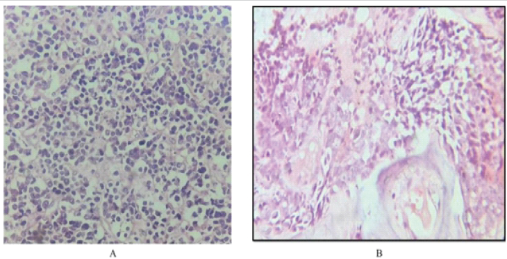
Figure 2: (a) Histopathology showing sheets of small round cells with round to oval nuclei, fine chromatin, and scant cytoplasm, (b) high-power view highlighting tightly packed uniform small round cells with centrally placed nuclei and minimal cytoplasm – features consistent with Ewing sarcoma morphology.
A 20-year-old female sought medical attention, reporting pain and increasing swelling in her right thigh that had developed over the preceding 6 months. On clinical examination, the swelling was diffuse, poorly demarcated, and tender to palpation. Radiographic evaluation revealed a permeative osteolytic lesion with a lamellated periosteal reaction located in the meta-diaphyseal region of the right femur (Fig. 3a). More workup in the form of CT, MRI, PET scan, and supportive laboratory tests was done. CT showed permeative lytic destruction with a wide zone of transition and lamellated periosteal reaction involving the proximal part of the right femur (Fig. 3b). MRI revealed abnormal marrow signaling involving the proximal shaft of the femur with cortical destruction and aggressive periosteal reaction and an associated soft-tissue mass surrounding the shaft of the femur (Fig. 3c). A PET scan revealed a metabolically active large expansile lytic lesion with permeative destruction and a large soft-tissue component involving the proximal and mid shafts of the right femur—likely Ewing sarcoma; metabolically active multiple lytic sclerotic lesions at the mentioned sites in the skeleton—skeletal metastatic lesions; and metabolically active few enlarged right external iliac and right inguinal lymph nodes—metastatic nodes (Fig. 3d).
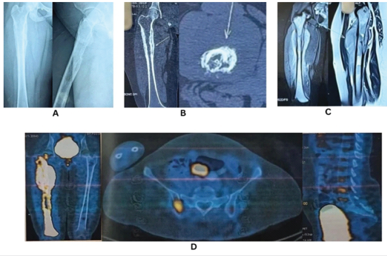
Figure 3: (a). X-ray showing a permeative osteolytic lesion with lamellated periosteal reaction in the meta-diaphyseal region of the right femur, (b) computed tomography demonstrating permeative lytic destruction with a wide zone of transition and lamellated periosteal reaction in the proximal right femur, (c) magnetic resonance imaging revealing abnormal marrow signal with cortical destruction, aggressive periosteal reaction, and surrounding soft-tissue mass in the proximal femoral shaft, (d) positron emission tomography-computed tomography showing a metabolically active expansile lytic lesion in the proximal and mid-shaft of the right femur with soft-tissue component, skeletal metastases, and metastatic right external iliac and inguinal lymph nodes.
An open mass biopsy was done, and biopsy samples of bone with overlying soft tissue were sent for histopathology. Histologically, there was a possibility of a small round cell tumor with differential diagnoses including Ewing sarcoma and small cell variant osteosarcoma (Fig. 2b). Immunohistochemistry was done, namely NKX2.2 (to rule out Ewing sarcoma) and SATB2 (to assess for osteogenic lineage), both of which were negative. Because the results were not conclusive, further sophisticated diagnostic modalities, such as molecular genetic analysis and gene sequencing, were believed to be necessary to characterize the lesion; however, these were not conducted in our center. Subsequent to this, the patient was referred to have the evaluation and planning of care done by a medical oncologist. The patient continues on palliative chemotherapy to date.
Ewing sarcoma is part of a spectrum of malignancies known as the Ewing Sarcoma Family of Tumors, which encompasses other related neoplasms such as peripheral primitive neuroectodermal tumors and Askin’s tumors. These tumors are grouped together due to their shared histologic features, underlying genetics, and characteristic chromosomal translocations involving the EWS gene and various ETS family transcription factors, most frequently producing the EWS-FLI1 fusion protein [2,3,4]. While Ewing sarcoma is the third most prevalent non-hematologic primary bone cancer in total, its relative frequency is several times higher in the young age groups. It is the second most frequent primary bone cancer, after osteosarcoma, in individuals under the age of 30. Furthermore, within the subgroup of children under the age of 10 years, Ewing sarcoma is the most frequent primary bone tumor encountered [5]. In alignment with these patterns, both individuals in our study were diagnosed during adolescence, within their second decade. Ewing sarcoma is rare in adult populations and predominates slightly among males, with a male-to-female ratio of nearly 1.5 to 1, as quoted. The prognosis is usually poorer in regional lymph node metastasis [6]. In our report, regional lymph node involvement was present in both cases, which carries a poor clinical prognosis. The diagnosis of Ewing sarcoma can be challenging due to its histologic similarity with other small round cell tumors. Microscopic presentation of Ewing sarcoma shows small cells packed tightly in clusters with a preserved round to oval shape. The nuclei are centrally placed, round, and contain a fine granular chromatin pattern and virtually unnoticeable, clear, or faintly eosinophilic cytoplasm. Uniform packing of cells and restricted cytoplasmic area are the most important distinguishing features of this malignancy. An interdepartmental approach incorporating clinical, radiologic, pathological, and cytogenetic information is needed to reach a conclusive diagnosis. CD99 is the most commonly reported IHC marker of Ewing sarcoma, and diffuse membranous positivity occurs in most cases [7,8]. CD99 is not specific but also expressed in other small round cell neoplasms, and hence, additional IHC and molecular investigation needs to be carried out [9]. Our study aimed to explore the difficulties that have arisen during the course of diagnosis of Ewing sarcoma from other small round blue cell tumors. In Case 1, the purulent-like discharge on biopsy provided a note of complexity, perhaps in favor of diverting toward infection [10]. Still, the biopsy report was suspicious of Ewing sarcoma, and IHC staining showed diffuse membranous positivity for CD99, and others were negative. On this basis, the pathologist suggested a few more markers, NKX2.2, to arrive at a conclusive diagnosis of Ewing sarcoma, and SATB2 to exclude small cell osteosarcoma [11,12,13]. Intensive diagnostic workup could not be completed because of the limited diagnostic resources, and the patient expired during palliative chemotherapy. In Case 2, both IHC markers NKX2.2 and SATB2 were successfully done but turned out to be negative in Case 1. This has resulted in a diagnostic challenge due to the discordance between the clinical-pathological features and the IHC results (Fig. 2). Following the diagnostic uncertainty, our pathology department suggested sophisticated diagnostic modalities in the form of gene sequencing and molecular genetic examination for the identification of EWSR1 gene rearrangements—these investigations could not be carried out due to limitations of resources in the region. This assumes special relevance in the background of the current World Health Organization (WHO) classification of undifferentiated small round cell sarcomas of bone and soft tissue (Table 1). 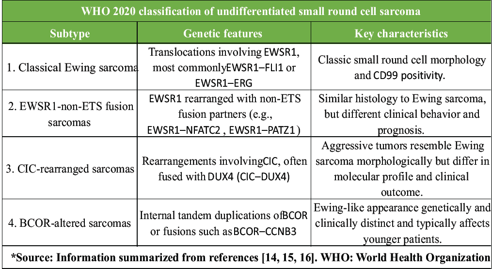 The new category now also acknowledges a more extensive range of genetically different entities aside from traditional Ewing sarcoma [14,15,16]. This new molecular taxonomy focuses on the limitations of using traditional diagnostic modalities such as radiographs and routine histopathologic evaluation, particularly when confronted with similarly appearing tumors. As shown in our case, a lack of molecular diagnostic ability can impede the potential for an absolute and specific diagnosis. Thus, it is critical for orthopedic surgeons to keep pace with emerging trends in tumor diagnosis and classification. Identification and incorporation of molecular genetic testing into standard diagnostic protocols will not only improve diagnostic accuracy but also result in more successful and targeted therapeutic interventions.
The diagnostic evaluation of Ewing sarcoma and the other round cell undifferentiated small cell sarcomas still poses significant challenges, especially without the aid of advanced molecular diagnostic technology. These cases brought to our notice have helped to highlight the shortcomings of using conventional imaging and histopathology. The evolving WHO classification emphasizes the necessity of genetic and molecular profiling to distinguish between the increasingly recognized subtypes, such as CIC-rearranged and BCOR-altered. In institutions without adequate infrastructure for molecular diagnosis, the physicians are compelled to resort to uncertainty that can impact treatment planning as well as prognosis. It becomes all the more necessary, therefore, to increase diagnostic facilities and encourage interdisciplinary consultation for integrative assessment.
An accurate diagnosis of Ewing sarcoma cannot always be obtained by imaging, histopathology, and immunohistochemistry alone. With the expansion of the WHO classification to genetically different Ewing-like sarcomas, additional molecular genetic examinations, such as the detection of EWSR1, CIC, or BCOR mutations, are needed. Orthopedic surgeons should be aware of the evolution of new disease classification, look for and suspect tumors in uncommon locations, and also include lymph node examination in their clinical examination. It is universal that uncommon presentations in such cases are usually associated with aggressive disease patterns and poor prognosis. Where direct advanced testing is not accessible, referral pathways and coordination with specialty centers must be emphasized to maximize patient outcomes.
References
- 1. Delattre O, Zucman J, Plougastel B, Desmaze C, Melot T, Peter M, et al. Gene fusion with an ETS DNA-binding domain caused by chromosome translocation in human tumours. Nature 1992;359:162-5. [Google Scholar] [PubMed]
- 2. Khoury J. Ewing sarcoma family of tumors. Adv Anat Pathol 2005;12:212-20. [Google Scholar] [PubMed]
- 3. Joo J, Christensen L, Warner K, States L, Kang HG, Vo K, et al. GLI1 Is a central mediator of EWS/FLI1 signaling in ewing tumors. PLoS One 2009;4:e7608. [Google Scholar] [PubMed]
- 4. Choi EK, Gardner JM, Lucas DR, McHugh JB, Patel RM. Ewing sarcoma. Semin Diagn Pathol 2014;31:39-47. [Google Scholar] [PubMed]
- 5. Azar FM, Terry Canale S, Beaty JH. Malignant tumors of bone, ewing sarcoma. In: Campbell’s Operative Orthopaedics. 14th ed., Ch. 27. Amsterdam: Elsevier; 2021. p. 1018. [Google Scholar] [PubMed]
- 6. Applebaum MA, Goldsby R, Neuhaus J, DuBois SG. Clinical features and outcomes in patients with ewing sarcoma and regional lymph node involvement. Pediatr Blood Cancer 2012;59:617-20. [Google Scholar] [PubMed]
- 7. Folpe AL, Goldblum JR, Rubin BP, Shehata BM, Liu W, Dei Tos AP, et al. Morphologic and immunophenotypic diversity in Ewing family tumors: A study of 66 genetically confirmed cases. Am J Surg Pathol 2005;29:1025-33 [Google Scholar] [PubMed]
- 8. Llombart-Bosch A, Machado I, Navarro S, Bertoni F, Bacchini P, Alberghini M, et al. Histological heterogeneity of Ewing’s sarcoma/PNET: An immunohistochemical analysis of 415 genetically confirmed cases with clinical support. Virchows Arch 2009;455:397-411. [Google Scholar] [PubMed]
- 9. Riggi N, Stamenkovic I. The biology of Ewing sarcoma. Cancer Lett 2007;254:1-10. [Google Scholar] [PubMed]
- 10. Azar FM, Terry Canale S, Beaty JH. Malignant tumors of bone, ewing sarcoma. In: Campbell’s Operative Orthopaedics. 14th ed. Ch. 27. Amsterdam: Elsevier; 2021. p. 1019. [Google Scholar] [PubMed]
- 11. Chen Y, Liu W, Zhong L, Liao D, Zhang R, Kang T, et al NKX2-2 suppresses osteosarcoma metastasis and proliferation by downregulating multiple target gene. J Cancer 2018;9:3067-77. [Google Scholar] [PubMed]
- 12. Lucas DR NKX2.2 is a useful immunohistochemical marker of Ewing sarcoma. Am J Surg Pathol 2011;35:1178-82. [Google Scholar] [PubMed]
- 13. Dragomir A. SATB2 in combination with cytokeratin 20 identifies over 95% of all colorectal carcinomas. Am J Surg Pathol 2014;38:1160-9. [Google Scholar] [PubMed]
- 14. Sbaraglia M, Bellan E, Dei Tos AP. The 2020 WHO classification of soft tissue tumours: News and perspectives. Pathologica 2021;113:70-84. [Google Scholar] [PubMed]
- 15. Choi JH, Ro YJ. The 2020 WHO classification of tumors of soft tissue and bone: An updated review. Adv Anat Pathol 2021;28:44-58. [Google Scholar] [PubMed]
- 16. Kao EY, Mantilla JG. What’s new in soft tissue and bone pathology 2022-updates from the WHO classification 5th edition. J Pathol Transl Med 2022;56:385-6. [Google Scholar] [PubMed]
- 17. WHO Classification of Tumours Editorial Board. Soft Tissue and Bone Tumours. 5th ed., Vol. 3. France: International Agency for Research on Cancer; 2020. [Google Scholar] [PubMed]




Related Articles in Journal of Orthopaedic Case Reports
 October 1, 2026 Management of Severe Knee Flexion Contracture of 140° with Fibrous Ankylosis in a Resource-Limited Setting: A Case Report
 October 1, 2026 Clinicopathological Spectrum and Management of Aneurysmal Bone Cysts in Atypical Anatomical Location: A Comprehensive Review of Demographics, Presentation, Diagnosis, and Treatment Outcomes
 September 1, 2026 Limb Salvage for Entire Tibial Ewing Sarcoma in a 4-year-Old Child Using Extracorporeal Irradiation and Free Flap Reconstruction: A Case Report and Surgical Technique
 September 1, 2026 Proximal Humerus Ewing Sarcoma: Technical Challenges and Strategies for Bone Defect Reconstruction