


Introduction
Total knee arthroplasty (TKA) is a widely performed surgical procedure for managing end-stage osteoarthritis, rheumatoid arthritis, and other debilitating knee joint conditions. While generally associated with excellent outcomes, complications such as metallosis, though rare, can lead to significant morbidity. Metallosis is defined as the deposition of metal debris in periprosthetic tissues, leading to an inflammatory response that may compromise joint function, cause pain, and necessitate revision surgery [1].
The etiology of metallosis after TKA is multifactorial, involving factors such as implant design, material wear, malalignment, or improper surgical technique. Nine different causes of metallosis (polyethylene liner dislocation, polyethylene liner wear, infectious failure, implant failure, metal-backed patellar failure, aseptic prosthesis loosening, saw blade debris, traumatic failure, and malalignment) have been identified in patients who have undergone TKA [2]. Metal-on-metal implants often cause progressive pain, swelling, and mobility issues. Imaging and histological findings reveal metallic debris deposits, cystic formations, and synovitis, which are crucial for diagnosis [3].
Extensive metallosis following TKA is less well documented. This paper presents a case of extensive metallosis following TKA, emphasizing its clinical presentation, diagnostic challenges, and management strategies. To the best of our knowledge, the degree of tibial component wear and bone loss in this case is among the more extensive reported, highlighting the need for vigilance in identifying early signs of metallosis and the role of surgical and non-surgical interventions in mitigating its progression.
Case Report
In 2008, a 72-year-old woman underwent successful bilateral total knee replacement with posterior-stabilized implants (DePuy Synthes/Johnson and Johnson) at a different hospital for osteoarthritis of both knees. A full range of motion was achieved during the index procedure and sustained postoperatively. The patient recovered well and remained healthy until 2021, when she sustained a twisting injury to her right knee, leading to pain and swelling. She was advised rest and oral medication by local physicians, and her symptoms improved within 2–3 weeks, allowing return to usual activities. Over the following year, she developed progressive bowing of the right knee and was advised to undergo revision surgery, which she deferred. In September 2023, she slipped and fell while rising from a chair, sustaining severe pain in the right knee that prompted her to seek medical attention.
On examination, a well-healed midline scar from the previous surgery was noted, with minimal joint effusion in the right knee. A varus deformity of 30° was measured, fully correctable on examination with a firm end-point and no valgus instability. Range of motion was 0–100° of flexion. Tenderness was noted along the lateral joint line. No distal neurovascular deficit was present.
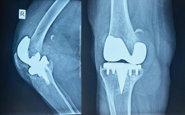
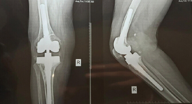
Radiographs revealed osteolysis affecting both the femur and tibia, with loosening of the tibial component. Amorphous, fluffy areas of increased density were seen within the periprosthetic soft tissues, along with bubble-like radiodensities outlining the supra- and retro-patellar capsule – findings characteristic of the ‘cloud and bubble sign’ associated with metallosis. A thin rim of linear increased density was also noted surrounding the infra- and retro-patellar regions. These imaging findings were consistent with advanced metallosis. Significant wear of the tibial component on the medial side was evident, occurring after complete wear-through of the medial polyethylene inlay (Fig. 1).
Laboratory evaluation showed hemoglobin of 12.3 g/dL, total leukocyte count of 6400/µL (within normal limits), C-reactive protein of 3.6 mg/L, and erythrocyte sedimentation rate of 16 mm/h.
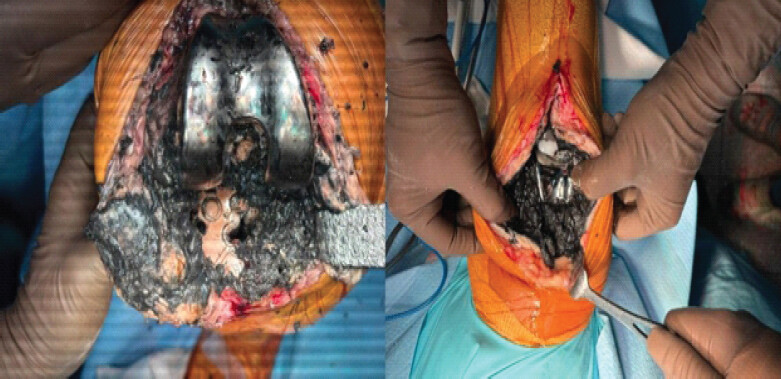
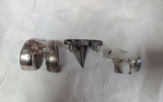
A diagnosis of metallosis-induced prosthetic loosening was confirmed, and a one-stage TKA revision was performed using a cemented semi-constrained modular revision knee system (Zimmer Legacy constrained condylar knee [CCK]). Intraoperatively, extensive black staining was observed in the synovium, joint fluid, and surrounding soft tissue (Fig. 2). Thorough debridement and lavage were performed. Significant wear of the medial tibial tray was noted following complete wear-through of the medial polyethylene inlay (Fig. 3). The components were removed using osteotomes; the femoral component showed aseptic loosening with severe distal femoral osteolysis, and there was significant bone loss posteriorly on the femur with metal fragments in the region of the femoral canal. Abnormal tissue was excised, and bone and soft-tissue samples were sent for histopathological and microbiological analysis. Residual cement was removed, and revision TKA was performed using press-fit diaphyseal stems and a constrained component with tibial wedges to reconstitute bone loss and restore the joint line (Fig. 4).
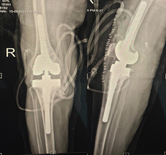
The patient had a smooth post-operative recovery and was allowed full weight-bearing. At 2 weeks, the wound had healed well, and she was mobilizing with a walker, with a range of motion of 0–100° and no instability. She was followed with regular physiotherapy and monthly clinical and radiographic assessment for the first 6 months. At 1 year, she had achieved full recovery — pain-free, full range of motion, improved functional scores, and independent ambulation. The latest radiographs showed a well-positioned prosthesis with no evidence of instability, fracture, or bone loss, closely matching the immediate post-operative films (Fig. 5).
Discussion
A 72-year-old woman developed metallosis 13 years after undergoing TKA, as presented in this case report. Metallosis is not exclusive to knee replacements and can also occur after hip, shoulder, elbow, and wrist joint replacement surgery. Its onset is thought to be triggered by mechanical factors, specifically abrasion between metal components or between metal and polyethylene surfaces, particularly in implants with a metal-backed patellar component. The likelihood of implant wear and metallosis is influenced by the materials used, with titanium components being more susceptible than cobalt-chromium components [4]. Metallic debris production is linked to the interaction between metal surfaces and polyethylene deformation; this debris infiltrates soft tissues such as the joint capsule and cavity, triggering a foreign-body reaction characterized by giant multinucleated cells. This infiltration also causes synovial fluid and surrounding tissue to appear blackish, as noted in previous studies [5]. Contemporary implants such as Smith and Nephew’s Oxinium, constructed from oxidized zirconium, are designed to reduce metal wear and may lower the future incidence of metallosis [6].
In this case, post-operative knee varus instability likely led to persistent impingement, releasing metallic debris and causing chronic synovitis. This synovitis exacerbated joint destruction, heightened instability, and accelerated wear in the medial compartment. On plain radiographs and computed tomography, three indicators – the “bubble sign,” “cloud sign,” and “metal-line sign” – have been described as useful in distinguishing metallosis from other complications following TKA, such as small-particle disease, infected prosthesis, or aseptic loosening [4, 7, 8]; osteolysis has also been noted in the context of metallosis. Metallosis can present with additional radiographic findings not observed in this case, such as reduced bone mineralization, fractures, and significant bone defects [8, 9]. These complications may be mitigated through careful debridement – synovectomy and excision of inflammatory tissue – together with revision arthroplasty [8].
Successful traditional TKA requires competent, functional collateral ligaments. In this patient, significant osteolysis due to metallosis was accompanied by notable bone deficiency on both the femoral and tibial sides. Revision surgery therefore used a CCK prosthesis incorporating wedges and stems to provide inherent medial and lateral stability [10, 11].
Conclusion
This case report emphasizes the importance of long-term monitoring after TKA to facilitate early identification of potential complications. Early detection of metallosis allows intervention through surgical excision of affected tissue and joint revision, mitigating the risk of further joint deterioration.
Clinical Message
Extensive metallosis should be considered in any patient presenting with progressive deformity, instability, or pain years after an otherwise uneventful total knee arthroplasty. Recognition of the radiographic “cloud and bubble sign” allows early diagnosis, and a constrained condylar revision system can achieve a stable, functional joint even in the presence of severe bone loss.
Conflict of Interest:
Nil
Source of Support:
Nil
Consent:
The authors confirm that informed consent was obtained from the patient for publication of this article
References
- Matusiewicz NH, Richter NM. Potentially toxic metallic wear nanoparticles and trace metal ions release from metal-on-metal orthopedic implants in the human biological specimens: An overview of in vivo and ex vivo clinical studies. World J Adv Res Rev 2020;8:242-92. [Google Scholar] | [PubMed]
- Goyal N, Hozack B, Parvizi J. Mechanical failure of modern total knee arthroplasty. Curr Orthop Pract 2011;22:236-40. [Google Scholar] | [PubMed]
- Xia Z, Ricciardi BF, Liu Z, Von Ruhland C, Ward M, Lord A. Nano-analyses of wear particles from metal-on-metal and non-metal-on-metal dual modular-neck hip arthroplasty. Nanomedicine 2016;13:1205-17. [Google Scholar] | [PubMed]
- Weissman BN, Scott RD, Brick GW, Corson JM. Radiographic detection of metal-induced synovitis as a complication of arthroplasty of the knee. J Bone Joint Surg Am 1991;73A:1002-7. [Google Scholar] | [PubMed]
- Essner A, Herrera L, Hughes P, Kester M. The influence of material and design on total knee replacement wear. J Knee Surg 2011;24:9-17. [Google Scholar] | [PubMed]
- Chew FS, Ramsdell MG, Keel SB. Metallosis after total knee replacement. AJR Am J Roentgenol 1998;170:1556. [Google Scholar] | [PubMed]
- Romesburg JW, Wasserman PL, Schoppe CH. Metallosis and metal-induced synovitis following total knee arthroplasty: Review of radiographic and CT findings. J Radiol Case Rep 2010;4:7-17. [Google Scholar] | [PubMed]
- Matsushita I, Morita Y, Gejo R, Kimura T. Severe bone defects and reduced mineralization caused by massive metallosis after total knee arthroplasty: Histopathologic and bone morphometric findings. Mod Rheumatol 2007;17:507-10. [Google Scholar] | [PubMed]
- Hernández-Vaquero D, Sandoval-García MA. Hinged total knee arthroplasty in the presence of ligamentous deficiency. Clin Orthop Relat Res 2010;468:1248-53. [Google Scholar] | [PubMed]
- Joshi N, Navarro-Quilis A. Is there a place for rotating-hinge arthroplasty in knee revision surgery for aseptic loosening? J Arthroplasty 2008;23:1204-11. [Google Scholar] | [PubMed]
© 2026 Journal of Orthopaedic Case Reports - Published by Indian Orthopaedic Research Group
About the Authors
-

Sharath Raj
-

Vivek P Ksheerasagar
-

Aditya Shah
-

Souvik Das
-

Vijay G Goni
Related Articles in Journal of Orthopaedic Case Reports
 January 1, 2026 Severe Metallosis-Induced Osteolysis and Polyethylene Liner Dissociation as a Cause of Total Knee Replacement Failure
 November 10, 2023 Oxidized Zirconium Bearing Surface Failure in Total Knee Arthroplasty: A Unique Case Report of Isolated Medial Compartment with Catastrophic Results
 October 1, 2026 Intraoperative Discovery of Incorrect Femoral Component Packaging during Bilateral Total Knee Arthroplasty: A Near-Miss Case Report
 October 1, 2026 Comparative Study of Effect of Periarticular Fentanyl and Dexmedetomidine Injection for Pain Management in Total Knee Arthroplasty a Randomized Control Trial