


Introduction
Hepatocellular carcinoma (HCC) typically metastasizes to the lungs, regional lymph nodes, and long bones. Metastasis to the oral cavity, particularly the mandible, is an exceptionally rare phenomenon [1,2,3]. In highly unusual instances, a metastatic jaw lesion can precede any clinical symptoms or diagnosis of the primary intrahepatic tumor [4]. Consequently, these lesions are frequently misdiagnosed as benign dental or maxillofacial conditions, such as periodontal disease or post-extraction complications [5]. This report details the clinical course and complex palliative management of a patient whose occult, Hepatitis C virus (HCV)-driven HCC first manifested as an aggressive mandibular mass.
Case Report
Initial evaluation and diagnostic timeline
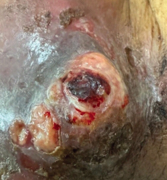
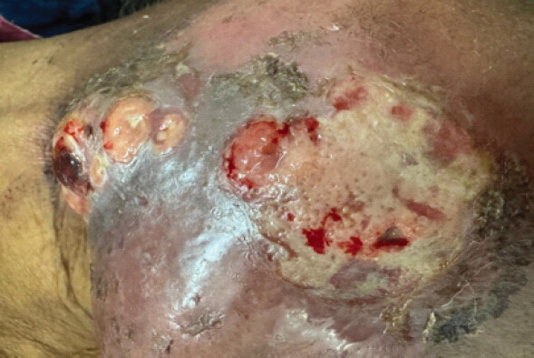
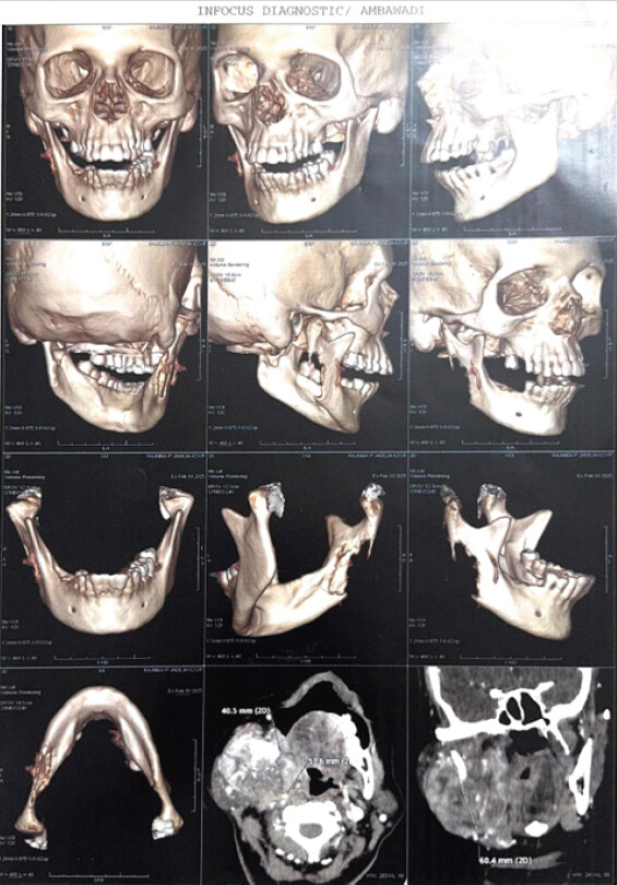
A 62-year-old female developed progressive right-sided facial and mandibular swelling shortly after a routine tooth extraction performed on January 12, 2025. Due to persistent growth of the mass, a right mandible tissue biopsy was performed on February 11, 2025 (Fig. 1, 2, 3).
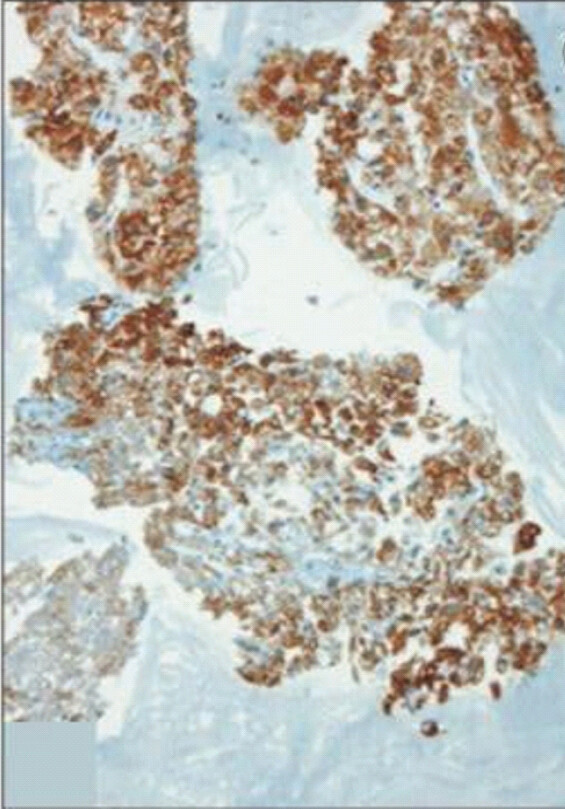
Histopathological evaluation of the deep tissue demonstrated fragments of bone and soft-tissue heavily infiltrated by trabeculae of neoplastic hepatoid cells with endothelial wrapping, moderate nuclear atypia, and prominent nucleoli. To definitively establish the tissue of origin, immunohistochemistry (IHC) was conducted. The tumor cells demonstrated strong positivity for Hepar-1 [6] (Fig. 4) and Glypican 3 [7,8], and weak positivity for alpha-fetoprotein (AFP) [9], while staying entirely negative for CK-7, Cdx-2, and PAX-8. This specific immunophenotype confirmed a diagnosis of metastatic HCC, Grade 2 [10].
Staging and underlying etiology
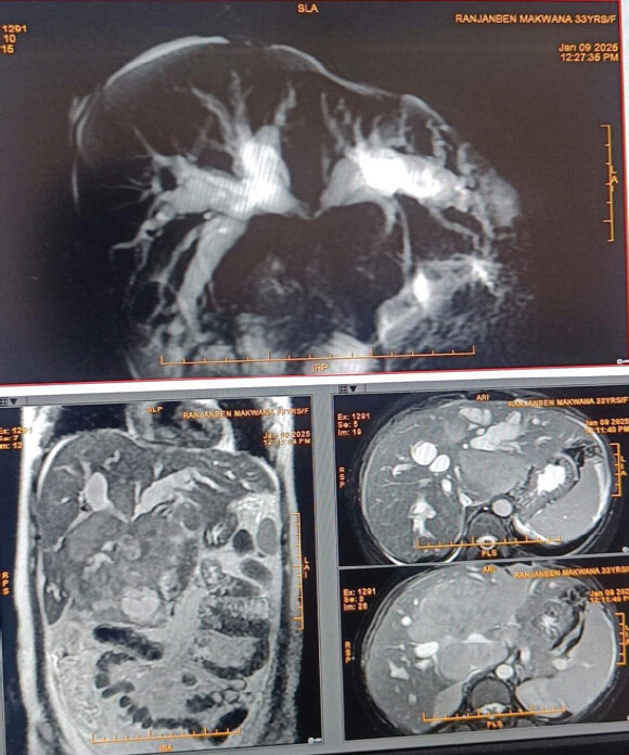
Baseline serum AFP was recorded at 172 ng/mL. A staging positron emission tomography-computed tomography scan on February 21, 2025, revealed a massive (66 × 59 × 60) mm osteolytic metastatic lesion in the right mandible (Fig. 3), a (43 × 44 × 24) mm primary lesion in the left lobe of the liver, and multiple satellite hepatic nodules (Fig. 5).
Subsequent gastroenterology workup revealed that the patient was positive for HCV, directly tracing back to a remote blood transfusion received in 1987. Clinical and sonographic features confirmed advanced underlying chronic liver disease, staged as Child-Pugh Class B [11], Barcelona clinic liver cancer (BCLC) Stage C [12,13], with a baseline MELD-Na score of 14 [14,15].
Treatment protocol and clinical progress
In this case, the discharging sinus has a perforated only outer cortex of the mandibular ramus. The inner cortex of the mandible is still intact, so the fracture is incomplete and stable. Hence, the surgical fixation of the mandible is not required as a palliative surgery, as the pain is very minimal at jaw.
Surgical resection was not planned in the present case because the patient was in the advanced stage of HCC (Child-Pugh Class B, BCLC Stage C, baseline MELD-Na score – 14, high), so the surgical risk was high. Treatment for HCC metastasis is typically palliative, and radiation therapy has been shown to be effective palliative therapy for bony metastasis in patients with HCC. In addition, targeted drugs for advanced HCC are also the standard treatment; Lenvatinib is a commonly used anti-angiogenic agent.
Systemic and local radiotherapy (February–March 2025)
In late February 2025, the patient was initiated on systemic targeted therapy with the tyrosine kinase inhibitor Lenvatinib (Tab. Lenvima). Concurrently, to address the severe local pain and tumor burden, she received palliative hypofractionated external beam radiotherapy three-dimensional conformal radiation therapy to the right mandibular region.
She completed a total dose of 3600 cGy in 12 fractions over 13 days (February 28–March 12, 2025). Local radiotherapy was well tolerated with only Grade 1 mucositis. However, concurrent systemic Lenvatinib had to be temporarily discontinued after 2 weeks due to severe dysphagia and debilitating oral ulcers, before being cautiously restarted at a modified dose following side-effect resolution.
Abdominal disease evolution (August 2025)
A follow-up abdominal ultrasound on August 19, 2025, documented significant intrahepatic tumor progression. The primary mass in segments II and III of the liver had increased in size to (7.5 × 4.6) cm up from (4.6 × 2.8) cm. Multiple separate, hypoechoic metastatic lesions across both hepatic lobes also exhibited an increase in size and structural number.
Late-stage complications and emergency presentation (November 2025)
In November 2025, the patient presented to the emergency department with a high fever, chills, severe localized back pain, progressive breathlessness, and recurrent episodes of active oral bleeding (Fig. 1 and 2). Physical examination revealed that the right mandibular tumor had eroded through the overlying skin layers, forming a large, fungating, ulcerated, and actively bleeding exophytic mass.
Emergency diagnostic imaging highlighted critical, multi-system progression:
-
Vascular tumor invasion: An abdominal ultrasound demonstrated advanced liver cirrhosis and a new left portal vein tumor thrombosis (Fig. 5), indicating direct vascular invasion by the malignancy.
-
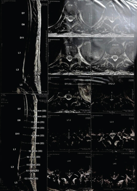
Spinal metastasis: A 3T dorso-lumbar spine magnetic resonance imaging (MRI) revealed an irregular area of altered signal intensity involving the left pedicle, lamina, and transverse process of the L3 vertebra, causing effacement of the left L3-L4 facet joint – consistent with a new skeletal metastasis (Fig. 6).
-
Neurological structural compression: The spine MRI also documented broad-based posterior disc protrusions at L3-L4 and L4-L5 causing direct compression of the thecal sac and nerve roots, alongside intramedullary T2 hyperintensities and mild cord expansion from D11-L2, highly suggestive of concurrent myelitis (Fig. 6).
Stabilization and discharge palliative care
Following emergency inpatient stabilization, severe hypokalemia was corrected. For her systemic vascular thrombosis, she was initiated on low-molecular-weight heparin (Inj. Fragmin 2500 IU S/C daily). Direct antiviral therapy for her underlying disease was started using Sofosbuvir/Velpatasvir (Tab. Velpanat). Tab Lenvatinib is commonly used as an anti-angiogenic agent, which is considered a targeted drug for advanced HCC. For multi-site pain control, she was stabilized on an aggressive palliative analgesic regimen, including scheduled base medications and short-acting narcotics (Tab. Morphine 10 mg SOS for breakthrough pain), before being transitioned to home-based supportive care.
Discussion
In 1998, Chin et al. published a review on metastatic tumors in the oral cavity. The majority of patients were male, with a male-to-female ratio of 1.6:1, and 90% of patients were over 50 years of age. The mandible was the most frequently involved site, with the anatomical distribution of cases reported as follows: Mandible: 40 cases, Maxilla: 8 cases, Tonsil: 1 case, Both Mandible and Maxilla: 1 case.
This case illustrates a highly unusual clinical trajectory where a hidden abdominal malignancy first declared itself via extrahepatic skeletal metastasis to the jawbone. Metastatic lesions in the maxillofacial region are notoriously difficult to diagnose early because their clinical and radiographic presentations closely mimic benign dental infections or standard post-extraction healing failures [5,16,17]. The immediate acceleration of facial swelling following her tooth extraction represents a classic example of a tumor capitalizing on the local inflammatory response and vascular remodeling of a healing dental socket to expand rapidly [18].
From a pathologic perspective, this case highlights the absolute necessity of IHC in clarifying tumors of unknown primary origins displaying “hepatoid” features. The diagnostic profiling – showing strong positivity for Hepar-1 [6] and Glypican 3 [7] – allowed the pathology team to confidently differentiate the mandibular mass from primary maxillofacial or clear-cell variants, bypassing the need for invasive liver biopsies in a patient with advanced background cirrhosis.
The final phase of this patient’s disease underscores the immense clinical challenge of managing advanced, simultaneous oncological complications. The development of a fungating, ulcerated facial mass represents a profound palliative hurdle, requiring highly specialized wound management to address constant capillary oozing, secondary bacterial colonization, and severe localized pain. Furthermore, the emergence of a portal vein tumor thrombosis simultaneously elevates the risk of internal variceal bleeding from exacerbated portal hypertension while demanding the cautious use of systemic anticoagulation.
Finally, the addition of an L3 spinal metastasis with structural thecal sac compression and underlying cord edema places the patient at extreme risk for rapid neurological decline, necessitating precise multi-specialty care.
Conclusion
-
Maintain high suspicion for unresolved jaw masses: Clinicians must consider metastatic malignancy in the differential diagnosis of any aggressive, non-healing, or rapidly expanding jaw lesion following a routine dental extraction, particularly in patients with a history of previous blood transfusions or unknown viral hepatitis status.
-
The long latency of chronic HCV: This case serves as a reminder of the multi-decade latency period of the HCV contracted through previous blood products before modern screening protocols, which silently drives liver cirrhosis and aggressive HCC.
-
Primacy of multidisciplinary palliative care: In advanced oncological states, systemic targeted therapies are restricted by severe side effects and liver dysfunction; the focus must shift entirely to integrated palliative care. Coordinated efforts between Medical Oncology, Radiation Oncology, Interventional Gastroenterology, and Palliative Wound Care are vital to optimize comfort, manage skin barrier integrity, titrate opioid analgesics safely, and preserve dignity at the end of life.
Clinical Message
Unresolved jaw pain, soft-tissue swelling, or persistent post-extraction socket healing failures require a high index of suspicion for metastatic bone disease. Comprehensive orthopedic evaluation – incorporating structural stability scoring and fracture risk assessment – is vital to determine whether prophylactic fixation or non-invasive palliation should be executed in patients with poor physiological reserve.
Conflict of Interest:
Nil
Source of Support:
Nil
Consent:
The authors confirm that informed consent was obtained from the patient for publication of this article
References
- Fernández-Ferreira R, Savage-Leyva R, Durán-Guerrero LF, Carranza-Sevilla MD, Zamorano-Vazquez C, Monroy-Godínez CF. Mandibular metastasis as the first manifestation of hepatocellular carcinoma: A case report. Case Rep Oncol 2023;16:88-95. [Google Scholar] | [PubMed]
- Du C, Feng Y, Li N, Wang K, Wang S, Gao Z. Mandibular metastasis as an initial manifestation of hepatocellular carcinoma: A report of two cases. Oncol Lett 2015;9:1213-6. [Google Scholar] | [PubMed]
- Mašulović D, Igić A, Filipović A, Zakošek M, Bulatović D, Mijović K. A rare case of isolated hepatocellular carcinoma metastasis in left mandibular region in a patient with hepatitis C virus liver cirrhosis diagnosed after the onset of COVID-19 infection. Medicina (Kaunas) 2023;59:1992. [Google Scholar] | [PubMed]
- Misra SR, Shankar YU, Rastogi V, Maragathavalli G. Metastatic hepatocellular carcinoma in the maxilla and mandible, an extremely rare presentation. Contemp Clin Dent 2015;6:S117-21. [Google Scholar] | [PubMed]
- Cho J. Metastatic hepatocellular carcinoma in the maxilla and temporal bone: A rare case report. J Korean Assoc Oral Maxillofac Surg 2021;47:224-8. [Google Scholar] | [PubMed]
- Koehne De Gonzalez AK, Salomao MA, Lagana SM. Current concepts in the immunohistochemical evaluation of liver tumors. World J Hepatol 2015;7:1403-11. [Google Scholar] | [PubMed]
- Kaseb AO, Hassan M, Lacin S, Abdel-Wahab R, Amin HM, Shalaby A. Evaluating clinical and prognostic implications of Glypican-3 in hepatocellular carcinoma. Oncotarget 2016;7:69916-26. [Google Scholar] | [PubMed]
- Pour AM, Masir N, Rose IM. Glypican-3 is useful but not superior to Hep Par 1 in differentiating hepatocellular carcinoma from other liver tumours. Pathology 2017;49:S79. [Google Scholar] | [PubMed]
- Timek DT, Shi J, Liu H, Lin F. Arginase-1, HepPar-1, and Glypican-3 are the most effective panel of markers in distinguishing hepatocellular carcinoma from metastatic tumor on fine-needle aspiration specimens. Am J Clin Pathol 2012;138:203-10. [Google Scholar] | [PubMed]
- Wang C, Shao X, Zhang X, Xie C, Yu J, Xu X. Diagnostic value of glypican-3 arginase-1 and hepatocyte paraffin antigen -1 in differentiating hepatocellular carcinoma from intrahepatic cholangiocarcinoma. Transl Cancer Res 2020;9:128-36. [Google Scholar] | [PubMed]
- Pugh RN, Murray-Lyon IM, Dawson JL, Pietroni MC, Williams R. Transection of the oesophagus for bleeding oesophageal varices. Br J Surg 1973;60:646-9. [Google Scholar] | [PubMed]
- Forner A, Reig M, Bruix J. Hepatocellular carcinoma. Lancet 2018;391:1301-4. [Google Scholar] | [PubMed]
- Reig M, Forner A, Rimola J, Ferrer-Fábrega J, Burrel M, Garcia-Criado Á. BCLC strategy for prognosis prediction and treatment recommendation: The 2022 update. J Hepatol 2022;76:681-93. [Google Scholar] | [PubMed]
- Kamath PS, Kim WR. Advanced Liver Disease Study Group. The model for end-stage liver disease (MELD). Hepatology 2007;45:797-805. [Google Scholar] | [PubMed]
- Kim WR, Biggins SW, Kremers WK, Wiesner RH, Kamath PS, Benson JT. Hyponatremia and mortality among patients on the liver-transplant waiting list. New Engl J Med 2008;359:1018-26. [Google Scholar] | [PubMed]
- Irani S. Metastasis to the Jawbones: A review of 453 cases. J Int Soc Prev Community Dent 2017;7:71-81. [Google Scholar] | [PubMed]
- Lyon PR, Trevathan S, Talukdar R, Nguyen DD, McPhaul CM, Rampisela D. A rare case of metastatic hepatocellular carcinoma to the hard palate. J Investig Med High Impact Case Rep 2022 Oct 27 10 23247096221133989 doi: 10.1177/23247096221133989. [Google Scholar] | [PubMed] | [CrossRef]
- Murgia MS, Orrù G, Casu C. Oral and maxillofacial metastasis of breast cancer: A case report and literature review. World Cancer Res J 2021;8:e1973. [Google Scholar] | [PubMed]
© 2026 Journal of Orthopaedic Case Reports - Published by Indian Orthopaedic Research Group
About the Authors
-

Varun R Mehta
-

Noopur R Mehta
-

Rajkumar Mehta
-

Sonal R Mehta
Related Articles in Journal of Orthopaedic Case Reports
 February 10, 2023 Massive Periprosthetic Osteolysis Spreads to the Soft Tissue and Pelvic Region after Primary Total Hip Replacement: A Case Report
 March 1, 2025 Prospective Study of Functional and Radiological Outcome after Operative Management of Supracondylar Fracture Humerus in Children
 April 10, 2016 Acromioclavicular Joint Dislocation with Ipsilateral Mid Third Clavicle, Mid Shaft Humerus and Coracoid Process Fracture – A Case Report
 January 10, 2017 Suspension Arthroplasty Combined with Ligament Reconstruction of the Thumb Carpometacarpal Joint to Salvage Two Failed Arthroplasties: A Case Report