


Introduction
The lumbosacral junction transfers axial loads from the spine to the pelvis and lower limbs, and the sacrum is the primary stabilizer of this functional complex. Structural failure of the sacrum therefore causes severe mechanical pain, reduced tolerance for sitting and standing, and rapid functional decline. The incidence of sacral insufficiency and fragility fractures has risen steadily, driven by population aging and by conditions that degrade bone quality, including osteoporosis, prior radiotherapy, and metastatic bone disease [1,2].
In patients with advanced cancer, sacral insufficiency fractures pose a particular challenge. Pathological bone, high lumbopelvic mechanical demands, and a compromised systemic state increase the risk of failure of conventional fixation strategies. In addition, prolonged immobilization in this group accelerates functional decline and increases morbidity and mortality, making immediate stabilization with minimal surgical insult a priority [3,4]. The neurologic-oncologic-mechanical-systemic (NOMS) framework integrates neurological, oncological, mechanical, and systemic criteria to guide treatment [5].
From a reconstructive standpoint, triangular fixation combines a vertical component (lumbar pedicle instrumentation extended to the pelvis) with a horizontal component (sacroiliac or transiliac-transsacral fixation) and is recognized as one of the most biomechanically stable configurations for complex sacral fractures and spinopelvic dissociation [6,7]. However, traditional open approaches carry considerable morbidity in oncologic patients due to extensive muscle dissection, greater blood loss, and a higher risk of wound complications [3,8].
Minimally invasive techniques produce stable constructs with minimal soft-tissue disruption. The S2-alar-iliac trajectory provides low-profile pelvic fixation that aligns coaxially with lumbar pedicle instrumentation, reducing the need for connectors and enabling percutaneous rod passage [9,10]. Polymethylmethacrylate (PMMA) augmentation, delivered through cannulated or fenestrated screws, improves screw purchase in osteoporotic and pathological bone and helps reduce loosening [11,12,13]. Minimally invasive techniques have been associated with less intraoperative bleeding, fewer complications, and earlier post-operative mobilization, even in frail patients or those with advanced systemic disease [3,8,13].
Despite these advances, evidence on the combined use of minimally invasive lumbopelvic fixation and percutaneous transiliac-transsacral fixation for metastatic sacral insufficiency fractures is scarce and limited to isolated reports with minimal technical detail and short follow-up [8,14]. We report the technical feasibility, perioperative findings, and 3-month course of a combined construct in a patient with a pathological sacral fracture secondary to metastatic breast cancer. This case is reported in accordance with the CARE guidelines [15].
Case Report
Background and presentation
A 53-year-old woman with stage IV triple-negative breast cancer (estrogen receptor/progesterone receptor/human epidermal growth factor receptor 2, breast cancer susceptibility gene-negative), diagnosed in 2021, initially presented with a bone metastasis from an unknown primary. At the time of the acute event, she was receiving fifth-line systemic therapy (sacituzumab govitecan, first cycle in October 2025), having previously received paclitaxel (2021), capecitabine (2021–2024), pegylated doxorubicin (2024–2025), and gemcitabine (2025). Relevant surgical history included cesarean section, gastric bypass, and a right hybrid total hip arthroplasty with a cemented femoral component. She was a non-smoker and reported allergies to metamizole and paclitaxel.
Ten days before admission, she sustained a ground-level fall from a seated position, with progressive low lumbar and right gluteal pain radiating to the sacroiliac region, unresponsive to oral analgesics. In the emergency department, she had severe pain (visual analog scale [VAS] 8/10), an intact neurological and neurovascular examination, a stable pelvis on palpation, and a compromised general condition. Concurrent urinary sepsis was identified at admission and treated with ceftriaxone.
Imaging
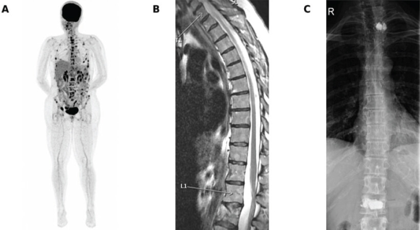
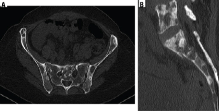
Computed tomography (CT) of the lumbar spine and pelvis showed multiple focal lytic, sclerotic, and mixed osseous lesions in both hemipelves, the lumbar spine, and the sacrum, consistent with secondary deposits (Fig. 1). A recent sacral fracture, not present on a prior study, showed longitudinal lines in both sacral alae and a transverse line at the S1 body, all impacted; the transverse line contacted the superior margin of both S1 neuroforamina, with mild narrowing of the right foramen (Fig. 2). Mild depression of the L1 superior endplate and an old L2 fracture with vertebroplasty material were also noted. Magnetic resonance imaging confirmed an H-shaped sacral fracture in pathological bone, without a soft-tissue mass in the neuroforamina and with regional marrow edema. The pattern corresponded to a bilateral Denis zone I fracture with a transverse component (Roy-Camille), category IV in the Rommens classification of fragility fractures of the pelvis.
Multidisciplinary evaluation and planning
The patient was assessed by medical oncology, spine surgery, hip and pelvis trauma, anesthesiology, and the acute pain unit. After 4 days of escalating multimodal analgesia (oral tapentadol, pregabalin, and paracetamol, followed by patient-controlled intravenous methadone supplemented with subcutaneous ketamine), pain remained refractory (VAS 4/10 at rest, 7/10 with movement), and the patient was unable to stand. Radiotherapy as a sole acute-phase measure was deferred by oncologic consensus, and isolated sacroplasty was considered insufficient to withstand loading in an H-shaped pattern on pathological bone with bilateral ala involvement and a transverse component [7,16]. A combined strategy was therefore planned, adding a percutaneous transiliac-transsacral screw to minimally invasive posterior lumbopelvic fixation (L5 pedicle and cement-augmented S2-alar-iliac screws), performed in two stages under a single anesthetic, to provide immediate rigid stability while minimizing soft-tissue morbidity. The patient and her husband received detailed information on alternatives, benefits, and risks, and provided written informed consent for the procedure and for publication of the case.
Surgical technique
The procedure was performed in two stages under a single general anesthetic, with multimodal analgesia and ceftriaxone prophylaxis. The patient was monitored with an arterial line, a pre-existing central venous catheter, and a urinary catheter.
First stage (supine) – transiliac-transsacral screw
With the patient supine on a radiolucent table and a bolster under the sacral region, adequate anteroposterior, inlet, and outlet fluoroscopic views of the sacral corridors were obtained. Under inlet and outlet guidance, the entry point was identified on the left iliac table for a transiliac-transsacral screw through the S1 corridor. After guidewire placement and progressive reaming, a full-threaded cannulated screw (7.0 × 165 mm; Zimmer Biomet) was inserted. An intraoperative CT scan (Cios Spin) confirmed satisfactory reduction and adequate screw placement. This screw was not cement-augmented.
Second stage (prone) – minimally invasive lumbopelvic fixation
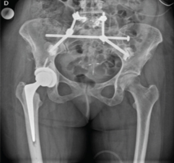
After repositioning to the prone position, the target levels (L5 and S2) were marked under fluoroscopy. Through a mini-open lateral paravertebral approach to the L5 pedicles, bilateral cannulated L5 pedicle screws (6.0 × 40 mm; Neo Medical) were placed over guidewires under biplanar fluoroscopy. Under anteroposterior, lateral, and obturator-outlet fluoroscopy, bilateral S2-alar-iliac screws (8.0 × 80 mm) were inserted at the S1–S2 level, directed across the sacroiliac joint, with position verified in three planes and with three-dimensional fluoroscopy. PMMA cement was delivered through the cannulated lumbopelvic screws and allowed to cure. Bilateral precontoured rods (60 × 5.5 mm) were connected and confirmed under biplanar fluoroscopy (Fig. 3). After copious saline irrigation, closure was performed in layers.
Consolidated perioperative parameters
Total operative time 325 min; total anesthetic time 400 min; estimated intraoperative blood loss <50 mL; 1,250 mL of lactated Ringer; intraoperative urine output 180 mL; and transient norepinephrine (maximum 0.04 µg/kg/min) was weaned early in the immediate post-operative period. Instrument and sponge counts were correct, and no intraoperative complications occurred. The patient was transferred to an intermediate care unit for monitoring.
Post-operative course
On the 1st post-operative day, the patient was hemodynamically stable, afebrile, and neurologically intact, with a VAS of 0/10 at rest on patient-controlled methadone. Assisted sitting was achieved at 24 h, and on the 4th post-operative day, she walked with a rollator without sciatic pain or signs of deep vein thrombosis. The wounds healed without signs of infection. She was discharged on the 5th post-operative day (the 11th day after admission), ambulating with two canes, on oral analgesics only, with instructions for progressive weight-bearing as tolerated and outpatient wound care. No surgical complications occurred during the 1st month.
Three-month follow-up
At the 3-month review, the patient had no lumbopelvic pain or sciatica, ambulated with a single cane for safety, and had no neurological deficit. Control radiographs showed the lumbopelvic fixation in an adequate position, with mild wedging of the inferior endplate of the vertebra immediately cephalad to the construct, without progressive displacement. Control positron emission tomography-CT showed no metabolic activity in the treated region, indicating a good response to concurrent systemic therapy (sacituzumab govitecan). The patient remained in active oncologic follow-up and was functionally independent at the close of the manuscript.
The clinical timeline is summarized in Table 1.
Discussion
This case illustrates the feasibility and mid-term clinical effectiveness of a combined, minimally invasive construct that adds a percutaneous transiliac-transsacral screw to cement-augmented lumbopelvic (L5–S2-alar-iliac) fixation for the management of a pathological sacral insufficiency fracture in a patient with advanced metastatic breast cancer. The procedure achieved immediate rigid stability with a remarkably low biological insult (estimated blood loss below 50 mL), allowing early mobilization and hospital discharge on the 5th post-operative day, in a clinical scenario usually associated with prolonged immobilization and accelerated functional decline.
Restoring mechanical stability of the lumbosacral junction is a fundamental objective in sacral insufficiency fractures, particularly when they occur in pathological bone of metastatic origin. Disruption of the axial load-transfer axis creates persistent micromotion that perpetuates pain and compromises ambulation. Isolated or lower-complexity fixation is often insufficient to resist shear, flexion, and rotational forces in the lumbopelvic region, increasing the risk of implant loosening or failure [3,6,16]. Biomechanical evidence supports triangular fixation as providing the greatest overall stability by combining vertical lumbopelvic support with a horizontal transsacral or sacroiliac anchor. This configuration is particularly relevant for complex patterns and severe bone compromise, where bone quality limits the holding capacity of isolated screws [1,6,7].
The minimally invasive, multidisciplinary approach to the procedure offers decisive advantages for oncologic patients. Previous minimally invasive lumbopelvic fixation series report blood loss frequently below 100 mL and a low rate of wound complications, which are critical in patients with limited physiological reserve or receiving concurrent systemic therapy [3,8,13]. Preservation of the paravertebral and gluteal musculature facilitates early mobilization, which is key to preventing the deleterious consequences of prolonged immobilization, including respiratory infection, venous thromboembolism, and functional decline [3].
Technically, S2-alar-iliac screws provide low-profile pelvic fixation that is coaxial with lumbar pedicle instrumentation, simplifying percutaneous rod insertion and reducing the need for additional connectors [9,10]. Adding a transiliac-transsacral screw provides a horizontal component that improves resistance to shear forces across an unstable sacrum, particularly with bilateral involvement [1,7]. Performing both components in two stages under a single anesthesia, with coordinated spine and pelvis teams, optimized resources and avoided a second general anesthesia in a frail patient. Intraoperative CT was used to confirm transsacral screw position within the S1 corridor.
For compromised bone quality, PMMA augmentation delivered through cannulated screws is an established adjunct to improve screw purchase in advanced osteoporosis or pathological bone [11,12,13]. In this case, cement was delivered through the cannulated lumbopelvic screws to enhance anchorage in diffusely metastatic bone, while the transiliac-transsacral screw was left unaugmented given the adequate compaction of the S1 corridor. Cement augmentation must be balanced against the risk of extravasation into neural foramina or vascular structures, a complication that is infrequent when the technique is performed under rigorous fluoroscopic control [11,16].
In the palliative oncologic setting, the main objective of surgical treatment is not bony union but the achievement of immediate rigid stability that allows effective pain control and rapid functional recovery, enabling the continuation of systemic therapy and preserving quality of life [5,14]. The NOMS framework helped guide this decision: Neurological integrity allowed wide decompression to be avoided, mechanical stability was the dominant factor (unstable sacral fracture and refractory mechanical pain), the systemic scenario was compatible with minimally invasive surgery, and radiation oncology could complement treatment without replacing mechanical stabilization [5].
A comparison with previous reports is summarized in Table 2. Existing reports mostly describe small series or isolated cases, with technical variability and heterogeneous follow-up. The present case provides a detailed description of a minimally invasive, cement-augmented lumbopelvic strategy in a clinically complex scenario, with a favorable midterm functional outcome.
Limitations
This is a single case, so the conclusions are not generalizable to all patients with metastatic sacral fractures. The 3-month follow-up, although informative about functional recovery, does not allow assessment of long-term construct durability or the risk of late loosening in the context of oncologic progression. No formal, validated functional instruments (for example, the Oswestry disability index, EQ-5D, or FACT-G) were used beyond standard clinical assessment and the VAS. Finally, the case was performed at a high-complexity center with advanced fluoroscopy and a multidisciplinary team, conditions that may not be replicated in all care settings.
Conclusion
In patients with sacral insufficiency fractures associated with metastatic disease, combining minimally invasive cement-augmented lumbopelvic fixation with a percutaneous transiliac-transsacral screw is a technically feasible strategy that restores spinopelvic stability with minimal biological impact. The present case documents early functional recovery, effective pain control, and no complications at 3 months, supporting the role of minimally invasive, multidisciplinary techniques in selected oncologic settings. Prospective series with longer follow-up and standardized functional outcomes are needed to define the precise role of this strategy in the treatment algorithm.
Clinical Message
For metastatic sacral insufficiency fractures with spinopelvic instability, the goal is rapid mechanical palliation. A combined construct that adds a percutaneous transiliac-transsacral screw to cement-augmented lumbopelvic (L5 and S2-alar-iliac) fixation can restore stability with minimal morbidity when non-operative management fails and the patient is fit for surgery, enabling early mobilization and continued oncologic care.
Conflict of Interest:
Nil
Source of Support:
Nil
Consent:
The authors confirm that informed consent was obtained from the patient for publication of this article
References
- Gewiess J, Albers CE, Keel MJ, Frihagen F, Rommens PM, Bastian JD. Chronic pelvic insufficiency fractures and their treatment. Arch Orthop Trauma Surg 2025;145:76. [Google Scholar] | [PubMed]
- Rommens PM, Hofmann A. Comprehensive classification of fragility fractures of the pelvic ring: Recommendations for surgical treatment. Injury 2013;44:1733-44. [Google Scholar] | [PubMed]
- Obid P, Conta A, Drees P, Joechel P, Niemeyer T, Schutz N. Minimally invasive lumbopelvic stabilization of sacral fragility fractures in immobilized geriatric patients: Feasibility and early return to mobility. Arch Orthop Trauma Surg 2021;141:1319-24. [Google Scholar] | [PubMed]
- Denis F, Davis S, Comfort T. Sacral fractures: An important problem. Retrospective analysis of 236 cases. Clin Orthop Relat Res 1988;227:67-81. [Google Scholar] | [PubMed]
- Laufer I, Rubin DG, Lis E, Cox BW, Stubblefield MD, Yamada Y. The NOMS framework: Approach to the treatment of spinal metastatic tumors. Oncologist 2013;18:744-51. [Google Scholar] | [PubMed]
- Schildhauer TA, Bellabarba C, Nork SE, Barei DP, Routt MLJr, Chapman JR. Decompression and lumbopelvic fixation for sacral fracture-dislocations with spino-pelvic dissociation. J Orthop Trauma 2006;20:447-57. [Google Scholar] | [PubMed]
- Roy-Camille R, Saillant G, Gagna G, Mazel C. Transverse fracture of the upper sacrum Suicidal jumper's fracture. Spine (Phila Pa 1976) 1985;10:838-45. [Google Scholar] | [PubMed]
- Hasan MY, Liu G. Minimally invasive dual iliac screw, dual rod fixation in a rare case of pathological sacral fracture from a paraganglionoma: A technique description. J Neurosurg Spine 2017;27:316-20. [Google Scholar] | [PubMed]
- Martin CT, Witham TF, Kebaish KM. Sacropelvic fixation: Two case reports of a new percutaneous technique. Spine (Phila Pa 1976) 2011;36:E618-21. [Google Scholar] | [PubMed]
- Sponseller PD, Zimmerman RM, Ko PS, Pull Ter Gunne AF, Mohamed AS, Chang TL. Low profile pelvic fixation with the sacral alar iliac technique in the pediatric population improves results at two-year minimum follow-up. Spine (Phila Pa 1976) 2010;35:1887-92. [Google Scholar] | [PubMed]
- Galbusera F, Volkheimer D, Reitmaier S, Berger-Roscher N, Kienle A, Wilke HJ. Pedicle screw loosening: A clinically relevant complication?. Eur Spine J 2015;24:1005-16. [Google Scholar] | [PubMed]
- Choma TJ, Pfeiffer FM, Swope RW, Hirner JP. Pedicle screw design and cement augmentation in osteoporotic vertebrae: Effects of fenestrations and cement viscosity on fixation and extraction. Spine (Phila Pa 1976) 2012;37:E1628-32. [Google Scholar] | [PubMed]
- Mendel T, Schenk P, Ullrich BW, Hofmann GO, Goehre F, Schwan S. Stability of cement augmented sacroiliac screw fixation in osteoporotic sacral fragility fractures: Biomechanical comparison of screw configurations. Eur Spine J 2021;30:2257-65. [Google Scholar] | [PubMed]
- Hartensuer R, Lodde MF, Keller J, Eveslage M, Stolberg-Stolberg J, Riesenbeck O. Safety, effect and feasibility of percutaneous SI-screw with and without augmentation-a 15-year retrospective analysis on over 640 screws. J Clin Med 2020;9:2660. [Google Scholar] | [PubMed]
- Gagnier JJ, Kienle G, Altman DG, Moher D, Sox H, Riley D. The CARE guidelines: Consensus-based clinical case reporting guideline development. Glob Adv Health Med 2013;2:38-43. [Google Scholar] | [PubMed]
- Frankel BM, Jones T, Wang C. Segmental polymethylmethacrylate-augmented pedicle screw fixation in patients with bone softening caused by osteoporosis and metastatic tumor involvement: A clinical evaluation. Neurosurgery 2007;61:531-7 discussion 537-8. [Google Scholar] | [PubMed]
© 2026 Journal of Orthopaedic Case Reports - Published by Indian Orthopaedic Research Group
About the Authors
-

Joaquín Zúñiga Soria
-

Gustavo Duarte Vallejos
-

Leonardo Salgado Herrera
-

Ratko Yurac Barrientos
Related Articles in Journal of Orthopaedic Case Reports
 February 1, 2026 Atraumatic Subtrochanteric Stress Fracture in a 45-Year-Old Housewife: Diagnostic Challenge and Management Strategy
 December 1, 2025 Delayed Sacral Insufficiency Fracture Following Oncologic Resection of a Chondrosarcoma: Emphasizing the Role of Biomechanical Risk Assessment
 June 10, 2024 Sacral Osteoblastoma in a Young Male Presenting as Radicular Pain: A Rare Case Report
 June 10, 2023 Conservative Management of Symptomatic Sacral Tarlov Cyst – A Rare Case Report