


Introduction
Irreparable supraspinatus tears (ISTs) are not common, but still remain a challenging clinical problem, particularly in active patients with minimal glenohumeral arthritis who wish to preserve their native shoulder joint. Various joint-preserving treatment options have been introduced, including partial repair, superior capsular reconstruction (SCR), subacromial spacer implantation, and tendon transfer [1,2,3,4,5]. The middle trapezius tendon (MTT) transfer has recently emerged as an anatomical solution for IST. Because the line of pull of the transferred MTT closely reproduces that of the native supraspinatus, the procedure provides dynamic stability of the glenohumeral joint while preserving the remaining intact rotator cuff [6,7,8,9]. Previous biomechanical studies [6,7,8] have demonstrated that MTT transfer effectively restores superior glenohumeral stability, and recent clinical studies [10,11,12,13] have reported significant improvements in pain, shoulder function, range of motion (ROM), and patient-reported outcome measures (PROMs) following arthroscopic-assisted MTT transfer for ISTs.
Bilateral rotator cuff disease is common, with previous studies demonstrating that patients presenting with a symptomatic rotator cuff tear frequently have pathology involving the contralateral shoulder [14,15,16]. Consequently, staged bilateral shoulder procedures, including bilateral arthroscopic rotator cuff repair and bilateral reverse total shoulder arthroplasty (RTSA), have been increasingly reported, with favorable outcomes in both shoulders and comparable results regardless of surgical order [17,18,19,20,21]. However, despite the recent interest in the MTT transfer, the outcomes of staged bilateral MTT transfer have not previously been described. Therefore, this case report shows clinical and structural outcomes of two patients who underwent staged bilateral arthroscopic-assisted MTT transfer for bilateral ISTs.
Case Report
Case description
Two patients (4 shoulders) with bilateral symptomatic ISTs underwent staged bilateral arthroscopic-assisted MTT transfer. The cohort consisted of one woman and one man with a mean age of 66.5 years (range, 65–68 years), and no shoulder had undergone previous rotator cuff repair. The mean follow-up duration was 27.5 ± 2.9 months (range, 24–31 months), and the interval between the first- and second-stage procedures was 4 months in both patients (Tables 1 and 2).
| Variables | Side | Age | Sex | BMI | DM | HTN | Follow-up period | Prior cuff repair | Goutaillier Grade | Variables | Side | Age |
|---|---|---|---|---|---|---|---|---|---|---|---|---|
| (year) | (month) | SSC | SSP | ISP | Tm | |||||||
| Patient 1 | Rt | 65 | Female | 23.4 | – | – | 31 | No | 1 | 4 | 2 | 1 |
| Lt | – | – | – | – | – | 27 | No | 1 | 4 | 2 | 1 | |
| Patient 2 | Rt | 68 | Male | 20.2 | – | – | 28 | No | 2 | 4 | 2 | 1 |
| Lt | – | – | – | – | – | 24 | No | 2 | 4 | 2 | 1 | |
| Mean±Standard | 66.5±2.1 | 21.8±2.2 | 27.5±2.9 | 1.5±0.6 | 4.0±0.0 | 2.0±0.0 | 1.0±0.0 |
BMI: Body mass index, DM: Diabetes mellitus, HTN: Hypertension, Rt: Right, Lt: Left, SSC: Subscapularis, SSP: Supraspinatus, ISP: Infraspinatus, Tm: Teres minor. Unless otherwise noted, values are mean±standard deviation
| Variables | Side | Interval between the first- and second-stage procedures (months) | Biceps | SSC repair | SSP repair | ISP repair | Type of graft usage |
|---|---|---|---|---|---|---|---|
| Patient 1 | Rt (First) | 4 | Intact | No | No | No | Achilles |
| Lt (Second) | Intact | No | No | Yes | Achilles | ||
| Patient 2 | Rt (First) | 4 | Complete tear with retraction | Yes | No | No | Achilles |
| Lt (Second) | Intact | Yes | No | Yes | Fascia lata |
SSC: Subscapularis, SSP: Supraspinatus, ISP: Infraspinatus, Rt: Right, Lt: Left. Unless otherwise noted, values are mean±standard deviation
Both patients presented with persistent shoulder pain, weakness, and difficulty performing overhead activities despite prolonged non-operative treatment, including activity modification, medication, physical therapy, and corticosteroid injections. Clinical examination demonstrated painful limitation of shoulder elevation and abduction with preserved external rotation, consistent with isolated supraspinatus dysfunction. Pre-operative functional assessment demonstrated a mean Visual Analog Scale (VAS) pain score of 5.0 ± 0.8, single assessment numeric evaluation (SANE) score of 47.5 ± 5.8, constant score of 45.0 ± 5.3, and American Shoulder and Elbow Surgeons (ASES) score of 51.2 ± 4.9. Mean active forward elevation and abduction were 102° ± 17° and 80° ± 24°, respectively, whereas external rotation at the side averaged 47° ± 3°. Mean shoulder strength was 31.3 ± 5.5 N in forward elevation, 28.3 ± 4.9 N in abduction, and 81.5 ± 19.3 N in external rotation (Tables 3, 4, 5).
| Variables | Side | FE (°) | ABD (°) | ER at side (°) | IR to back (°)* | ||||
|---|---|---|---|---|---|---|---|---|---|
| Pre | Post | Pre | Post | Pre | Post | Pre | Post | ||
| Patient 1 | Rt* | 120 | 110 | 100 | 80 | 45 | 40 | 8 | 6 |
| Lt | 100 | 160 | 70 | 140 | 50 | 60 | 6 | 6 | |
| Patient 2 | Rt | 80 | 150 | 50 | 120 | 50 | 50 | 4 | 8 |
| Lt | 110 | 160 | 100 | 140 | 45 | 60 | 6 | 8 | |
| Mean±Standard | 102±17 | 145±23 | 80±24 | 120±28 | 47±3 | 52±9 | 6.0±1.6 | 7.0±1.1 | |
Rt: Right, Lt: Left, FE: Forward elevation, ABD: Abduction, ER: External rotation, IR: Internal rotation. *Internal rotation was measured as the level that could be reached by the thumb; 0, greater trochanter; 2, buttock; 4, lumbosacral junction; 6, L3; 8, T12; and 10, T7; *, partial retear of the graft; Unless otherwise noted, values are mean±standard deviation
| Variables | Side | FE(N) | ABD(N) | ER at side(N) | IR to back(N)* | ||||
|---|---|---|---|---|---|---|---|---|---|
| Pre | Post | Pre | Post | Pre | Post | Pre | Post | ||
| Patient 1 | Rt* | 32.4 | 38.6 | 29.6 | 34.1 | 84.8 | 80.9 | 99.6 | 97.7 |
| Lt | 27.6 | 86.9 | 23.8 | 81.2 | 61.4 | 91.5 | 102.6 | 104.8 | |
| Patient 2 | Rt | 26.5 | 91.4 | 25.2 | 83.8 | 106.7 | 102.5 | 84.9 | 101.5 |
| Lt | 38.7 | 107.5 | 34.7 | 99.6 | 72.9 | 91.7 | 88.4 | 99.8 | |
| Mean±Standard | 31.3±5.5 | 81.1±29.7 | 28.3±4.9 | 74.7±28.2 | 81.5±19.3 | 91.7±8.8 | 93.9±8.5 | 101.0±3.0 | |
Rt: Right, Lt: Left, FE: Forward elevation, ABD: Abduction, ER: External rotation, IR: Internal rotation, N: Newton. *Internal rotation was measured as the level that could be reached by the thumb; 0, greater trochanter; 2, buttock; 4, lumbosacral junction; 6, L3; 8, T12; and 10, T7; *, partial retear of the graft; Unless otherwise noted, values are mean±standard deviation
| Variables | Side | VAS | SANE | Constant | ASES | ||||
|---|---|---|---|---|---|---|---|---|---|
| Pre | Post | Pre | Post | Pre | Post | Pre | Post | ||
| Patient 1 | Rt* | 4 | 4 | 54 | 48 | 51 | 45 | 57 | 50 |
| Lt | 5 | 1 | 47 | 77 | 45 | 74 | 51 | 82 | |
| Patient 2 | Rt | 6 | 2 | 40 | 76 | 38 | 72 | 45 | 78 |
| Lt | 5 | 1 | 49 | 80 | 46 | 77 | 52 | 85 | |
| Mean±Standard | 5.0±0.8 | 2.0±1.4 | 47.5±5.8 | 70.2±14.9 | 45.0±5.3 | 67.0±14.8 | 51.2±4.9 | 73.7±16.1 | |
Rt: Right, Lt: Left, VAS: Visual Analog Scale, SANE: Single Assessment Numeric Evaluation, ASES: American Shoulder and Elbow Surgeons; *, partial retear of the graft; Unless otherwise noted, values are mean±standard deviation
Pre-operative radiographs demonstrated preserved glenohumeral joint space without advanced arthritic changes in all shoulders. Magnetic resonance imaging (MRI) confirmed isolated irreparable supraspinatus tendon tears with Goutallier et al. [22] Grade 4 fatty infiltration of the supraspinatus in every shoulder. The infraspinatus and teres minor muscles demonstrated preserved muscle quality with a mean Goutallier et al. [22] grade of 2.0 ± 0.0 and 1.0 ± 0.0, respectively, whereas the subscapularis muscle demonstrated only mild fatty infiltration (mean grade, 1.5 ± 0.6) (Table 1).
Surgical procedure
All procedures were performed by a single senior surgeon (C.H.B.) with the patient in the lateral decubitus position and followed techniques previously described in the literature [13]. Arthroscopic evaluation was initially performed to assess the rotator cuff tear configuration and confirm irreparability of the supraspinatus tendon. A tear was considered irreparable when the tendon could not be reduced to its native footprint despite adequate release and mobilization. Concomitant pathology involving the long head of the biceps tendon, subscapularis, and infraspinatus was examined.
After debridement of the supraspinatus remnant, the supraspinatus footprint was prepared to create a bleeding bone bed. To bridge a gap between the MTT and greater tuberosity of the humerus, an interpositional graft (Achilles tendon allograft or fascia lata autograft) was prepared. An Achilles tendon allograft was prepared by removing the bone section and reinforced with #2 free sutures at each end. In the case of the fascia lata autograft, a 5 cm × 15 cm section was harvested from the patient’s ipsilateral thigh, folded in half, and also reinforced with #2 free sutures at each end.
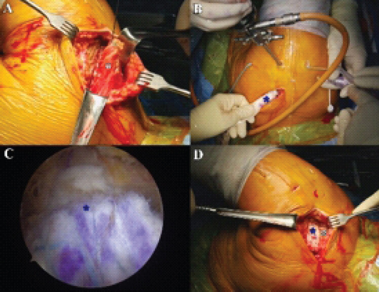
To harvest MTT, a transverse skin incision was then made over the scapular spine to expose the insertion of the MTT. The distal portion of MTT was carefully released from its insertion, while avoiding injury to the posterior part of the acromioclavicular joint (Fig. 1a). The harvested MTT was secured using a #2 free suture at each edge. The prepared graft was passed through a supraspinatus outlet to bridge the gap between the MTT and the greater tuberosity (Fig. 1b). The distal portion of the graft was secured to the supraspinatus footprint using a double-row suture-bridge configuration with the triple-loaded two medial-row anchors and three knotless lateral-row anchors (Fig. 1c). The graft was secured with the arm positioned in approximately 45° of shoulder abduction and neutral rotation. The proximal end of the graft was attached to the harvested MTT using a continuous running locking suture technique (Fig. 1d). Final arthroscopic inspection confirmed stable fixation of the reconstructed tendon without impingement throughout passive shoulder motion.
Post-operative rehabilitation
The operated shoulder was immobilized in a 45° abduction brace for 4 weeks to protect the tendon transfer during the early healing phase. During immobilization, active motion of the elbow, wrist, and hand was encouraged, and passive shoulder motion was initiated using a continuous passive motion machine as tolerated. After 4 weeks, the abduction brace was removed, and active-assisted shoulder ROM exercises were initiated, followed by progressive active ROM. Strengthening exercises were introduced at 3 months postoperatively and gradually advanced according to individual functional recovery. Heavy labor, repetitive overhead activities, and sports were prohibited until at least 6 months after surgery.
Post-operative outcomes
At a mean follow-up of 27.5 ± 2.9 months (range, 24–31 months), three of the four shoulders demonstrated substantial improvements in pain, PROMs, ROM, and muscle strength. Mean VAS pain score improved from 5.0 ± 0.8 preoperatively to 2.0 ± 1.4 at final follow-up. Likewise, mean SANE, Constant, and ASES scores improved from 47.5 ± 5.8 to 70.2 ± 14.9, 45.0 ± 5.3 to 67.0 ± 14.8, and 51.2 ± 4.9 to 73.7 ± 16.1, respectively (Table 5). Mean forward elevation increased from 102° ± 17° to 145° ± 23°, mean abduction from 80° ± 24° to 120° ± 28°, and mean external rotation at the side from 47° ± 3° to 52° ± 9°. Internal rotation improved from a mean score of 6.0 ± 1.6 to 7.0 ± 1.1 (Table 3). Post-operative shoulder strength demonstrated marked recovery in most shoulders. Mean forward elevation strength increased from 31.3 ± 5.5 N to 81.1 ± 29.7 N, while mean abduction strength improved from 28.3 ± 4.9 N to 74.7 ± 28.2 N. External rotation strength increased from 81.5 ± 19.3 N to 91.7 ± 8.8 N, and internal rotation strength improved from 93.9 ± 8.5 N to 101.0 ± 3.0 N (Table 4).
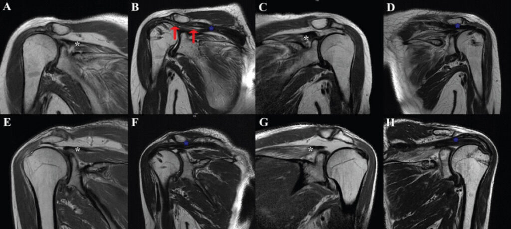
Post-operative MRI demonstrated intact healing of the transferred tendon construct in three shoulders (Fig. 2). One shoulder (Patient 1, first-operated right shoulder) demonstrated partial retear of the tendon transfer (Fig. 2b). This patient continued to experience persistent pain and limited functional improvement, with deterioration of post-operative shoulder PROM and ROM compared with the pre-operative evaluation. In contrast, the contralateral shoulder of the same patient, which underwent staged MTT transfer 4 months later, demonstrated intact tendon healing and substantial improvement in pain, PROMs, ROM, and strength. Similarly, both shoulders in Patient 2 demonstrated intact structural healing with favorable clinical recovery at final follow-up. No post-operative infection, neurologic complication, wound problem, or additional surgical procedure was required during the follow-up period.
Discussion
The most important finding of the present case report is that staged bilateral MTT transfer provided satisfactory clinical and structural outcomes in three of four shoulders with bilateral ISTs. Despite one partial retear in the first-operated shoulder of one patient, the contralateral shoulder in the same patient healed successfully and demonstrated substantial improvements in pain, PROMs, ROM, and muscle strength. Similarly, both shoulders of the second patient achieved favorable clinical recovery with intact tendon healing. These findings suggest that staged bilateral MTT transfer may be a feasible joint-preserving treatment option for carefully selected patients with bilateral ISTs.
Bilateral rotator cuff pathology is common, and many patients who initially present with unilateral symptoms subsequently develop symptomatic disease in the contralateral shoulder [14,15,17,18, 19,20,21]. Although staged bilateral arthroscopic rotator cuff repair and bilateral RTSA have previously been reported to provide satisfactory outcomes, to our knowledge, no study has described the outcomes of staged bilateral tendon transfer for irreparable rotator cuff tears [17,18,19, 20, 21]. The present report therefore expands the current clinical application of MTT transfer by demonstrating that satisfactory functional recovery can be achieved in both shoulders following staged reconstruction.
MTT transfer has recently gained attention as an anatomical tendon transfer for isolated ISTs [6,7,8]. Unlike other tendon transfers, the transferred MTT closely reproduces the native line of pull of the supraspinatus, thereby restoring the superior force couple while preserving the remaining intact rotator cuff musculature [6,7,8]. Previous biomechanical studies have demonstrated restoration of superior glenohumeral stability following MTT transfer, and recent clinical studies have reported significant improvements in pain, PROMs, ROM, and strength [6, 7, 8, 9, 10,11,13]. The favorable outcomes observed in three of the four shoulders in the present report are consistent with these previous findings and further suggest that satisfactory outcomes can be achieved even when both shoulders require staged reconstruction. An important observation from this report is that partial retear of the first-operated shoulder did not adversely influence the outcome of the second-stage procedure. Patient 1 experienced partial retear following the initial MTT transfer, resulting in limited functional improvement. Nevertheless, after undergoing MTT transfer on the contralateral shoulder, the patient achieved excellent pain relief, restoration of shoulder function, and intact tendon healing. This finding suggests that partial retear after unilateral MTT transfer may not necessarily preclude consideration of contralateral reconstruction when appropriate surgical indications remain. Instead, each shoulder should be evaluated independently based on patient symptoms, functional demands, and remaining rotator cuff integrity.
This report has several limitations. First, it describes only two patients with four treated shoulders, limiting the generalizability of the findings. Second, the follow-up period was limited to approximately two years, and longer-term structural durability remains unknown. Third, different grafts were used in different shoulders. Finally, the absence of a comparison group precludes direct comparison with alternative joint-preserving procedures or arthroplasty. Nevertheless, the rarity of bilateral staged MTT transfer makes this report valuable as the first description of its clinical and structural outcomes and may provide a foundation for future studies involving larger patient cohorts.
Conclusion
Staged bilateral MTT transfer may be a feasible joint-preserving treatment option for selected patients with bilateral ISTs. Favorable clinical and structural outcomes were achieved in three of four shoulders, with substantial improvements in pain, shoulder function, ROM, and muscle strength. Partial retear in one shoulder did not preclude successful reconstruction of the contralateral shoulder. Further studies with larger cohorts and longer follow-up are warranted to establish the reproducibility and long-term durability of bilateral MTT transfer.
Clinical Message
Staged bilateral MTT transfer can restore satisfactory shoulder function in selected patients with bilateral irreparable supraspinatus tendon tears while preserving the native glenohumeral joint.
Conflict of Interest:
Source of Support:
Nil
Consent:
The authors confirm that informed consent was obtained from the patient for publication of this article
References
- Campbell RE, Lee D, Day LM, Dixit A, Freedman KB, Tjoumakaris FP.Management of massive rotator cuff tears without arthropathy. Orthopedics 2023;46:e1-e12. [Google Scholar] | [PubMed]
- Oh JH, Park MS, Rhee SM.Treatment strategy for irreparable rotator cuff tears. Clin Orthop Surg 2018;10:119-34. [Google Scholar] | [PubMed]
- Rho JY, Kwon YS, Choi S.Current concepts and recent trends in arthroscopic treatment of large to massive rotator cuff tears: A review. Clin Shoulder Elb 2019;22:50-7. [Google Scholar] | [PubMed]
- Yoon JY, Kim PS, Jo CH.Clinical and radiological results after arthroscopic superior capsular reconstruction in patients with massive irreparable rotator cuff tears. Clin Shoulder Elb 2018;21:59-66. [Google Scholar] | [PubMed]
- Kim DH, Jung YS, Kim KR, Yoon JP.The best options in superior capsular reconstruction. Clin Shoulder Elb 2021;24:114-21. [Google Scholar] | [PubMed]
- Moroder P, Akgün D, Lacheta L, Minkus M.Middle trapezius transfer for treatment of irreparable supraspinatus tendon tears- anatomical feasibility study. J Exp Orthop 2021;8:5. [Google Scholar] | [PubMed]
- Baek G, Kim JG, Baek GR, Hui AT, McGarry MH, Baek CH.Middle trapezius tendon transfer using Achilles allograft for irreparable isolated supraspinatus tendon tears effectively restores the superior stability of the humeral head without restricting range of motion: A biomechanical study. J Shoulder Elbow Surg 2024;33:1366-76. [Google Scholar] | [PubMed]
- Kandeel AA.Rotator cuff irreparability or failure of repair (re-tear): Technical note on middle trapezius tendon transfer for reproduction of supraspinatus function. J Exp Orthop 2021;8:105. [Google Scholar] | [PubMed]
- Moroder P, Lacheta L, Danzinger V, Thiele K, Ellermann S, Akgün D.Arthroscopic middle trapezius transfer for treatment of irreparable superior rotator cuff tendon tears. Arthrosc Tech 2021;10:e581-6. [Google Scholar] | [PubMed]
- Kandeel AA.Middle trapezius tendon transfer for augmentation of in situ superior capsular reconstruction-reinforced partial rotator cuff repair: Short-term outcomes of a prospective cohort study. Orthop J Sports Med 2023;11:23259671221147537. [TPS2.1]. [Google Scholar] | [PubMed]
- Baek CH, Kim JG.Outcomes of arthroscopic-assisted middle trapezius tendon transfer for isolated irreparable supraspinatus tendon tears: Minimum 2-year follow-up. Arch Orthop Trauma Surg 2023;143:2547-56. [Google Scholar] | [PubMed]
- Kim JG, Lim C, Kim BT, Baek CH.Superior capsular reconstruction versus middle trapezius tendon transfer for isolated irreparable supraspinatus tendon tears: Static versus dynamic reconstruction. Am J Sports Med 2024;52:431-40. [Google Scholar] | [PubMed]
- Baek CH, Kim BT, Kim JG, Kim SJ.Short-term clinical results of middle trapezius tendon transfer with Achilles tendon allograft for irreparable supraspinatus tears. J Shoulder Elbow Surg 2025;34:1407-16. [Google Scholar] | [PubMed]
- Smith KM, Clinker CE, Cutshall ZA, Lu CC, Joyce CD, Chalmers PN.Progression of symptomatic bilateral rotator cuff disease. JSES Int 2023;7:586-91. [Google Scholar] | [PubMed]
- Davis G, Zareef U, Perez A, Paul RW, Givner D, Erickson BJ.In patients with bilateral rotator cuff tear, staged surgery is superior to simultaneous surgery: A systematic review. Arthrosc Sports Med Rehabil 2025;7:101033. [Google Scholar] | [PubMed]
- Minagawa H, Yamamoto N, Abe H, Fukuda M, Seki N, Kikuchi K.Prevalence of symptomatic and asymptomatic rotator cuff tears in the general population: From mass-screening in one village. J Orthop 2013;10:8-12. [Google Scholar] | [PubMed]
- Fliegel BE, Hall A, Patel A, Lee D, Cohen SB, Freedman K.Patient preferences in short-interval staged bilateral rotator cuff repair. Orthopedics 2023;46:e347-52. [Google Scholar] | [PubMed]
- Aleem AW, Syed UA, Wascher J, Zoga AC, Close K, Abboud JA.Functional outcomes after bilateral arthroscopic rotator cuff repair. J Shoulder Elbow Surg 2016;25:1668-73. [Google Scholar] | [PubMed]
- Kim HJ, Kee YM, Kim JY, Rhee YG.Safe time frame of staged bilateral arthroscopic rotator cuff repair. J Shoulder Elbow Surg 2019;28:1707-15. [Google Scholar] | [PubMed]
- Polascik BA, Chopra A, Hurley ET, Levin JM, Rodriguez K, Stauffer TP.Outcomes after bilateral shoulder arthroplasty: A systematic review. J Shoulder Elbow Surg 2023;32:861-71. [Google Scholar] | [PubMed]
- Daher M, Fares MY, Koa J, Singh J, Abboud J.Bilateral reverse shoulder arthroplasty versus bilateral anatomic shoulder arthroplasty: A meta-analysis and systematic review. Clin Shoulder Elb 2024;27:196-202. [Google Scholar] | [PubMed]
- Goutallier D, Postel JM, Bernageau J, Lavau L, Voisin MC.Fatty muscle degeneration in cuff ruptures. Pre- and post-operative evaluation by CT scan. Clin Orthop Relat Res 1994;304:78-83. [Google Scholar] | [PubMed]
© 2026 Journal of Orthopaedic Case Reports - Published by Indian Orthopaedic Research Group
About the Authors
-

Dr. Chang Hee Baek
-

Dr. Bo Taek Kim
-

Dr. Jung Gon Kim
-

Dr. Chaemoon Lim
Related Articles in Journal of Orthopaedic Case Reports
 January 1, 2026 Dual Reconstruction, Combined Anterior Latissimus Dorsi with Teres Major and Middle Trapezius Tendon Transfer, Reduces Anterior Superior Humeral Subluxation: Case Report
 October 1, 2026 Posterior Fibular Entrapment in a Bosworth Fracture-Dislocation: A Case Report and Review of Diagnostic and Operative Considerations
 October 1, 2026 Recurrent Ventral Scapular Osteochondroma Presenting with Pseudo-winging in an Adolescent: A Case Report and Follow-up of a Previously Published Case
 October 1, 2026 Development of a Non-ossifying Fibroma in the Same Femur 6 Years after Limb-Salvage Surgery for Osteosarcoma: A Case Report